
Case Report
Austin J Otolaryngol. 2015;2(1): 1026.
Prolonged Survival in a Nasopharyngeal Carcinoma (NPC) Patient with Metastatic Disease: A Case Report
De Meulenaere A1, Deron P2, Duprez F3, Ferdinande L4, Verbeke L5, De Vuyst M6 and Rottey S1*
1Department of Medical Oncology, Ghent University Hospital, Belgium
2Department of Head and Neck Surgery, Ghent University Hospital, Belgium
3Department of Radiotherapy, Ghent University Hospital, Belgium
4Department of Pathology, Ghent University Hospital, Belgium
5Department of Radiotherapy, OLV Hospital, Belgium
6Department of Pathology, ASZ Hospital, Belgium
*Corresponding author: Rottey Sylvie, Department of Medical Oncology, Ghent University Hospital, De Pintelaan 185, 9000 Ghent, Belgium
Received: December 13, 2014; Accepted: January 17, 2015; Published: January 19, 2015
Abstract
Nasopharyngeal carcinoma (NPC) arises from the epithelial lining of the nasopharynx. Stage I-IIa disease can be cured by radiotherapy alone. Locally or locoregionally advanced disease (stage III, IVA, and IVB) is treated with a combination of radiotherapy with concurrent chemotherapy (CRT). This treatment modality provides 5-year overall survival rates of 50–70%.
We present the case of a 28-year old woman; diagnosed with UNPC cT4cN2M0, stage IVa. After primary cisplatin-based CRT with curative intent, there was a complete locoregional remission. However, within six months after completion of primary treatment, bone metastases became apparent. The patient was treated repeatedly with radiotherapy at metastatic sites to avoid additional chemotherapy (renal insufficiency present). Eventually, the patient died at the age of 35, seven years after primary diagnosis.
Keywords: Nasopharyngeal carcinoma (NPC); Metastases; Survival
Introduction
Nasopharyngeal carcinoma (NPC) arises from the epithelial lining of the nasopharynx. It represents 75-95% of the malignancies originating from the nasopharynx. The incidence and prevalence of NPC is high in South East Asia, while it is lower in Europe. In Belgium, approximately 60 new diagnoses are made every year [1].
From pathological point of view, three types of NPC are described: keratinising squamous cell carcinoma (Type I), nonkeratinising differentiated carcinoma (Type II) and non-keratinising undifferentiated carcinoma (Type III) [2]. Epstein- Barr virus (EBV) is postulated to play a key-role in the pathophysiology of type II and III NPC. In addition, genetics and environmental factors such as tobacco use, alcohol consumption and intake of salt-preserved food have been identified as possible risk factors [3].
Diagnosis of NPC is usually based on symptoms related to tumor growth into surrounding structures. Endoscopic examination is mandatory to confirm diagnosis. Staging is based on physical examination combined with imaging studies, i.e. computed tomography (CT) scan or magnetic resonance imaging (MRI) [4].
NPC is highly curable in early stage disease. The treatment of choice for stage I-IIa NPC is radiotherapy with 5-year overall survival rates up to 95%. Therapeutic options available for more advanced disease include concurrent chemoradiotherapy (CRT) associated with (neo) adjuvant chemotherapy. Metastatic disease is known to be chemosensitive, but remains essentially incurable [5].
In case of treatment with curative intent, rigorous patient followup is required, with a primarily clinical focus. Imaging by CT or MRI might be useful in follow-up of patients diagnosed with T3 or T4 NPC [4].
Case Presentation
In August 2006, a 28-year old woman was admitted to the department of Head and Neck Surgery at the Ghent University Hospital. A history of sinusitis, tracheitis and an eye correcting surgery at the age of three was withheld. The patient did not smoke or used alcohol. At the moment of admission she took ibuprofen (400mg a day) and paracetamol (1000mg 1 to 4 a day). She was suffering from nasal obstruction and intermittent epistaxis since a few weeks. Moreover, the patient mentioned a cervical nodular mass on the right (region II), diplopia, cervical discomfort and fatigue. She also noticed a weight loss of 15 kilogram over the past six months. Except from the nodular mass, clinical investigation was normal. Endoscopic inspection showed a large nasopharyngeal mass. On histological examination of the biopsies, diagnosis of undifferentiated NPC (UNPC) was made (Figure 1). In-situ-hybridization for EBV-DNA was strongly positive (Figure 2). Additionally, a CT scan of the neck and the chest and MRI of the brain were performed. No arguments for metastatic disease were retained. CT scan did show a large tumor mass extending to the sphenoidal sinus/ the pterygopalatine fossa with bilateral multifocal pathological lymph nodes. Based on these findings, the patient was diagnosed with UNPC cT4cN2M0; stage IVa according to the Union for International Cancer Control (UICC) [6]. During the multidisciplinary team meeting, cisplatinum based CRT followed by combination chemotherapy, was proposed as the best treatment option for this patient.
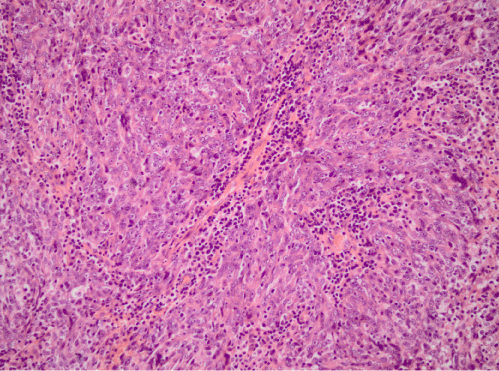
    Figure 1:  Undifferentiated nasopharyngeal carcinoma.
Neoplastic cell nests with syncytial growth pattern and interspersed nonneoplastic
lymphoid component. Tumor cells have round to oval nuclei,
prominent eosinophilic nucleoli and scant cytoplasm. (Hematoxylin and eosin
stain [HE], magnification x200)
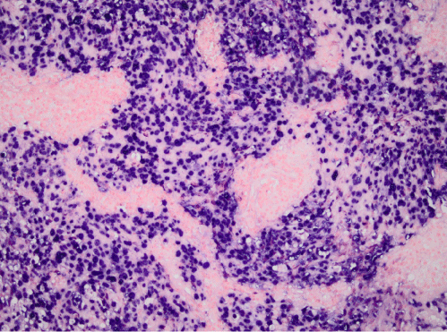
    Figure 2:  In-situ-hybridization for EBV-DNA.
EBV chromogenic in-situ-hybridization reveals the presence of EBV DNA in
the tumor cells (blue staining). (magnification x200)
Radiotherapy (64Gy in 2 Gy fractions) with concomitant threeweekly cisplatinum was administrated in a curative setting. The patient received three cycles of cisplatinum 100 mg/m² during radiotherapy. In the adjuvant setting, only one cycle (instead of the planned three) of cisplatinum+5-fluorouracil could be administrated due to severe side-effects (grade III nausea and vomiting, grade IV dysphagia, grade III erythema in the radiation field, neutropenic fever with pseudomonas sepsis and grade III renal insufficiency). Dysphagia and vomiting persisted, with requirement of total parenteral nutrition (TPN). TPN needed to be continued on a chronic basis. Complete clinical remission was obtained.
Within six months after the last administration of chemotherapy, the patient noticed some articular discomfort on the level of the left knee, irradiating towards the ipsilateral hip. Bone scintigraphy highlighted multifocal metastatic lesions in the left femur, the seventh rib and the facial skeleton. Pathological evaluation (open biopsy at the left femur) confirmed the presence of metastatic bone lesions of the NPC. Surgical fixation was applied to stabilize the left hip after which radiotherapy was performed on the hip (36Gy throughout 12 fractions). Given the presence of renal impairment (GFR 49ml/ min/1.73m²) secondary to prior cisplatinum chemotherapy and a Karnofsky score of 60% at that time, chemotherapy was not initiated.
Eighteen months later, CT and PET-CT scan revealed lymphonodular bulging with invasion of the left psoas muscle for which the patient was treated with local radiotherapy (50Gy in 2 Gy fractions), resulting in adequate pain management.
Another six months later the patient felt pain of the left shoulder. Radiological evaluation could not show metastatic disease so physiotherapy and analgesics were recommended. CT scan of chest and abdomen at that time revealed progression of the metastatic disease with growing lymph nodes in the lower neck. Still, because the patient did not experience any discomfort other than the above mentioned pain that was controlled with the proposed interventions, watchful waiting was preferred. However, the shoulder pain persisted and seemed to be caused by lymphonodular enlargement (cervical region III) compressing the brachial plexus for which local palliative antalgic radiotherapy, (30Gy in 2 Gy fractions) was performed.
The patient’s condition remained stable over a period of two years. In April 2013, 71 months after diagnosis of metastatic disease, her condition deteriorated significantly. She felt exhausted and showed progressive weight loss. CT imaging showed diffuse metastatic disease: two hypodense lesions in the right liver, two micronodular lesions in the lower lobe of the right lung, multiple pathological lymph nodes in the mediastinum and the mesothelium, pleural effusion on both sides, pericardial effusion, a limited amount of fluid in the perineum and a solitary lesion on the left adrenal gland.
The option of palliative chemotherapy was discussed with the patient again. Because of the renal impairment and the poor general condition of the patient and according to her own preference, no chemotherapy was initiated. Eventually, the patient died at the age of 35 due to the disease progression.
An overview of the patient’s case is summarized in Table 1.
![]()
Date
Aug 17th, 2006
Sept 8th, 2006
-
Sept 11th, 2006
Sept 28th, 2006
-
Febr 9th, 2007
May 3th, 2007
Aug 1st, 2007
Nov 20th,� 2009
Aug 23rd, 2010
March 18th,� 2011
April 2nd, 2013
June 18th, 2013
Event
Diagnosis NPC
Nausea
Vomitus
AKI
- nausea+vomitus
- mucositis gr. IV
- AKI
- neutropenic fever
- anemia
- hyponatri�mia
Articular discomfort
Diagnosis bone metastasis:
-prox femur
-7th rib
-facial massif
Enlarged abdominal lymphnodes
Progressive disease
Symptomatic supra-clavicular mass
Progressive disease
Decease
-CKI
-anemia
Intervention
Cisplatinum-based
Chemo-
radiotherapy
Supportive care
- anti-emetics
- TPN
- morphine, IV
- AB
- blood Tx
- hypotonic fluids
Surgery left femur
+ RT 36Gy over 12 fractions
RT 50Gy over 25 fractions
Best supportive Care
RT 30Gy over 15 fractions
Best supportive Care
Table 1: Laboratory values at hospital admission.
Discussion
The incidence of NPC is usually two to three times higher in male patients. Also, incidence increases with age in most low-risk populations [2,7]. Therefore, our patient does not match the typical profile one would expect from a patient diagnosed with NPC. EBV infection might only partially explain the development of this rare type of cancer. Therefore it is assumed underlying genetic mechanisms play an important role in the pathophysiology of NPC [7].
The overall European population taken into count, the relative survival for NPC is 76% at one year and 50% at five years [8,9]. Age has a significant impact on survival. Survival at five years is 72% for those patients ranging from 15 to 45 years and 36% in the eldest group of patients (65-74 years). Metastatic disease is an important cause of death in NPC and is seen more often in patients with NPC than in other patients diagnosed with malignancies arising in the head and neck region [10,11].
Our patient survived 71 months from the time metastatic disease was diagnosed and almost seven years after primary diagnosis. In this context, several factors have been identified as predictors for longtime survival in case of metastasis: initial diagnosis before the age of 40, absence of local recurrence and complete response to aggressive multimodal therapy. Short disease-free survival after initial therapy and diffuse metastatic disease on the other hand were inversely correlated with long-time survival. Also, performance status should be taken into account [10,12]. Our patient maintained a Karnofsky performance status of 60 over a long period of time. We suggest that continuous administration of TPN after the first therapeutic intervention may have positively influenced patient’s outcome and contributed to prolonged survival [13].
Another unusual feature noted in this case was the fact that our patient did not receive chemotherapy for recurrent metastatic disease. NPC is characterized by its high chemosensitivity [12]. However, clinical burden was small at the time metastasis was diagnosed. Moreover, the patient suffered from renal insufficiency secondary to prior cisplatinum chemotherapy making further application of chemotherapy more complicated. Therefore, it was decided not to start palliative chemotherapy but the patient was treated repeatedly with radiotherapy at metastatic sites to control symptoms. In this context it is important to mention that ionizing radiation not only offers local benefits but can involve systemic immune activation inducing out-of-field antitumor reactions (= “abscopal effect”) [14,15]. However difficult to address in this specific case, this abscopal effect might have resulted in regression of lesions that are outside the radiation field. We can conclude that radiotherapy, together with best supportive care, analgesics and TPN, kept quality of life as high as possible and may have influenced survival in this patient.
Conclusion
We presented the case of a 28-year old woman with stage IVa UNPC, initially treated with curative intent with CRT. Metastatic disease was diagnosed only six months after treatment. Although she never received systemic treatment for her metastases, she survived 71 months with only radiotherapy and antalgic medication.
References
- https://www.kankerregister.org/default.aspx?url=Statistieken_tabellen_jaarbasis.
- Barnes L, Eveson J, Reichart P, Sidransky D. Pathology and genetics of head and neck tumors. World Health Organization Classification of Tumours. 2005.
- Spano JP. Nasopharyngeal carcinoma: an update. European Journal of Cancer. 2003; 39: 2121-2135.
- Chan ATC, Grégoire V, Lefebvre JL, Licitra L, Hui EP, Leung SF, et al. Nasopharyngeal cancer: EHNS–ESMO–ESTRO Clinical Practice Guidelines for diagnosis, treatment and follow-up. Annals of Oncology. 2013; 23: 83-85.
- Rottey S, Madani I, Deron P, Van Belle S. Modern treatment for nasopharyngeal carcinoma: current status and prospects. Curr Opin Oncol. 2011; 23: 254-258.
- Jemal A, Bray F, Center MM, Ferlay J, Ward E, Forman D. Global cancer statistics. CA Cancer J Clin. 2011; 61: 69-90.
- Chang ET, Adami HO. The enigmatic epidemiology of nasopharyngeal carcinoma. Cancer epidemiology, biomarkers & prevention: a publication of the American Association for Cancer Research, cosponsored by the American Society of Preventive Oncology. 2006; 15: 1765-1777.
- Ferlay J, Shin HR, Bray F, Forman D, Mathers C, Parkin DM. Estimates of worldwide burden of cancer in 2008: GLOBOCAN 2008. Int J Cancer. 2010; 127: 2893-2917.
- Curado M, Edwards B, Shin H, Storm H, Ferlay J. Cancer incidence in five continents. IARC scientific publications. 2007; 160.
- Teo PM, Kwan WH, Lee WY, Leung SF, Johnson PJ. Prognosticators determining survival subsequent to distant metastasis from nasopharyngeal carcinoma. Cancer. 1996; 77: 2423-2431.
- Lee AW, Poon YF, Foo W, Law SC, Cheung FK, Chan DK, et al. Retrospective analysis of 5037 patients with nasopharyngeal carcinoma treated during 1976-1985: overall survival and patterns of failure. International journal of radiation oncology, biology, physics. 1992; 23: 261-270.
- Ma BB, Hui EP, Chan AT. Systemic approach to improving treatment outcome in nasopharyngeal carcinoma: current and future directions. Cancer Sci. 2008; 99: 1311-1318.
- Prevost V, Grach MC. Nutritional support and quality of life in cancer patients undergoing palliative care. Eur J Cancer Care (Engl). 2012; 21: 581-590.
- Gaipl US, Multhoff G, Scheithauer H, Lauber K, Hehlgans S, Frey B, et al. Kill and spread the word: stimulation of antitumor immune responses in the context of radiotherapy. Immunotherapy. 2014; 6: 597-610.
- Frey B, Rubner Y, Wunderlich R, Weiss EM, Pockley AG, Fietkau R, et al. Induction of abscopal anti-tumor immunity and immunogenic tumor cell death by ionizing irradiation - implications for cancer therapies. Curr Med Chem. 2012; 19: 1751-1764.