Case Report
Austin J Otolaryngol. 2015;2(2): 1030.
Middle Turbinate Osteoma Causing Migraine like Symptoms
Ciftci Z1, Deniz M1, Celik O2*, Oznur M3 and Gultekin E1
1Department of Otorhinolaryngology, Namik Kemal University, Turkey
2Department of Otorhinolaryngology, Maltepe University, Turkey
3Department of Pathology, Namik Kemal University, Turkey
*Corresponding author: Çelik Oner, Department of Otorhinolaryngology, Maltepe University, Feyzullah cad. No 39 34843, Maltepe/Ýstanbul, Turkey
Received: November 19, 2015; Accepted: February 14, 2015; Published: February 17, 2015
Abstract
Objective: To report an extremely rare case of an osteoma localized in a pneumatized middle turbinate and to emphasize the importance of a detailed otorhinolaryngological examination and imaging in headache patients, even in the presence of a previously diagnosed headache etiology.
Features of the Case: A 50-year-old female patient presenting with symptoms of progressive headache on one side of the head, facial pain and nasal obstruction and attended to neurology department with a suspicion of migraine. Cranial MRI was performed with a report of normal cranial investigation and a mass in the middle turbinate. Therefore, patient was referred to otorhinolaryngology clinic.
Treatment and Prognosis: The patient underwent endoscopic excision of the tumor and histopathological diagnosis confirmed the diagnosis of ‘osteoma’. She reported that she quitted taking analgesic medications following the operation.
Conclusion: Sinonasal osteomas should be taken into consideration when dealing with a patient presenting with headache. Detailed otorhinolaryngological examination along with imaging in suspected cases may prevent misdiagnoses and unnecessary use of medications.
Keywords: Osteoma; Migraine; Misdiagnosis; Pneumatized middle turbinate
Introduction
Pneumatization of the middle turbinates is a common anatomical variation in paranasal region and an important etiology of headache. In symptomatic patients, they trigger pain response either by impeding the drainage and ventilation of other sinuses or creating contact points in the nasal cavity [1].
Presence of an osteoma in the nasal cavity is a rarely encountered situation. Its incidence was reported to be as low as 0.6% in the literature [2]. Infrequently, the osteomas may be confined to middle turbinates. They may either be incidentally found in asymptomatic patients or discovered when they become symptomatic. They may cause symptoms like headache, facial and orbital pain and nasal obstruction [3]. Diagnosing an osteoma localized within a cavity in the paranasal region is a real challenge for the physician because conventional physical examination methods may not be helpful in establishing a diagnosis. Furthermore, the pain caused by the space occupying lesion in the paranasal region – also referred to as ‘sinus headache’ – usually may overlap or trigger other primary headache syndromes such as migraine [4].
Co-existence of the abovementioned pathologies, in other words ‘presence of an osteoma in a pneumatized middle turbinate’, is an extremely rare condition. In this article, the symptoms and findings of a 50-year-old female patient with bilateral pneumatized middle turbinates and an osteoma in the left middle turbinate are presented. The diagnostic and therapeutic options of the paranasal sinus osteomas are also discussed under the scope of the literature with a special emphasis on the importance of a detailed endoscopic examination and imaging in approach to patients presenting with headache.
Case Presentation
A 50 year old woman admitted to neurology department with symptoms of progressive headache on one side of head, facial pain and nasal obstruction. At that time cranial magnetic resonance imaging (MRI) was performed with a report of normal cranial investigation and a mass in the middle turbinate. Therefore, patient was referred to otorhinolaryngology clinic. Her medical history was insignificant and she reported that she did not attend to any ear nose throat surgeon before. Physical examination of the patient was normal except a slightly deviated septum on the right side. Nasal endoscopy revealed bilateral hypertrophic middle turbinates. No further pathology was observed. Computed tomography (CT) scan of the paranasal sinuses was taken and bilateral pneumatized middle turbinates and a hyperdense enlargement in the left middle turbinate were observed (Figure 1). The lesion was reported to be a possible osteoma. Aeration patterns and structures of the other regions were reported to be normal.

Figure 1: Hyperdense solid mass in the pneumatized middle turbinate
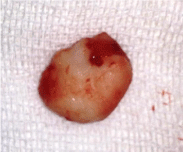
Under general anesthesia, the lateral portions of the pneumatized middle turbinates were excised using 0-degree rod telescopes. The tumor in the middle turbinate was removed using gentle blunt and sharp dissection. Care was given to keep the middle turbinate stable during the procedure to avoid any cranial base problems. The mass was totally excised. And macroscopic examination revealed a solid mass with a smooth surface (Figure 2). It measured 12x7.5 mm and histopathological diagnosis confirmed the diagnosis of ‘osteoma’ (Figure 3).

Figure 2: Macroscopic appearance of the osteoma measuring 12x7.5 mm.

    Figure 3:  The diagnosis of osteoma was confirmed histologically (x100
magnification).
In the first postoperative day her nasal packs were removed and no complications were observed. In follow up examinations 1 month, 3 months and 6 months later, no recurrence was observed. No complaint was reported by the patient during the follow up period. She reported that she quitted taking analgesics for headache following the operation.
Discussion
Osteomas are benign and slowly growing lesions with a male predilection. They may occur in any bone but usually the appendicular skeleton is affected. Although less frequent, bones of face and calvarium may also be involved. The incidence of paranasal sinus osteomas was reported to be 3% [5]. The most affected sinus was reported to be the frontal sinus followed by ethmoidal, maxillary and less commonly sphenoidal sinuses. Osteomas of the paranasal region are usually asymptomatic and found incidentally. The patients are usually affected between 2nd and 5th decades [6].
The turbinates, although rare, were reported to be affected by osteomas. Literature review revealed that Ishimaru [7], Viswanatha [8], Mesolella [2] and Kumar [9] reported superior and inferior turbinate osteomas. Infrequently, the tumor may also be localized in the middle turbinate. The literature review revealed that only 8 cases of osteoma in the middle turbinate were reported [3,8,10,11]. Moreover, to our knowledge ‘presence of an osteoma in a pneumatized middle turbinate’ was only reported in one case so far by Migirov et al. [12]. They reported that, their patients suffering from headache refused surgical treatment and they could not delineate the exact origin of the headache.
Cases that were previously diagnosed with another disease, such as migraine, may create a confusing clinical picture, as observed in our case. In this confusion, it may be hard to find out the concealed pathology. The medical history of our patient harbored an interesting and noteworthy detail. Assuming that she had migraine headache, the patient did not attend to an otorhinolaryngology clinic despite her progressive headache. In the literature there still exists an ongoing debate to scan or not to scan migraine patients. It was proposed that ‘a positive history of migraine does not obviate the need for a thorough ENT workup and possibly CT scanning’ [13]. The case presented in this report also supports this opinion. Index of suspicion should be heightened while approaching such patients and in suspected cases, scanning should be performed to find out another or maybe the sole underlying pathology. The pain perceived by the patient was thought to arise from irritation of the trigeminal nerve secondary to presence of contact points between septum and middle turbinate.
Many theories were proposed to explain the pathogenesis of paranasal sinus osteomas, including embryological, traumatic or infectious theories [14]. The osteoma in our patient was localized in the middle turbinate but not in the junction of embryonic membranous frontal bones and cartilaginous ethmoid bones as proposed in embryological theory. She did not have a history of either trauma or chronic sinusitis. The only remarkable finding was presence of bilateral pneumatized turbinates. In the literature, no association was reported between the pathogenesis of osteoma formation and middle turbinate pneumatization. However, although it is hard to allege such an association, the effects of pneumatization on a normally non-pneumatized bone may be questioned.
The treatment strategies for paranasal sinus osteomas should be planned according to the patient’s symptoms and localization of the neoplasm. For asymptomatic patients with relatively small lesions, a conservative approach and follow up of the patient at regular intervals were recommended [15]. Another interesting fact about osteomas is the increasing number of reported cases in the literature. In our opinion, as the nasal endoscopic examination and imaging methods become more accessible, the number of reported cases may even further increase.
Surgical excision is warranted in symptomatic patients and in the presence of rapidly growing lesions. A lesion should be surgically excised if it is localized adjacent to nasofrontal duct or orbita and if it extends beyond the boundaries of the sinus. In cases presenting with chronic sinusitis or signs of chronic mucosal inflammation, facial deformities, intracranial complications and headache of unexplained origin, surgical excision of the tumor is suggested [16]. Although the growth rate of osteoma in our case could not be assessed, the presence of progressive headache refractory to medical treatment led us to perform a surgical intervention.
External, endoscopic and combined approaches may be chosen for surgical excision of the tumor. The type of the procedure is defined by the localization of the tumor and the experience of the surgeon. Surgical approaches are divided into external, endoscopic drill-out, and combined endoscopic and external procedures. External procedures, including lateral rhinotomy and osteoplastic flap technique, were commonly used to treat paranasal osteomas. Endoscopic and less invasive techniques have replaced the classical ones within the last three decades and proved to be successful [17]. The tumor found in our case was amenable to endoscopic resection so we used endoscopic approach for surgery.
Conclusion
In conclusion, sinonasal osteomas should be taken into consideration when dealing with a patient presenting with headache. Detailed otorhinolaryngological examination and use of rigid telescopes for visualization of the nasal cavity are mandatory. Imaging should be performed when clinical suspicion arises. This approach may prevent misdiagnoses and unnecessary use of medications for treatable pathologies. Establishing the diagnosis earlier may also prevent troublesome surgeries and possible complications.
References
- Harrison L, Jones NS. Intranasal contact points as a cause of facial pain or headache: a systematic review. Clin Otolaryngol. 2013; 38: 8-22.
- Mesolella M, Galli V, Testa D. Inferior turbinate osteoma: a rare cause of nasal obstruction. Otolaryngol Head Neck Surg. 2005; 133: 989-991.
- Daneshi A, Jalessi M, Heshmatzade-Behzadi A. Middle turbinate osteoma. Clin Exp Otorhinolaryngol. 2010; 3: 226-228.
- Patel ZM, Kennedy DW, Setzen M, Poetker DM, DelGaudio JM. "Sinus headache": rhinogenic headache or migraine? An evidence-based guide to diagnosis and treatment. Int Forum Allergy Rhinol. 2013; 3: 221-230.
- Earwaker J. Paranasal sinus osteomas: a review of 46 cases. Skeletal Radiol. 1993; 22: 417-423.
- Eller R, Sillers M. Common fibro-osseous lesions of the paranasal sinuses. Otolaryngol Clin North Am. 2006; 39: 585-600, x.
- Ishimaru T. Superior turbinate osteoma: a case report. Auris Nasus Larynx. 2005; 32: 291-293.
- Viswanatha B. Middle turbinate osteoma. Indian J Otolaryngol Head Neck Surg. 2008; 60: 266-268.
- Kumar KK, Sumathi V, Sundhari V. Inferior Turbinate osteoma - rare cause of nasal obstruction. Indian J Otolaryngol Head Neck Surg. 2010; 62: 332-334.
- Kutluhan A, Salviz M, Bozdemir K, Deger HM, Culha I, Ozveren MF. Middle turbinate osteoma extending into anterior cranial fossa. Auris Nasus Larynx. 2009; 36: 702-704.
- Yadav SP, Gulia JS, Hooda A, Khaowas AK. Giant osteoma of the middle turbinate: a case report. Ear Nose Throat J. 2013; 92: E10-12.
- Migirov L, Drendel M, Talmi YP. Osteoma in an aerated middle nasal turbinate. Isr Med Assoc J. 2009; 11: 120.
- Mehle ME, Kremer PS. Sinus CT scan findings in "sinus headache" migraineurs. Headache. 2008; 48: 67-71.
- Smith ME, Calcaterra TC. Frontal sinus osteoma. Ann Otol Rhinol Laryngol. 1989; 98: 896-900.
- Boffano P, Roccia F, Campisi P, Gallesio C. Review of 43 osteomas of the craniomaxillofacial region. J Oral Maxillofac Surg. 2012; 70: 1093-1095.
- Strek P, Zagolski O, Wywial‚ A, Sacha E, Pasowicz M. Osteoma of the sphenoid sinus. B-ENT. 2005; 1: 39-41.
- Akmansu H, Eryilmaz A, Dagli M, Korkmaz H. Endoscopic removal of paranasal sinus osteoma: a case report. J Oral Maxillofac Surg. 2002; 60: 230-232.