Case Report
Austin J Otolaryngol. 2015;2(3): 1033.
Retro-auricular Approach in a Middle Ear Drop Weld. An Unusual Approach to an Unusual Injury
Canale A, Arciello F, Dagna F* and Albera R
ENT Division, University of Turin, Italy
*Corresponding author: Federico Dagna, ENT Division, University of Turin, via Genova 3, 10100 Torino, Italy
Received: December 09, 2014; Accepted: February 24, 2015; Published: February 26, 2015
Abstract
Objective: To describe a rare case of middle ear foreign metal body and its management.
Patient: A 47 years old male welder presenting to Emergency Department with left othalgia and mild hearing impairment after a metallic spark has entered his ear. The patient underwent clinical evaluation, pure-tone audiometry and HRCT scan, that showed inferior eardrum perforation with purulent discharge, left moderate mixed type hearing loss and the presence of a foreign body in the protympanum near the carotis.
Results: After one month of local and systemic therapy the patient underwent surgery although his ear was still discharging. The metallic spark was removed with a retro-auricular approach and underlay myringoplasty performed. After 12 weeks from surgery the eardrum was intact and air-bone gap partially closed.
Conclusion: Time and technique of surgery are the key-factors and early single-step surgical intervention should be the treatment of choice in order to get better results. Using earplugs must be recommended in order to avoid this kind of accidents.
Keywords: Middle ear; Myringoplasty; Foreign bodies
Introduction
Foreign bodies in the ear are common in ENT, especially in urgencies, but they are almost exclusively located in the external auditory meatus and they rarely affect the middle ear. We here present a case of a male welder with a foreign metallic body localized in the middle ear, result of an occupational accident, treated with a retroauricular approach in order to remove the spark and to reconstruct the eardrum in a single surgical intervention.
Case Presentation
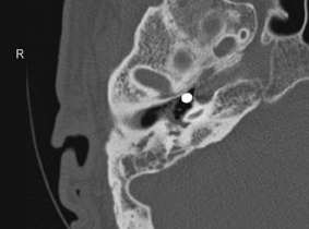
A 47 year-old man was referred to our ENT Unit by the Emergency Department presenting left otalgia associated with mild hearing impairment. Under a more detailed interview, the patient referred the use of electric welding and the eventuality of an accident with a metal drop falling in his left ear. Inspection showed a swollen external ear canal (EAC), filled with a yellowish purulent discharge and the eardrum was not clearly visible. Since micro-otoscopy was not feasible due to secretions and oedema, the patient was treated both with oral and local antibiotics and corticosteroids. CT scan and pure-tone audiometry were prescribed. One week later, under a follow-up visit, the patient referred persistence of pain, hearing loss and sense of “fullness”. Othorrea, initially ameliorated, relapsed, however the EAC was less oedematous. Micro-otoscopy, now possible, revealed an inferior perforation of the tympanic membrane. Pure-tone audiometry showed left moderate mixed hearing loss with a sensorineural drop at the high frequencies. Mean air (AC) and bone conduction (BC) thresholds at 0.5-1-2-4 kHz were respectively 67.5 dB and 37.5 dB. The CT scan highlighted a metallic foreign body in the middle ear cavity (Figure 1-2). Ossicular chain was otherwise normal.

Figure 1: Axial CT scan.

Figure 1: Coronal CT scan.
In prevision of surgery the patient underwent topical medications twice per week in order to dry his ear up. Nevertheless after one month patient’s left ear was still discharging. A surgical intervention was then performed: we chose a retro-auricular approach to remove the foreign body, whose position was so deeply pro-tympanic to be unreachable for a more traditional transcanal technique. Also, a retro- auricular approach allowed us to perform underlay myringoplasty in the same surgical momentum.
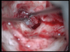
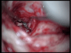
Under general anaesthesia and after skin incision, we harvested a graft from the temporal muscle. Right after, with a retro-auricular approach we reached the meatus: the eardrum was thickened and an inferior perforation evident. As seen in the CT scan, the foreign body was in the protympanum, surrounded by granulation tissue (Figure 3-4): once removed, a routine underlay myringoplasty was performed with the graft previously harvested. Healing process took more time than usual (about 8 weeks), however 12 weeks after surgery the patient’s eardrum was still intact and in place. Postoperative pure-tone audiometry showed a partial air-bone gap closure: mean AC and BC thresholds at 0.5-1-2-4 kHz were 53.75 dB and 37.5 dB respectively.

Figure 3: Middle ear granulation tissue enclosing ossicles.

Figure 4: Removing the foreign body.
The patient gave his informed consent prior to the publication of this report.
Discussion
This case was an occasion for us to review the literature about weld spark in the middle ear, though very few cases are reported and in even fewer a retro-auricular approach was adopted in order to remove the foreign body. As in all other cases reported in the English literature [1-8], the accident was caused by patient’s disregard for basic occupational rules. Such accidents determine tympanic perforation, hearing loss – from mild to deafness – and often facial nerve injury. In our case, the position of the foreign body near the Eustachian tube prevented injuries both to the facial nerve and the ossicles. For this same reason, we had no choice but a retroauricular approach in order to get the proper angle to reach the protympanic space [3]: this way we could perform also myringoplasty in a single stage procedure, avoiding an unnecessary second surgery.
Eventually, beside the chosen surgical approach, in the removal of a weld spark is important to remember the thermal nature of the accident: the burn scar may delay healing of the eardrum and raise the risk of recurrence of the perforation [1]. Moreover waiting for better local conditions before performing surgery is useless as othorrea is particularly resistant to therapy and it is hard to get a dry ear. For these reasons, in our opinion, time and technique of surgery are the key-factors and early single-stage surgical intervention should be the treatment of choice in order to get better results.
Last but not least transtympanic traumas in welders, even if less frequent than eyes injuries, are avoidable accidents, whose prevention is strictly connected to the use of earplugs [9]. Their use and those of other protective devices must be always firmly recommended.
References
- Keogh IJ, Portmann D. Drop weld thermal injuries to the middle ear. Rev Laryngol Otol Rhinol (Bord). 2009; 130: 317-319.
- Eleftheriadou A, Chalastras T, Kyrmizakis D, Sfetsos S, Dagalakis K, Kandiloros D. Metallic foreign body in middle ear: an unusual cause of hearing loss. Head Face Med. 2007; 3: 23.
- Ribeiro Fde A. Foreign body in the Eustachian tube: case presentation and technique used for removal. Braz J Otorhinolaryngol. 2008; 74: 137-142.
- Panosian MS, Dutcher PO Jr. Transtympanic facial nerve injury in welders. Occup Med (Lond). 1994; 44: 99-101.
- Kupisz K, Klatka J, Hryciuk-Umer E, Klonowski S. Injuries of the middle ear with a welding spark. Ann Univ Mariae Curie Sklodowska Med. 2000; 55: 401-404.
- Robertson MW, Morris DP. Welding injury to the ear: looking beyond the perforation. J Otolaryngol Head Neck Surg. 2011; 40: E15-18.
- Panosian MS, Dutcher PO Jr. Transtympanic facial nerve injury in welders. Occup Med (Lond). 1994; 44: 99-101.
- Stage J, Vinding T. Metal spark perforation of the tympanic membrane with deafness and facial paralysis. J Laryngol Otol. 1986; 100: 699-700.
- Fisher EW, Gardiner Q. Tympanic membrane injury in welders: is prevention neglected? J Soc Occup Med. 1991; 41: 86-88.