Research Article
Austin J Otolaryngol. 2015;2(3): 1036.
Long-term Outcomes for Children with Middle Ear Disease in Western Australia
Prunty SL1*, Spilsbury K2, Kadhim AL3, Semmens JB2, Coates HL1,4 and Lannigan FJ1,5
1Department of Otolaryngology, Head & Neck Surgery, Princess Margaret Hospital for Children, Australia
2Centre for Population Health Research, Curtin University of Technology, Australia
3Department of Otolaryngology, Head & Neck Surgery, St John of God Hospital Bunbury, Australia
4School of Paediatrics and Child Health, University of Western Australia, Australia
5Department of Otolaryngology, Head & Neck Surgery, University of Western Australia, Australia
*Corresponding author: Sarah L Prunty, Department of Otolaryngology, Head & Neck Surgery, Princess Margaret Hospital for Children, Roberts Road, Subiaco, 6008, WA, Australia
Received: August 30, 2014; Accepted: March 19, 2015; Published: March 21, 2015
Abstract
Introduction: Because of the tortuous nature of the EAC, a broken earplug may become impacted and result in reduced hearing along with other symptoms. Our aim is to raise awareness that impacted earplugs can be a cause of both acute and chronic hearing loss.
Materials and Methods: We have managed 15 patients presenting with hearing loss secondary to an impacted earplug in the EAC over a 3 years period. Prospectively, the information was collected on demographics, symptomatology, indication for earplug use, laterality of the affected ear, examination findings, earplug type, prior attempts to remove the earplug and the outcome of our interventions. Thirteen patients became aware of the hearing loss soon after the earplug impaction. The earplug was removed by an experienced otolaryngologist and the patients reported resolution of their symptoms. Two of the patients complaining about ongoing hearing loss affecting one ear were unaware of the fact that they had part of the earplug impacted in the external canal.
Discussion: Once a patient is found to have an earplug impaction, an experienced otolaryngologist should attempt its removal as their removal can be difficult without adequate equipment which includes microscope and fine ear instruments. The dough like consistency of these plugs means that they are difficult to remove with forceps or suction.
Conclusion: Broken earplugs forgotten in the EAC can lead to chronic hearing loss and may pose a diagnostic challenge. If suspected to have an impacted earplug referral to an otolaryngologist would be appropriate.
Keywords: External auditory canal; Silicone; Ear plugs; Microscope
Introduction
Earplugs are devices often used by patients to block the external auditory canal (EAC) to avoid annoying noise. Earplugs are also used to prevent water intrusion in the ears during swimming. They may be broadly grouped into three categories based on the material used to make it: foam plugs, silicone plugs and wax and cotton fibre plugs. Silicone earplugs can be mouldable or pre-moulded and are available in different colours. These offer a noise reduction rating (NRR) of 22 to 33, which is effective in most demanding noise situations. Because of the tortuous nature of the EAC, a broken earplug may become impacted and result in reduced hearing along with other symptoms. The general practitioners unfamiliar with silicone ear plugs may not recognize the impacted plugs in the ear canal. Similarly, an attempted removal of an impacted silicone ear plug without the use of microscope and fine ear instruments may lead to complications. So the aim of this educational article is to raise awareness among general practitioners that impacted earplugs can be a cause of both acute and chronic hearing loss. We also recommend that an experienced otolaryngologist should remove a deeply impacted earplug to minimize trauma to the EAC.
Materials and Methods
The authors have managed 15 patients presenting with hearing loss secondary to an impacted earplug in the EAC over a 3 years period (Table 1). Prospectively, the information was collected on demographics, symptomatology, indication for earplug use, laterality of the affected ear, examination findings, earplug type, prior attempts to remove the earplug and the outcome of our interventions. The data was collected and analysed using Microsoft Excel. We describe some of the cases in detail below.
    Serial No.
    Age
    Gender
    Side
    Previous attempt at plug removal
    GA needed
    Type of plug
    Inflammation of EAC
    Topical antibiotics


    24
    F
    R
    Yes
    No
    Foam
    Yes
    Yes


    25
    M
    R
    Yes
    No
    Silicone
    Yes
    Yes


    28
    M
    L
    Yes
    No
    Wax
    Yes
    Yes


    29
    F
    L
    Yes
    Yes
    Silicone
    Yes
    Yes


    34
    F
    L
    Yes
    Yes
    Silicone
    No
    No


    35
    M
    R
    Yes
    No
    Silicone
    No
    No


    56
    F
    R
    Yes
    No
    Silicone
    No
    No


    59
    F
    L
    Yes
    No
    Wax
    No
    No


    23
    M
    R
    Yes
    No
    Silicone
    No
    No


    30
    M
    R
    Yes
    No
    Silicone
    No
    No


    24
    F
    L
    Yes
    No
    Wax
    No
    No


    20
    F
    L
    No
    No
    Silicone
    Yes
    Yes


    66
    F
    R
    No
    No
    Silicone
    Yes
    Yes


    59
    F
    L
    No
    No
    Silicone
    No
    No


    13
    M
    R
    No
    No
    Wax
    No
    No
Table 1: Summary of patients with earplug impaction in the ear (EAC: External auditory canal).
Thirteen patients became aware of the hearing loss soon after the earplug impaction. Three patients also complained of slight dizziness. The earplug was removed by an experienced otolaryngologist and the patients reported resolution of their symptoms.
Two of the patients attended their GP complaining about hearing loss affecting one ear, believing some sinister cause for their problem. They were unaware of the fact that they had part of the earplug impacted in the external canal.
One young lady presented with a 2 months history of hearing loss which had failed to improve even after the use of over the counter ear drops aimed at softening the ear wax. On otoscopic examination the GP identified a red foreign body in the EAC of this patient. The impacted silicone earplug was removed under microscopic guidance leading to resolution of her hearing loss.
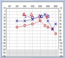
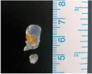
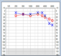
The second patient had been struggling with impaired hearing loss for 4 months; she had used ear drops to soften suspected wax and undergone syringing with no beneficial effect on hearing loss. When reviewed by her GP, the tympanic membrane was recorded as greyish, featureless and protuberant in the canal. A middle ear or mastoid pathology was suspected. This patient was routinely referred to our otology clinic where she was found to have moderate conductive hearing loss on pure tone audiometry (PTA) (Figure 1). On otoscopy, a transparent silicone earplug was identified completely occluding the EAC. The earplug was removed in the clinic under microscopic guidance (Figure 2). The patient reported improvement in her hearing, which was confirmed on repeat PTA (Figure 3).

    Figure 1:  Pure tone audiogram showing moderate conductive hearing loss
affecting the right ear.

    Figure 2:  Transparent silicone earplug removed from the external auditory
canal.

    Figure 3:  Pure tone audiogram confirming normal hearing threshold in the
right ear after earplug removal.
Discussion
Acute conductive hearing loss in adults can occur secondary to foreign body impaction in the EAC and commonly reported objects include button batteries, cotton buds, and insects in the EAC. Chronic conductive hearing loss most commonly occurs because of the middle ear pathology. We present a unique aural foreign body “earplug” which caused acute and chronic conductive hearing loss in our series of patients. The majority of impactions are related to silicone plugs which are increasingly used by patients in western hemisphere. All these cases originated in the primary care where our colleagues struggled to identify the underlying pathology but very rightly they referred these patients to our department. Some of these patients had undergone attempted removal of the impacted ear plugs without any success but led to increased patient discomfort, EAC bruising and further displacement of the ear plugs into the deeper part of the ear canal.
A retained earplug in the canal can produce an occlusive effect, leading to conductive hearing loss. This has been demonstrated in a recent study on the effects of foam earplugs inducing a mild, frequency-dependent, conductive hearing loss of 10-38 dB [1]. Unfortunately, 2 of our patients suffered for few months before the right diagnosis of ear plug impaction in the ear canal was made. Both of them reported improvement in hearing after ear plug removal and we believe that an earlier diagnosis and timely intervention would have avoided this unnecessary suffering for our patients.
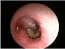
Not surprisingly, impacted material may push ear wax and other debris deeper into the EAC, creating a favourable environment for otitis externa (Figure 4).

    Figure 4:  Endoscopic view of the right ear showing wax impaction against the
tympanic membrane seen after removal of an earplug.
Most of our patients were aware of earplug impaction giving rise to hearing impairment but two of our patients did not recall having had any problems removing their earplugs. Hence our assertion that patients presenting with hearing impairment with no previous history of ear problems should undergo otoscopic examination and use of earplugs should be established during consultation as some patient might not divulge history of earplug use unless asked specifically.
The majority of earplugs in our cohort of patients were made of silicone. Silicone earplugs can be transparent or coloured (Figure 5). If the earplug is coloured, it is easier to identify during otoscopic examination compared to the transparent earplug.

    Figure 5:  Endoscopic view of a pink silicone and a blue wax earplug lodged
in the external auditory canals.
Once a patient is found to have an earplug impaction, an experienced otolaryngologist should attempt its removal as their removal can be difficult without adequate equipment. The dough like consistency of these plugs means that they are difficult to remove with forceps or suction. Most of the time it will be possible to remove the earplug in an ENT clinic setting but the patient should be counseled about the possible need for general anaesthetic [2].
Ideally, untrained personnel should not attempt removing the aural foreign body. If there have been previous failed attempts at removal patients with an aural foreign body should be referred directly to an otolaryngologist as they are more likely to be managed without general anaesthesia [3]. Another study had reported a complication rate of as high as 77% and 10% of the cases required a general anaesthetic for aural foreign body removal in patients treated by health care professionals other than otolaryngologists [4].
The patients normally report resolution of hearing impairment after earplug removal, as was the case in our cohort study. No other treatment is required following removal of the earplug. However, if
there is inflammation of the EAC or tympanic membrane, a short course of topical antibiotic/steroid treatment may be required; 6 of our patients needed topical treatment. All patients settled without any long term sequelae.
Conclusion
Earplugs are commonly used to block the EAC. The silicone and wax earplugs develop a dough-like consistency because of the increased temperature of the EAC. At times, these earplugs can become lodged in the EAC and lead to acute hearing loss along with other symptoms. Broken earplugs forgotten in the EAC can lead to chronic hearing loss and may pose a diagnostic challenge to a physician unfamiliar with the possible complication. If suspected to have an impacted earplug referral to an otolaryngologist would be appropriate.
Acknowledgement
We are grateful to our colleagues in the Audiology department for providing us with pure tone audiograms.
References
- Stuart J. Guidelines on the management of paediatric middle ear disease. Med J Aust. 1994; 160: 451.
- Daly KA, Hunter LL, Giebink GS. Chronic otitis media with effusion. Pediatr Rev. 1999; 20: 85-93.
- Bluestone CD. Modern management of otitis media. Pediatr Clin North Am. 1989; 36: 1371-1387.
- Economics A, Coates H, GlaxoSmithKline. The Cost Burden of Otitis Media in Australia. Access Economics. 2008.
- Chant K ME, Lyle D. The NSW Health Department Working Party. Guidelines on the management of paediatric middle ear disease. Med J Aust. 1993; 159: S1-S8.
- Maw AR, Herod F. Otoscopic, impedance, and audiometric findings in glue ear treated by adenoidectomy and tonsillectomy. A prospective randomised study. Lancet. 1986; 1: 1399-1402.
- Casselbrant ML. Chapter 194 Acute Otitis Media & Otitis Media with Effusion. Cummings Otolaryngology: Head & Neck Surgery 5th Edition. 2010; 3: 2761-2777.
- Maw AR, Parker A. Surgery of the tonsils and adenoids in relation to secretory otitis media in children. Acta Otolaryngol Suppl. 1988; 454: 202-207.
- Gates GA. Adenoidectomy for otitis media with effusion. Ann Otol Rhinol Laryngol Suppl. 1994; 163: 54-58.
- Kay DJ, Nelson M, Rosenfeld RM. Meta-analysis of tympanostomy tube sequelae. Otolaryngol Head Neck Surg. 2001; 124: 374-380.
- Golz A, Goldenberg D, Netzer A, Westerman LM, Westerman ST, Fradis M, et al. Cholesteatomas associated with ventilation tube insertion. Arch Otolaryngol Head Neck Surg. 1999; 125: 754-757.
- Herdman R, Wright JL. Grommets and cholesteatoma in children. J Laryngol Otol. 1988; 102: 1000-1002.
- Vlastarakos PV, Nikolopoulos TP, Korres S, Tavoulari E, Tzagaroulakis A, Ferekidis E, et al. Grommets in otitis media with effusion: the most frequent operation in children. But is it associated with significant complications? Eur J Pediatr. 2007; 166: 385-391.
- Semmens JB, Lawrence-Brown MM, Fletcher DR, Rouse IL, Holman CD. The Quality of Surgical Care Project: a model to evaluate surgical outcomes in Western Australia using population-based record linkage. Aust N Z J Surg. 1998; 68: 397-403.
- Holman CD BA, Rouse IL, Hobbs MS. Population-based linkage of health records in Western Australia: development of a health services research linked database. Aust N Z J Public Health. 1999; 23: 453-459.
- Desai SN, Kellner JD, Drummond D. Population-based, age-specific myringotomy with tympanostomy tube insertion rates in Calgary, Canada. Pediatr Infect Dis J. 2002; 21: 348-350.
- Arason VA, Sigurdsson JA, Kristinsson KG, Gudmundsson S. Tympanostomy tube placements, sociodemographic factors and parental expectations for management of acute otitis media in Iceland. Pediatr Infect Dis J. 2002; 21: 1110-1115.
- Kvaerner KJ, Nafstad P, Jaakkola JJ. Otolaryngological surgery and upper respiratory tract infections in children: an epidemiological study. Ann Otol Rhinol Laryngol. 2002; 111: 1034-1039.
- Rob MI, Westbrook JI, Taylor R, Rushworth Rl. Increased rates of ENT surgery among young children: have clinical guidelines made a difference? J Paediatr Child Health. 2004; 40: 627-632.
- Australian Social Trends. Canberra, Australia; Australian Bureau of Statistics. 2001; Report 4102.0.
- Paradise JL, Rockette HE, Colborn DK, Bernard BS, Smith CG, Kurs-Lasky M, et al. Otitis media in 2253 Pittsburgh-area infants: prevalence and risk factors during the first two years of life. Pediatrics. 1997; 99: 318-333.
- Maw AR. Chronic otitis media with effusion and adeno-tonsillectomy - a prospective randomized controlled study. Int J Pediatr Otorhinolaryngol. 1983; 6: 239-246.
- Rosenfeld RM, Culpepper L, Doyle KJ, Grundfast KM, Hoberman A, Kenna MA, et al. Clinical Practice Guidelines: Otitis Media with Effusion. Otolaryngology Head and Neck Surgery. 2004; 130: S95-118.
- Spilsbury K, Miller I, Semmens JB, Lannigan FJ. Factors associated with developing cholesteatoma: a study of 45,980 children with middle ear disease. Laryngoscope. 2010; 120: 625-630.