Case Report
Austin J Otolaryngol. 2015;2(4): 1039.
Keratosis Obturans: A Rare Cause of Facial Nerve Palsy
Ahmad Nasrat Al-Juboori*
Department of Otorhinolaryngology, Head and Neck Surgeon, Al-Iraqia University, Iraq
*Corresponding author: Ahmad Nasrat Al-Juboori, Department of Otorhinolaryngology, Head and Neck Surgeon, Ibn Sina College of Medicine, Al-Iraqia University, Baghdad, Iraq
Received: February 10, 2015; Accepted: March 31, 2015; Published: April 02, 2015
Abstract
Keratosis obturans is a rare condition characterized by the accumulation of desquamated keratin material in the bony portion of the external auditory canal (EAC). It is thought that keratosis obturans is due to abnormal epithelial migration of ear canal skin. Classically, it is reported to present with severe otalgia, conductive deafness and global widening of the canal. The frequency of keratosis obturans has been estimated as 4-5 in 1000 new otological cases. Extensive erosion of the bony meatus, with exposure of the facial nerve, has been previously reported as one of the complications, on Medline the 1st published such complication in 2006. The purpose of this case report was to presents a rare and probably the first reported keratosis obturans in Iraq that caused facial palsy.
Keywords: Keratosis obturans; External auditory canal; Cholesteatoma
Introduction
The presence of a keratin plug occluding the deep external auditory canal was first noted and documented in the 19th century. Keratosis obturans is a rare condition characterized by the accumulation of desquamated keratin material in the bony portion of the external auditory canal (EAC) [1]. The keratin squames are shed from the complete circumference of the deep ear canal forming a lamina (onion skin) arrangement. It is thought that keratosis obturans is due to abnormal epithelial migration of ear canal skin [2]. It may be associated with sinusitis and bronchiectesis. The frequency of keratosis obturans has been estimated as 4-5 in 1000 new otological cases [2]. Classically, it is reported to present with severe otalgia, conductive deafness and global widening of the canal [3]. CT scan used for the diagnosis, it shows typical ballooning of the bony external meatus [2]. It has subsequently been proposed that two different diseases can be responsible for the presence of this type of obstruction within the deep meatus, keratosis obturans and external auditory canal cholesteatoma (EACC). Extensive erosion of the bony meatus, with exposure of the facial nerve, has been previously reported as one of the complications, on Medline the 1st published such complication in 2006 [3,4]. The pain and deafness associated with keratosis obturans means that is necessary to remove it, often under general anesthesia. Canalplasty has been suggested for recurrent keratosis obturans. The purpose of this case report was to presents a rare and probably the first reported keratosis obturans that caused facial nerve palsy in Iraq.
Case Presentation
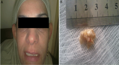
A 42-year-old female patient from Al-Fallujah city presented to us on December 2012 complaining from recurrent left sided earache for the last three months, this complaint was associated with hearing impairment on the same side there was no associated ear discharge, and she consulted ENT clinics without benefits. On examination there was facial asymmetry with flattening of the left nasolabial fold, examination of the facial nerve revealed left sided facial nerve weakness mainly on the lower half of the face (Figure 1a). Otoscopic examination showed a very large piece of keratinous material filling the left EAC which was removed completely (Figure 1b).

    Figure 1:  (a) Showed left sided facial weakness, (b) showed the mass of
keratin removed from the left ear.
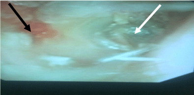
EAC further examined and magnified by endoscopic photograph of the ear, the picture showed a widening of the EAC with anteriorly located granulation tissue; the tympanic membrane was thick but intact (Figure 2). Right ear was normal, no associated sensorineural hearing loss, taste examination of the tongue, remaining ENT and chest examination were normal.

    Figure 2:  Endoscopic photograph showed thickening of the left tympanic
membrane (white arrow) and global widening of the EAC with anteriorly
located granulation tissue (black arrow).
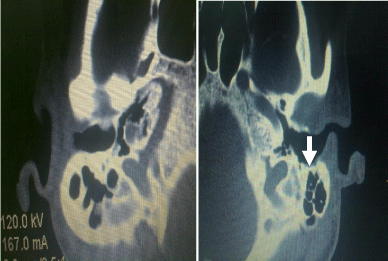
CT scan of both temporal bones showed difference in the diameter of the external auditory meatus between the right (7.4mm) and the left (10.1mm), in picture called ballooned ear canal. The case report was approved by Al-Anbar Health Directorate, and an informed consent was taken from the patient (Figure 3).

    Figure 3:  CT scan of both temporal bones showed difference in the diameter
of the external auditory meatus between (a) the right (7.4mm) and (b) the left
(10.1mm) in picture called ballooning of the ear canal (white arrow).
Discussion
Keratosis obturans is a rare condition of the bony part of external ear canal caused by hyperkeratosis of epidermis and disorders of migration process. Hyper accumulation of desquamated epidermis in the external ear canal leads to hearing loss, earache and inflammation of ear canal skin [5]. Keratosis obturans and EACC have been considered as separate entities for the last 20 years, after being regarded as variations of the same disease for at least 87 years. While both disorders are distinct, they do have some overlapping characteristics which may make it difficult to reach a definite diagnosis [6,7]. In EACC there is otorrhoea with normal hearing, the keratin in random pattern and there is localized osteitis or erosion of the external auditory canal with sequestration of the bone [8], its therapeutic principle should be a thoroughly clearance of cholesteatoma sac, degeneration tissues and sequestrum and the improvement of healing of lesions [9]. Extensive erosion of the bony meatus due to keratosis obturans with exposure of the facial nerve has been previously reported, but no case of facial nerve palsy has as yet been published, till Glynn et al. report the first published case of a unilateral facial nerve palsy secondary to neglected keratosis obturans [3]. This case presents a rare and probably the first reported keratosis obturans in Iraq that caused facial palsy. No reported case of keratosis obturans in Iraq had been published in Medline.
Acknowledgment
I want to express my acknowledgment to radiologist Dr. Ali Qadooree for his great help and comments on CT scan.
References
- Persaud R, Chatrath P, Cheesman A. Atypical keratosis obturans. J Laryngol Otol. 2003; 117: 725-727.
- Tristram HJ. Keratosis obturans and primary auditory canal cholesteatoma. Scott-Brown’s Otorhinolaryngology, Head and Neck Surgery. 7th edn. London (UK): Hodder Arnold. 2008; 3342-3344.
- Glynn F, Keogh IJ, Burns H. Neglected keratosis obturans causing facial nerve palsy. J Laryngol Otol. 2006; 120: 784-785.
- Saunders NC, Malhotra R, Biggs N, Fagan PA. Complications of keratosis obturans. J Laryngol Otol. 2006; 120: 740-744.
- Kuczkowski J, Izycka-Swieszewska E. [Keratosis obturans of the external auditory canal]. Otolaryngol Pol. 2007; 61: 675-679.
- Persaud RA, Hajioff D, Thevasagayam MS, Wareing MJ, Wright A. Keratosis obturans and external ear canal cholesteatoma: how and why we should distinguish between these conditions. Clin Otolaryngol Allied Sci. 2004; 29: 577-581.
- McCoul ED, Hanson MB. External auditory canal cholesteatoma and keratosis obturans: the role of imaging in preventing facial nerve injury. Ear Nose Throat J. 2011; 90: E1-7.
- Dubach P, Mantokoudis G, Caversaccio M. Ear canal cholesteatoma: meta-analysis of clinical characteristics with update on classification, staging and treatment. Curr Opin Otolaryngol Head Neck Surg. 2010; 18: 369-376.
- Liu X. [The preliminary analysis of the clinical characteristics and misdiagnosis of external auditory canal cholesteatoma]. Lin Chuang Er Bi Yan Hou Ke Za Zhi. 2005; 19: 792-793.