Case Report
Austin J Otolaryngol. 2015;2(4): 1040.
Submandibular Gland Sialolithiasis Presenting as Fistula in the Neck- A Case Report
Singh R1, Bhagat S1*, Bhagat R2 and Singh B1
1Department of ENT, Government Medical College, India
2Department of Pathology, Government Medical College, India
*Corresponding author: Bhagat S, Department of ENT, Government Medical College, 11-D Rajindra Hospital, Patiala, (Punjab) 147001, India
Received: February 16, 2015; Accepted: April 08, 2015; Published: April 10, 2015
Abstract
Although sialolithiasis of submandibular gland is common, the occurrence of fistula in the neck secondary to sialolithiasis is rare. Sialolithiasis should be kept as a differential diagnosis in evaluating a patient, presenting with discharging fistula in the neck. We presents a rare case of fistulae in submandibular region associated with submandibular gland stone.
Keywords: Salivary gland fistulae; Submandibular gland; Sialolithiasis
Introduction
Salivary gland fistulae are uncommon. Submandibular gland fistula may arise from infection resulting from trauma, actinomycosis, tuberculosis, syphilis, salivary calculi and malignancies [1,2]. Sialolithiasis usually arises in the Sub-mandibular gland in more than 80% of patients. It is less common in parotid glands [-10%] and rare in sublingual and minor salivary glands [3]. Long-term sialolithias with recurrent sialodochitis and/or sialoadenitis may occasionally cause spontaneous elimination of the sialoliths or opening of “new ductal course” or fistula. There are very few case reports of salivary gland sialolithiasis resulting in orocutaneous fistula [4,5]. We report a rare case of discharging fistula in the neck secondary to submandibular calculus.
Case Presentation
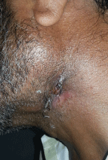
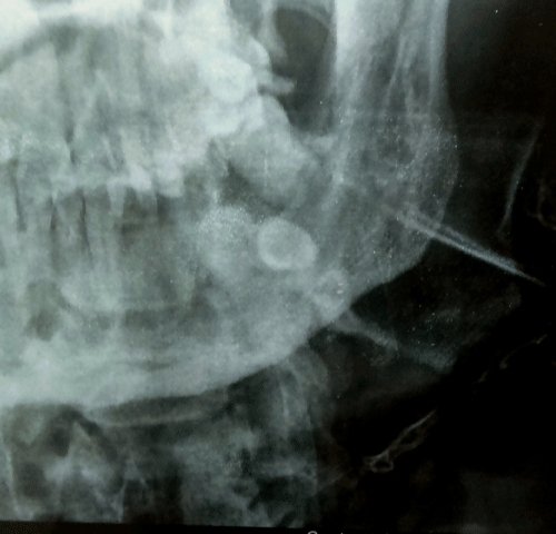
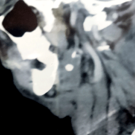
A 53 year old male presented with history of discharging sinus present on the left side of neck since 3 months. Discharge increased during meals. the patient had not undergone any surgical procedure and there was no history of trauma. On examination, fistulous opening 2cm x 1cm with irregular margins was present on left submandibular region (Figure 1). Watery discharge with pus could be expressed on pressure over the submandibular region. On bimanual examination palpation of left submandibular gland, the gland was enlarged and nontender and no stone could be palpated. Examination of oral cavity was normal. Submandibular duct opening was normally visulised. Blood investigations revealed that the patient was HCV positive. All other routine investigations were normal. Considering the clinical possibility of tuberculosis, in this case, the biopsy was taken from the margin of ulcer which revealed chronic inflammation only. X-ray fistulogram showed stone in the submandibular region (Figure 2). CECT neck also showed 1x1 cm stone in submandibular gland (Figure 3). A fistulogram was done which showed fistulous tract was commnicating with submandibular gland up to the stone. There was no communication with oral cavity. The diagnosis of sialolithiasis with cutaneous fistula communicating with submandibular tract was made excision of the submandibular gland and the fistulous tract was carried out under general anesthesia. A lot of fibrous tissue was present along the tract and adjoining submandibular gland. The tract was found to be communicating with the submandibular gland and a small stone was presented just distal to the point of communication between the fistulous tract and the submandibular gland. Biopsy was sent which showed chronic sialadenitis with sialolithiasis. Biochemistry examination of the stone revealed phosphate in stone. Postoperative recovery of the patient was uneventful.

    Figure 1:  Patient showing fistulous opening 2cm x 1cm with irregular marginst
on left submandibular region.

    Figure 2:  X-ray fistulogram shows fistula tract extending from skin to stone in
the submandibular region.

Figure 3: CECT neck showing stone in submandibular gland.
Discussion
Salivary calculi are common and are found in 1.2 % of population at necroscopy [6]. Salivary gland sinus or fistulae are usually caused by trauma, surgery and infections [7]. There are few cases reported in the literature, of salivary fistula secondary to submandibular gland calculus. Kishore kumar R V, et al. [2] evaluated 200 cases of cutaneous sinuses of cervicofacial region to identify the focus of infection and found submandibular calculus producing fistula in 1 patient (0.5%) only.
The patient of sialolithiasis usually presents with intermittent pain during meals or may remain asymptomatic when the size of stone is small. Since our patient, did not have any symptoms of sialadenitis, so the possibility of tubercular sinus was kept and biopsy was taken, which ruled out tuberculosis. The occlusal radiograph is the most reliable method of viewing the submandibular sialolith [8,9]. High Resolution Ultrasonography should be the first screening imaging tool followed by sialography, if required. CT is the mainstay of imaging in sialolithiasis while MRI is more optimal for neoplastic processes with associated invasion [10]. Later on X-ray and CT scan revealed stone in the submandibular gland.
Submandibular sialadenectomy is indicated when the gland has been damaged by recurrent infection and fibrosis or calculi have formed with in the gland [11]. In our case, submandibular gland was fibrosed, and the stone was present in the parenchyma of the gland, communicating with the fistula tract, so was removed along with the fistula tract. Sub-mandibular fistula as a consequence of submandibular gland calculus is rare and hence reported.
Conclusion
Salivary fistula can rarely present as discharging lesions in the neck. Clinical suspicion of calculous should always be kept in mind, when the patient presents with discharging fistula, without any swelling in the submandibular gland.
References
- Cherrick HM, Wood N. Pits, fistulae and draining lesions. In: Wood N, Goaz PW, editors. Differential diagnosis of oral & maxillofacial lesions, 5th edn. Mosby, Missouri. 1997; 217–224.
- Kishore Kumar RV, Devireddy SK, Gali RS, Chaithanyaa N, Chakravarthy C, Kumarvelu C. Cutaneous sinuses of cervicofacial region: a clinical study of 200 cases. J Maxillofac Oral Surg. 2012; 11: 411-415.
- Mario A, Luna MD. Pathology of Tumors of salivary glands, Chapter 53, Comprehensive management of head and neck tumours, 2nd edition. 1143.
- Asfar SK, Steitiyeh MR, Abdul-Amir R. “Giant salivarycalculi: an orocervical fistula caused by a submandibular gland calculus,” Can J Surg.1989; 32: 295–296.
- Druez W, De Wolf JP, Tant L. [Cutaneous fistula caused by submandibular lithiasis]. Acta Stomatol Belg. 1982; 79: 269-271.
- Rauch S, Gorlin RJ. Disease of the salivary glands. In: Gorlin RJ, Goldman HM editors. Thoma’s oral pathology. 6th edn. St Louis: Mosby. 1970; 997-1003.
- Barrowman RA, Rahimi M, Evans MD, Chandu A, Parashos P. Cutaneous sinus tracts of dental origin. Med J Aust. 2007; 186: 264-265.
- Grases F, Santiago C, Simonet BM, Costa-Bauzá A. Sialolithiasis: mechanism of calculi formation and etiologic factors. Clin Chim Acta. 2003; 334: 131-136.
- Rangappa VB, Soumya MS, Sreenivas V. Salivary Fistula with a Calculus!! Int J Head Neck Surg. 2014; 5: 96-98.
- Rastogi R, Bhargava S, Mallarajapatna Gj, Singh SK. Pictorial essay: Salivary gland imaging. Indian J Radiol Imaging. 2012; 22: 325-333.
- Siddique SJ. Sialolithiasis: An unusually large submandibular salivary stone. British Dental Journal. 2002; 193: 89-91.