Review Article
Austin J Otolaryngol. 2015;2(5): 1043.
Medial Migration of Tympanostomy Tubes: The Why and What to do? Case report and Review of Literature
Shraddha Mukerji*
Department of Pediatric Otolaryngology, University Boulevard, USA
*Corresponding author: Shraddha Mukerji,Department of Pediatric Otolaryngology, University Boulevard, Galveston, Texas, USA
Received: August 25, 2014; Accepted: June 01, 2015; Published: June 03, 2015
Abstract
Bilateral myringotomy with tube insertion (BMT) is the most common surgery performed in the pediatric population. Many common complications after surgery have been widely discussed in the literature and include otorrhea, early extrusion of the tube, retained myringotomy tube, granuloma formation and even residual perforation. Majority of the complications after surgery are minor or resolve with topical antibiotic drops. A residual perforation may require myringoplasty if associated with infection and/or hearing loss. Medial migration of tympanostomy tube is a rare complication following tube insertion where the tube is displaced behind an intact tympanic membrane instead of following the natural path of extrusion towards the ear canal. Our case report and review of literature discuss the likely causes and pathogenesis of this unlikely complication following tube insertion. We also highlight preventative techniques especially during surgery that may prevent the development of this condition. Finally, management and follow up protocol of these patients is also discussed.
Keywords: Tympanostomy; Medial Migration; TT; ETD
Introduction
A 2 year old male child was referred for chronic otitis media with effusion (OME), Eustachian tube dysfunction (ETD) and speech articulation problems. Pre-operative audiogram revealed type B tympanograms bilaterally with mild conductive hearing loss for at least one ear on sound field testing. His history was otherwise unremarkable. He underwent an uneventful bilateral myringotomy and tube (BMT) surgery. Exam of the ears at a routine post-operative visit one month later revealed a patent left ear tube and the right tube was found to be behind the right tympanic membrane. Audiogram showed type B tympanograms bilaterally with slight conductive hearing loss for the right ear and normal hearing for the left. The patient had complained of right otalgia for a week prior presentation to our clinic that resolved spontaneously.
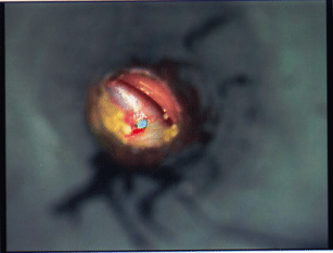
A decision was made to remove the tube from the right middle ear. At the time of surgery a small incision was made on the right tympanic membrane overlying the position of the tube in the middle ear (Figure1). The Armstrong tube was removed gently through this incision site using an alligator forceps and a blunt ball probe. A new Armstrong tube was placed through the same incision site.

Figure 1: Incision over a green medially migrated tube during surgery.
The patient did well at one month follow up and, at 2 years follow up, both tubes were noted to have fallen out and the drums had healed bilaterally. Audiogram revealed normal hearing with bilateral type A tympanograms.
Discussion
Tympanostomy tube (TT) placement in children is one of the most common pediatric surgeries performed. TT surgery is quick, carries minimal morbidity and improves quality of life in both parents and patients [1]. A meta-analysis of TT sequelae performed by Kay et al. in 2001 showed that common, non-severe sequelae after surgery included tube otorrhea, focal atrophy of tympanic membrane and tympanosclerosis [2]. They also reported that cholesteatoma formation and medial migration of TT occurred rarely after surgery. Some complications, such as cholesteatoma formation, residual permanent perforation and tube otorrhea, are more common with long standing tubes.
We discuss a rare complication after TT surgery where the tube migrate medial to the drum instead of following normal epithelial migration towards the canal. Information on this rare complication is reported as isolated case reports in the literature [3]. A study done by Groblewski et al. in 2006 is the most widely cited study discussing this rare complication [4]. Another case report discusses formation of a perilymph fistula following medial displacement of TT [5].
The exact etiology of this complication is unknown. The most implicated theory is chronic ETD, which is very common in children having TT surgery [1,6]. Under normal conditions, the direction of epithelial migration from the outer surface of the drum is towards the canal and, as a result, the tube tends to fall outwards into the canal after sufficient epithelial debris has built up under the medial flange of the tube. It has been theorized that constant negative middle ear pressure may generate enough force to counteract the lateral movement of the tube towards the canal. The negative pressure may become greater if the TT tube is blocked due to debris or cerumen. Groblewski, in their 2006 study, reported that all TT removed from the middle ear space were found to be blocked at the time of the surgery.
Chronic ETD theory, however, fails to explain why this complication occurs so rarely. Negative middle ear pressure is very common in children undergoing TT surgery and may persist after surgery in some children [2]. Blocked TT tube is not uncommon, but most of these children do not have medial migration of tubes. There has been no correlation between this complication and the type of tubes used (long lasting versus short lasting) during surgery.
In a recent letter to the editor, Mehmet Eken [6] raises the possibility of the role of biofilms contributing to this rare complication. Biofilm is a microbially derived sessile community of cells that are attached irreversibly to each other or a substratum, embedded in a matrix of extracellular substances that they have produced [7]. Biofilms have been noted to adhere to all types of TT and a recent study reported that biofilms will form on each type of TT currently used [8]. Biofilms prefer moist surfaces and can clog the medial aspect of TT, likely negating the outward epithelial force and making it more difficult for the tube to follow the natural course of migration. The etiology of this condition is more complex and multifactorial than currently understood. Further studies are required to elucidate the role and interaction of ETD, biofilms and other factors.
Groblewski et al, in their study, attempted to distinguish between primary and secondary medial displacement based on the timing of the complication. Primary displacement is usually due to faulty technique including incorrect positioning of the incision or too large of an incision. As a result medial migration of the tube is usually noted at the first post-operative visit within a month of surgery. It is likely that ours was a case of primary displacement since it occurred at the first post-operative visit. Secondary displacement usually occurs later and is more difficult to explain. The fact that incorrect surgical technique may contribute to the medial displacement of a tube emphasizes the importance of meticulous and accurate placement of incision during surgery.
Majority of the children with a medially displaced tube are asymptomatic though otalgia, otorrhea and mild hearing loss may occur. The diagnosis is confirmed by otoscopic examination showing the reflection of the TT behind the drum (Figure2). Audiogram should be done to assess the type and degree of hearing loss if any. Though there is no consensus as to how to proceed in an asymptomatic child with a medially displaced tube, many authors agree that a medially displaced tube acts like a foreign body in the middle ear and should be removed as soon as possible.

    Figure 2:  Medially migrated green tympanostomy tube behind the tympanic
membrane.
Surgical management involves making a small incision on the drum overlying the TT. Care should be taken to make an adequate incision and retrieve the tube with a small blunt probe and alligator forceps. A separate incision may be made for the new tube and the original incision can be closed with a small piece of gelfoam with good results. All patients should be followed closely in the post-operative period to ensure correct position of the tube. Our patient did not have any complication after insertion of a new tube and did well at 2 years follow up.
Conclusion
TT surgery is very common in the pediatric population but rare complications like medial migration of the tube may occur. The exact etiology of this condition is likely multifactorial, though ETD and biofilms have been implicated. The condition is usually asymptomatic, but many authors agree that, once a diagnosis has been made, the tube should be removed from the middle ear. Surgery is quick, safe and has good results.
References
- American Academy of Pediatrics Subcommittee on Management of Acute Otitis Media. Diagnosis and management of acute otitis media. Pediatrics. 2004; 113: 1451-1465.
- Kay DJ, Nelson M, Rosenfeld RM. Meta-analysis of tympanostomy tube sequelae. Otolaryngol Head Neck Surg. 2001; 124: 374-380.
- Derkay CS, Carron JD, Wiatrak BJ, Choi SS, Jones JE. Postsurgical follow-up of children with tympanostomy tubes: results of the American Academy of Otolaryngology-Head and Neck Surgery Pediatric Otolaryngology Committee National Survey. Otolaryngol Head Neck Surg. 2000; 122: 313-318.
- Groblewski JC, Harley EH. Medial migration of tympanostomy tubes: an overlooked complication. Int J Pediatr Otorhinolaryngol. 2006; 70: 1707-1714.
- Hajiioannou JK, Bathala S, Marnane CN. Case of perilymphatic fistula caused by medially displaced tympanostomy tube. J Laryngol Otol. 2009; 123: 928-930.
- Eken M. The etiology of medial migration of tympanostomy tubes. Int J Pediatr Otorhinolaryngol. 2007; 71: 678.
- Costerton JW, Montanaro L, Arciola CR. Biofilm in implant infections: its production and regulation. Int J Artif Organs. 2005; 28: 1062-1068.
- Wang JC, Hamood AN, Saadeh C, Cunningham MJ, Yim MT, Cordero J. Strategies to prevent biofilm-based tympanostomy tube infections. Int J Pediatr Otorhinolaryngol. 2014; 78: 1433-1438.