
Clinical Image
Austin J Otolaryngol. 2015;2(5): 1044.
Destructive Middle Ear Cholestatoma Without Complication
Demir MG1* and Aydin S2
¹ENT Department, Celal Ertug Etimesgut State hospital, Turkey
²ENT Department, Lutfi Kirdar Kartal Education and Research Hospital, Turkey
*Corresponding author: Demir Mehmet Gökhan,ENT Department, Celal Ertug Etimesgut State Hospital, Ankara, Turkey
Received: May 31, 2015; Accepted: June 11, 2015; Published: June 13, 2015
Clinical Image
Eighteen years old boy presented with recurrent drainage of his left ear during the last 5 years. The patient was examined and diagnosed as chronic supurative otitis media with cholestatoma. The clinical image was obtained from the mastoidectomy operation. When the cortical part of the mastoid bone was elevated, we had recognised a huge cavity. Lateral semicircular canal is destructed with cholestatoma (black star) and also fascial canal is destructed on the mastoid segment of the nerve (black arrow).
Question: What is the anatomic structure seen in black square?
Answer: This was a very agressive type of chronic infection with cholestatoma. The disease destructed fascial canal, lateral semisurcular canal. The cavity seen in black square belongs to jugular bulb and sigmoid sinus. There is only a thin bony lamel surronding these vascular structures. Here we just emphesize the devastating disease without complication.
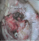
Figure 1: Text here.