Case Report
Austin J Otolaryngol. 2015;2(5): 1045.
Management of Acute Pinna Haematoma using Modified Nasal Splints
Beegun I¹*, Fox R² and Ahmed TS³
¹Department of ENT Surgery, Royal Free Hospital, UK
²Department of ENT Surgery, Northwick Park Hospital, UK
³Department of ENT Surgery, Basingstoke and North Hampshire Hospital, UK
*Corresponding author: Issa Beegun, Department of ENT Surgery, Royal Free Hospital, Pond Street, London,NW3 2QG, UK
Received: June 05, 2015; Accepted: June 22, 2015; Published: June 24, 2015
Background
Acute haematoma of the pinna refers to a collection of blood deep to the perichondrial layer of the pinna [1]. It is commonly caused by blunt trauma and if untreated will ultimately result in a deformity known as ‘cauliflower’ or ‘wrestler’s’ ear [1]. Various treatments have been described in order to relieve the haematoma and surgical drainage is often advocated. No consensus exists, however, on optimum management to minimise the possibility of haematoma recurrence [1].
We present a simple technique using commonly available materials that in our experience effectively prevents re-accumulation after surgical evacuation.
Case Presentation
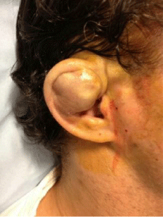
Our case is that of a 36 year old healthy male who sustained an acute right pinna haematoma following blunt trauma to the ear during a play fight with his friend. The patient presented twelve hours after the initial injury. Figure 1 demonstrates the appearance at presentation.

Figure 1: Pre-operative: large pinna haematoma.
Management
After appropriate preparation with povidone iodine topical wash, 2.2 ml of Lignospan Special® (2% Lidocaine hydrochloride with 2% Adrenaline 1: 80 000) was infiltrated and an incision in the helical crease was made to allow haematoma evacuation.
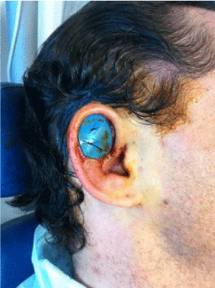
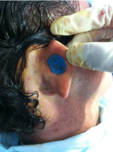
Exmoor® nasal silicone splints were cut to size and the pinna was sandwiched between two such modified splints applied to the medial and lateral surfaces. This allowed appropriate pressure to be distributed evenly to the pinna surfaces, thereby preventing haematoma recollection.
The pinna-silicone splint sandwich was secured using 3/0 Ethilon® sutures. Large bites were taken to minimise the risk of pressure necrosis. A seven day course of oral antibiotics was prescribed postoperatively (Co-amoxiclav 250/125).
Figures 2 and 3 demonstrate the medial and lateral modified silicone splints in situ.

    Figure 2:  Placement of lateral splint secured with two through-and-through
Ethilon® sutures.

Figure 3: Placement of medial splint.
Outcome
The splints were removed in the outpatient clinic after seven days. The patient was reviewed and discharged after a subsequent week with no evidence of haematoma recurrence.
Conclusion
This case highlights a simple and effective method for acute surgical management of pinna haematomas using readily available materials.
References