Clinical Image
Austin J Otolaryngol. 2015;2(6): 1050.
Huge Sialolith in Submandibular Wharton Canal
Demir MG¹*, Aydin S² and Demir HO³
¹Department of ENT, Prof. Dr. Celal Ertug Etimesgut State hospital, Turkey
²Department of ENT, Dr. Lutfi Kirdar Kartal education and research hospital, Turkey
³Department of Anesthesiology, Yildirim beyazit university, Turkey
*Corresponding author: Demir Mehmet Gökhan, Department of ENT, Prof. Dr. Celal Ertug Etimesgut State Hospital, Ankara-Turkey
Received: July 23, 2015; Accepted: August 05, 2015; Published: August 07, 2015
Clinical Image
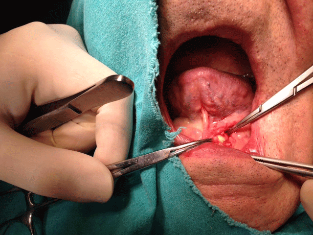
Thirty three years old male patient attended to Otorhinolaryngology outpatient clinic with a complaint of swelling on his left submandibular region. According to physical examination a huge canalicular calculi which is palpated, also detected on ultrasonography.
Question: What can be the treatment method for this patient?
Answer: Although the calculi is huge and obstructed the canal, we perform intraoral sialolithotomy. This surgical procedure can be done via incision to floor of the oral mucosa and taking out the calculi by manipulation (Figures 1,2). This method can be done under local anesthesia and is gland sparing [1]. We encourage this method in wharton canal calculi safely.
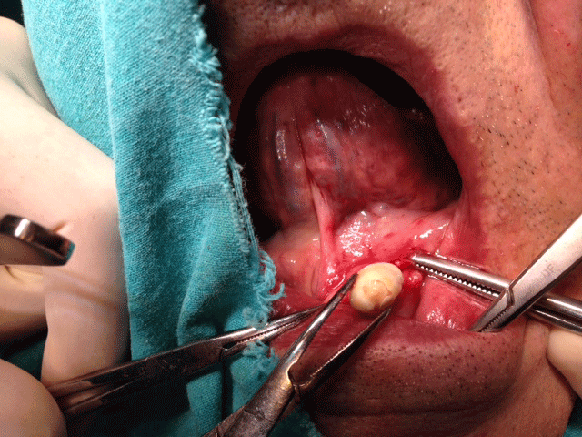
Figure 1: