Research Article
Austin J Otolaryngol. 2015; 2(7): 1056.
Carotid Artery Surgery for Cervical Lymph Node Invasion: Limitations of Neck Dissection
Furusaka T1,2*, Asakawa T¹, Hasegawa H¹, Shigihara S¹ and Matsuda H²
¹Department of Otolaryngology-Head and Neck Surgery, Nihon University School of Medicine, Japan
²Laboratory of Veterinary Molecular Pathology and Therapeutics, Division of Animal Life Science, Tokyo University of Agriculture and Technology, Japan
*Corresponding author: Tohru Furusaka, Department of Otolaryngology-Head and Neck Surgery, Nihon University School of Medicine, 30-1 Oyaguchi-kami-cho, Itabashi-ku, Tokyo 173-8610, Japan
Received: December 01, 2014; Accepted: September 05, 2015; Published: September 07, 2015
Abstract
Objectives/Hypothesis: To evaluate reconstructive surgery of the common carotid artery; and to investigate the indications for this treatment, the limitations of neck dissection as treatment, and the feasibility of concurrent chemoradiation therapy (CCRT).
Study Design: Retrospective observational study.
Methods: Forty-four patients underwent reconstructive surgery of carotid artery concurrently in radical neck dissection. Twelve patients did not undergo carotid artery reconstruction, 7 underwent reconstruction using a polytetrafluoroethylene (PTFE) graft, 11 underwent reconstruction using an autologous great saphenous vein graft, and 14 underwent vein patch angioplasty after partial resection of the carotid artery.
Results: All 7 patients who received PTFE grafts and 11 patients who received saphenous vein grafts died within 2 years after surgery. Four of the 12 patients who did not undergo carotid artery reconstruction and 5 of the 14 patients who underwent vein patch angioplasty survived for 5 years.
Conclusion: Patients who underwent neck dissection with reconstructive surgery of the carotid artery had a poor prognosis. Patients who underwent carotid artery resection without reconstruction followed by postoperative radiation therapy survived. CCRT without surgery should be considered in patients with N3 cases.
Keywords: PTFE graft; Great saphenous vein; Resection without reconstruction; Patch angioplasty; Concurrent chemoradiation therapy
Introduction
In patients with advanced head and neck cancer, surgical excision of the primary site often causes severe functional and aesthetic damage. We have achieved high rates of organ preservation and overall survival by treatment of these patients with chemotherapy in combination with complete regional lymph node dissection. Our chemotherapy regimen includes two cycles of intra-arterial docetaxel and cisplatin plus continuous intravenous infusion of 5-fluorouracil for 5 days starting on day 2, followed by two cycles of concurrent chemoradiation therapy (CCRT) [1-4].
The ability to control cervical lymph node metastasis significantly affects the prognosis of patients with head and neck cancer. The current technique of radical neck dissection including resection of the internal jugular vein, sternocleidomastoid muscle, and accessory nerve was established in 1933 [5]. To our knowledge, however, there have been no reports addressing the optimal extent of dissection. In 1905, Crile, who first described systematic neck dissection, recommended temporary clamping of the carotid artery to reduce blood loss, but did not describe the results of this technique [6]. We often encounter invasion of metastatic lymph nodes into the common carotid artery, but the optimal management of such invasion is currently unclear.
We reviewed outcomes in patients with invasion of metastatic lymph nodes into the common carotid artery who underwent carotid artery resection during radical neck dissection, according to the carotid artery reconstruction technique used. The indications for carotid artery reconstruction surgery are reviewed, and the optimal treatment of patients with N3 disease is discussed.
Materials and Methods
Patients
This study included 44 patients with primary squamous cell carcinoma of the head and neck (Table 1). The patients were 35 males and 9 females with a mean age of 58.6 years (median 59 years, range 39–69 years). The primary site was the oropharynx in 16 patients, hypopharynx in 16 patients, and larynx in 12 patients. Preoperative treatment was by radiation therapy alone in 11 patients, chemotherapy alone in 11 patients, and chemoradiation therapy in 22 patients.
    Characteristic
    Value


    Sex


    Male
    35


    Female
    9


    Mean age and age range (years)
    58.6 (39–69)


    Median age (years)
    59


    Tissue type


    Squamous cell carcinoma
    44


    Primary site


    Oropharynx
    16


    Hypopharynx
    16


    Larynx
    12


    Subject status


    Radiation therapy alone
    11


    Chemotherapy alone
    11


    Chemoradiation therapy
    22
Table 1:  Characteristics of the 44 Patients who underwent Carotid Artery
Resection.
According to the Union for International Cancer Control classification (2009), 2 patients had T2 disease, 4 had T3 disease, 30 had T4a disease, 8 had T4b disease, and all had N3 disease. Table 2 shows the classifications for all 44 patients.
    N0
    N1
    N2a
    N2b
    N2c
    N3


    T1a


    T1b


    T2


    2


    T3


    4


    T4a


    30


    T4b


    8
Table 2:  TNM Classifications of the 44 Patients who underwent Carotid Artery
Resection.
Methods
All 44 patients underwent radical neck dissection including resection of the common carotid artery for the treatment of squamous cell carcinoma at Nihon University Hospital and affiliated hospitals between 1980 and 2007. Twelve patients did not undergo carotid artery reconstruction, 7 underwent reconstruction using a 6- or 8-mm polytetrafluoroethylene (PTFE) interposition graft, 11 underwent reconstruction using an autologous great saphenous vein interposition graft, and 14 underwent vein patch angioplasty after partial resection of the carotid artery (Table 3).
Surgical procedure
n
5-year survival
Died
Cause of death
1
PTFE interposition graft placement
7
0
7
•Failure to control cervical lymph node metastasis in 3 cases
•Distant metastasis in 4 cases2
Autogenous great saphenous vein interposition graft placement
11
0
11
•Failure to control the primary site in 2 cases
•Failure to control cervical lymph node metastasis in 4 cases
•Distant metastasis in 4 cases
•Other cause in 1 case3
Resection without reconstruction
12
4
8
•Failure to control the primary site in 2 cases
•Failure to control cervical lymph node metastasis in 2 cases
•Distant metastasis in 3 cases
•Other cause in 1 case4
Patch angioplasty with vein
after partial resection
14
5
9
•Failure to control the primary site in 2 cases
•Failure to control cervical lymph node metastasis in 1 case
•Distant metastasis in 4 cases
•Other cause in 1 case
•Unknown cause in 1 case
Table 3: Postoperative Outcomes According to Common Carotid Artery Reconstruction Technique.
For resection of the common carotid artery without reconstruction, the artery was ligated at both ends before resection. For vein patch angioplasty, the common carotid artery was reconstructed using a venous valve after resection of approximately one-third of the artery.
Results
All seven patients who received PTFE grafts died within 2 years after surgery because of failure to control cervical lymph node metastasis (three patients) or distant metastasis (four patients).
All 11 patients who received great saphenous vein grafts died within 2 years after surgery because of failure to control the primary cancer (2 patients), failure to control cervical lymph node metastasis (4 patients), distant metastasis (4 patients), and another cause (1 patient).
Four of the 12 patients who did not undergo carotid artery reconstruction survived for at least 5 years. The other eight patients died within 2 years after surgery because of failure to control the primary cancer (two patients), failure to control cervical lymph node metastasis (two patients), distant metastasis (three patients), and another cause (one patient).
Five of the 14 patients who underwent vein patch angioplasty after partial resection of the carotid artery survived for at least 5 years. The other nine patients died within 2 years after surgery because of failure to control the primary site (two patients), failure to control cervical lymph node metastasis (one patient), distant metastasis (four patients), and an unknown cause (one patient) (Table 3).
Discussion
Crile, an American surgeon, established the concept of systematic neck dissection in 1906 [7]. In 1951, Martin proposed radical neck dissection, including dissection of all lymph nodes from levels I to V, and resection of the internal jugular vein, sternocleidomastoid muscle, accessory nerve, omohyoid muscle, submandibular gland, tail of the parotid gland, and most of the cervical nerve plexus [8]. In 1975, Conley proposed that radical neck dissection is essential [9].
However, Bocca proposed modified neck dissection in 1967 and described functional neck dissection in 1980 [10,11]. Medina proposed a new classification system to describe the extent of neck dissection in 1989 [12], and Robbins et al. proposed an official classification system based on modification of Medina’s system at the Academy’s Committee for Head and Neck Surgery and Oncology in 1991 [13]. Hasegawa et al. subsequently proposed revisions to further clarify the classification system [14]. However, the indications for resection of the common carotid and internal carotid arteries have not been described.
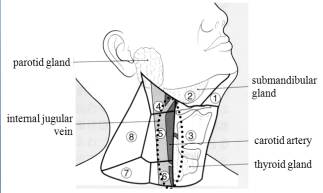
There is no doubt that control of cervical lymph node metastasis is important for improving the prognosis of patients with head and neck cancer. Extended surgery may include mastoidectomy, mandibulectomy, thyroidectomy, laryngectomy, hypopharyngectomy, claviculectomy, resection of the trapezius muscle, and resection of overlying skin. The most challenging surgical issue is the treatment of invasion of the common carotid artery and scalene muscles (Figure 1) [15].

Figure 1: Structures surrounding the carotid artery and internal jugular vein.
This study evaluated the indications and contraindications for surgical resection of metastatic cervical lymph nodes invading the common carotid artery in patients with N3 stage squamous cell carcinoma. None of the 7 patients who received PTFE grafts or the 11 patients who received great saphenous vein grafts survived for 5 years after surgery. No complications associated with vascular reconstructive surgery such as air embolism, thrombosis, or infections were observed. PTFE grafts are associated with relatively low risks of thrombosis and infection, because PTFE is highly antithrombogenic, hydrophobic, and not biodegradable. However, autologous great saphenous vein grafts are considered to be superior to artificial grafts. We achieved a 5-year patency rate of 70% after reconstruction of atherosclerotic common carotid arteries with a diameter of =5 mm using autologous great saphenous vein grafts, with no cases of air embolism, thrombosis, or infection, indicating that this technique provides good long-term outcomes.
To reduce the risk of postoperative complications, we sutured the arterial anastomoses at intervals of approximately 1 mm, ensuring that the vascular adventitia was not exposed on the inner aspect of the vessel. We also removed air from the graft by releasing the proximal forceps before the distal forceps. As anastomotic bleeding can be stopped by application of gentle pressure to the anastomotic site for approximately 3 minutes, which slightly exceeds the bleeding time, extra sutures were minimized to avoid complications.
Vascular grafting was performed under general anesthesia at normal body temperature. It has been reported that the carotid artery can be occluded for up to 20 minutes without damage to the brain, and Inouchi et al. reported that occlusion for a mean time of 16 minutes caused no adverse effects [16]. In our patients, the mean time of carotid artery occlusion was 14 minutes, and no adverse effects were observed.
Some of the patients who underwent vein patch angioplasty after partial resection of approximately one-third of the common carotid artery survived for at least 5 years. In these patients, adequate dissection was achieved without resection of the scalene muscles.
Four of the 12 patients who did not undergo carotid artery reconstruction survived for at least 5 years. All four of these patients received postoperative radiation therapy including irradiation of the scalene muscles. None of these 12 patients had abnormal findings on the Matas test [17] or electroencephalography, but one patient developed hemiplegia. Common carotid artery resection without reconstruction should therefore only be performed with informed consent from the patient and their family, and only if magnetic resonance angiography or computed tomography angiography findings show adequate communication between the vertebral and internal carotid arteries.
Adequate dissection of the scalene muscles is required prior to common carotid artery resection. If dissection of the scalene muscles is not possible, CCRT may be more useful than carotid artery resection. In this study, all postoperative deaths occurred within 2 years, indicating that carotid artery reconstruction does not improve the prognosis in patients with invasion of the scalene muscles.
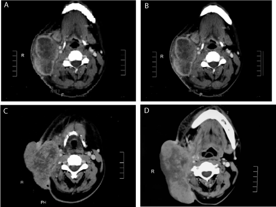
We also treated a 39-year-old male with Stage IVB oropharyngeal squamous cell carcinoma (poorly differentiated type, T2N3M0) and a giant cervical lymph node metastasis who underwent CCRT without surgery, at his request. CCRT included chemotherapy with cisplatin 50 mg/m², docetaxel 50 mg/m², and 5-fluorouracil (600 mg/m², continuous infusion for 5 days starting on day 2), and radiotherapy with a total dose of 66 Gy. After CCRT, he had a complete response at the primary site and a partial response at the site of cervical lymph node metastasis (reduction from 110 × 100 mm to 50 × 42 mm). The changes in cervical lymph nodes over time are shown in Figure 2. His cervical lymph nodes started to increase in size approximately 1 year after CCRT. He was hospitalized with anemia due to bleeding from the neck lesion at 2 years after CCRT, and received palliative care for pain control. He eventually died as a result of bleeding from the neck lesion. He survived for 2 years and 6 months after CCRT, and continued his job with maintenance of a relatively good quality of life (QOL) until the last 2 months.

    Figure 2:  (A) Immediately after CCRT (cisplatin 50 mg/m², docetaxel 50 mg/
m², 5-fluorouracil 600 mg/m²/day by continuous infusion for 5 days, concurrent
radiation therapy at 2 Gy/day to a total dose of 66 Gy). The cervical lymph
nodes had reduced in size and density. (B) Six months after CCRT, showing
minimal change. (C) Thirteen months after CCRT, showing progression of
disease. (D) Nineteen months after CCRT, showing further progression of
disease.
Although common carotid artery reconstruction using a PTFE or great saphenous vein graft could be performed even in patients with recurrent tumor, some of these patients experienced further recurrence, despite postoperative three-dimensional contrastenhanced computed tomography showing vascular patency and complete resection of the tumor. In all these cases, tumor invasion of the scalene muscles may not have been adequately resected. Unless vein patch angioplasty after partial resection of the common carotid artery is possible, resection of the carotid artery should only be considered if postoperative radiotherapy can be applied to the scalene muscles. CCRT without surgery should be considered in patients with N3 disease, because this may prolong survival while maintaining QOL even if the patient does not have a complete response to therapy. As QOL is not significantly affected by cervical lymph node metastasis in patients with a complete response at the primary site, some patients prefer to avoid neck dissection. We have experienced several such cases since the introduction of CCRT, but the number of these patients remains small and comparison with conventional treatment still needs to be performed. After carotid artery reconstruction using a PTFE or great saphenous vein graft, most patients survived for approximately 1 year, and none survived for longer than 2 years. Reconstructive surgery may be difficult and requires technical proficiency, because the carotid artery may be fragile in these patients.
We have achieved good results by preferentially using CCRT for treatment of the primary lesion with the aim of functional preservation, and aggressive treatment of cervical lymph node metastasis with neck dissection [1-4]. Invasion of the scalene muscles limits the ability to completely resect metastatic disease, and further treatment of scalene muscle invasion is important in these patients.
In summary, resection of the common carotid artery is indicated only if communication between the vertebral and internal carotid arteries has been confirmed, informed consent has been obtained from the patient and their family, and radiation therapy of the scalene muscles is feasible. Other patients with N3 disease should be treated with CCRT.
Conclusion
Radical neck dissection should be performed according to the techniques described by Martin et al. and Conley [8,9]. In selected patients, extended surgery should be performed including mastoidectomy, mandibulectomy, thyroidectomy, laryngectomy, and hypopharyngectomy, as appropriate.
When the common carotid artery is surrounded by primary tumor or lymph node metastasis, the artery can be resected without reconstruction if invasion of the scalene muscles can be controlled. However, carotid artery resection should be performed only if postoperative radiation therapy is feasible, communication between the vertebral and internal carotid arteries has been confirmed, and informed consent has been obtained from the patient and their family. If these conditions are not met, CCRT without involving the carotid artery should be considered.
References
- Furusaka T, Asakawa T, Tanaka A, Matsuda H, Ikeda M. Efficacy of multidrug superselective intra-arterial chemotherapy (docetaxel, cisplatin, and 5-fluorouracil) using the Seldinger technique for tongue cancer. Acta Otolaryngol. 2012; 132: 1108-1114.
- Furusaka T, Matsuda A, Tanaka A, Matsuda H, Ikeda M. Superselective intra-arterial chemotherapy for laryngeal preservation in carcinoma of the anterior oropharyngeal wall. Acta Otolaryngol. 2013; 133: 194-202.
- Furusaka T, Matsuda A, Tanaka A, Matsuda H, Ikeda M. Laryngeal preservation in advanced piriform sinus squamous cell carcinomas using superselective intra-arterial chemoradiation therapy with three agents. Acta Otolaryngol. 2013; 133: 318–326.
- Furusaka T, Matsuda A, Tanaka A, Matsuda H, Ikeda M. Superselective intra-arterial chemoradiation therapy for functional laryngeal preservation in advanced squamous cell carcinoma of the glottic larynx. Acta Otolaryngol. 2013; 133: 633–640.
- Blair VP, Brown JB. The treatment of cancerous or potentially cancerous cervical lymph-nodes. Ann Surg. 1933; 98: 650-661.
- Crile GW. On the surgical treatment of cancer of the head and neck. With a summary of one hundred and twenty-one operations performed upon one hundred and five patients. Trans South Surg Gynecol Assoc. 1905; 18: 108–127.
- Crile G. Landmark article Dec 1, 1906: Excision of cancer of the head and neck. With special reference to the plan of dissection based on one hundred and thirty-two operations. By George Crile. JAMA. 1987; 258: 3286-3293.
- Martin H, Del Valle B, Ehrlich H, Cahan WG. Neck dissection. Cancer. 1951; 4: 441-499.
- Conley J. Radical neck dissection. Laryngoscope. 1975; 85: 1344-1352.
- Bocca E, Pignataro O. A conservation technique in radical neck dissection. Ann Otol Rhinol Laryngol. 1967; 76: 975-987.
- Bocca E, Pignataro O, Sasaki CT. Functional neck dissection. A description of operative technique. Arch Otolaryngol. 1980; 106: 524-527.
- Medina JE. A rational classification of neck dissections. Otolaryngol Head Neck Surg. 1989; 100: 169-176.
- Robbins KT, Medina JE, Wolfe GT, Levine PA, Sessions RB, Pruet CW. Standardizing neck dissection terminology. Official report of the Academy's Committee for Head and Neck Surgery and Oncology. Arch Otolaryngol Head Neck Surg. 1991; 117: 601-605.
- Hasegawa Y, Saikawa M. Update on the classification and nomenclature system for neck dissection: revisions proposed by the Japan Neck Dissection Study Group. Int J Clin Oncol. 2010; 15: 5-12.
- Furusaka T. Clinical anatomy for neck dissection. J Otolaryngol Head & Neck Surg. 2008; 24: 505–511.
- Inokuchi K, Yagi H. Vascular reconstruction for radical neck dissection system for neck dissection. Otologia Fukuoka. 1967; 13: 97–104.
- Matas R. Personal experiences in vascular surgery: a statistical synopsis. Ann Surg. 1940; 112: 802-839.