Case Report
Austin J Otolaryngol. 2015; 2(8): 1057.
Neurotologic Symptoms as the Initial Presentation of Nasopharyngeal Carcinoma
Celis-Aguilar E¹*, Mayoral-Flores HO¹, Burgos- Paez A¹ and Caballero-Rodríguez CB²
¹Department of Otolaryngology, Universidad Autónoma de Sinaloa, México
²Department of Pathology, Universidad Autónoma de Sinaloa, México
*Corresponding author: Erika Celis-Aguilar, Department of Otolaryngology, Centro de Investigación y Docencia en Ciencias de la salud (CIDOCS), Universidad Autónoma de Sinaloa (UAS), Eustaquio Buelna No. 91 Col. Gabriel Leyva C.P. 80030, Culiacán, Sinaloa, México
Received: July 13, 2014; Accepted: September 09, 2015; Published: September 15, 2015
Abstract
Nasopharyngeal carcinoma is a rare malignant neoplasm that arises from nasopharyngeal epithelium. Although globally rare, is endemic in Southeast Asia particularly south China. One fifth of these patients develop cranial nerve dysfunction and headache.
Neurotologic symptoms are inadequately diagnosed since only 20% develop these symptoms at diagnosis. Tinnitus, serous otitis media and hearing loss are the most frequent neurotologic symptoms. Cranial nerve palsies are rare and most frequently affect V and VI nerves. Patients face referral and diagnostic problems due to a myriad of symptoms, requiring high suspicion by the medical team. Furthermore, otolaryngology referral and endoscopic visualization are paramount for diagnosis.
We report a case that developed right nasal obstruction, epistaxis, otitis media, headache, neck pain and diplopia. The patient was referred due to nasal symptoms and erosion of sphenoid sinus as an incidental finding on CT scan. We highlight the clinical symptoms at presentation, particularly neurotologic symptoms.
Keywords: Nasopharyngeal carcinoma; Neurotologic symptoms
Abbreviations
CN: Cranial Nerve; CT: Computed Tomography; OME: Otitis Media with Effusion
Introduction
Nasopharyngeal carcinoma is a malignant neoplasm more common in south china and Taiwan [1-4]. The incidence in the rest of the world is 1 per 100,000 [1]. The clinical presentation is subtle, frequently misdiagnosed. Eighty percent of patients develop cervical adenopathy. Unfortunately nasal symptoms such as epistaxis and nasal obstruction are non-specific.
Neurotologic symptoms such as cranial neuropathy affect 7.8- 30% of patients [2-5]. The sixth and fifth cranial nerves are the most frequently affected [3-5]. Cranial nerve palsies are the result of direct tumor extension to the cavernous sinus and skull base. We present a case with neurotologic symptoms as the initial presentation.
Case Presentation
We report a 24 year old male with cervical pain and headache, diplopia, nasal obstruction, bilateral ear fullness and occasional epistaxis. The patient was referred from the ophthalmology and neurology department for nasal symptoms and sphenoid erosion as an incidental finding on Computed tomography (CT) scan. Diplopia started 3 months ago and was observed by the ophthalmology department with no clear diagnosis. The patient at the first visit only complained of diplopia, headache and cervical pain, the rest of symptoms where obtained by direct interrogation and were considered by the patient as subtle or irrelevant to his current medical condition.
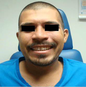
Physical exploration revealed right six cranial nerve (CN) palsy (Figure 1) and mild asymmetry at the right inferior facial third (Figure 2). Accumetry revealed bilateral conductive hearing loss. The rest of cranial nerves were normal.

Figure 1: Sixth cranial nerve palsy.

Figure 2: Mild facial asymmetry (inferior third).
Bilateral otoscopy showed otitis media with effusion (OME). Nasal endoscopy found a submucosal hypervascular irregular lesion on right nasopharynx. Bilateral multiples cervical lymphadenopathies were palpated on nodal level III, IV, V.
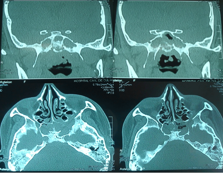
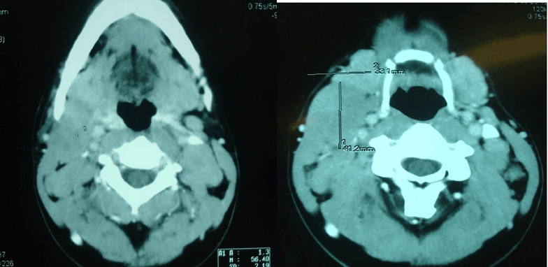
CT scan showed nasopharynx occupation and lateral and inferior sphenoid erosion (Figure 3). Additionally CT scan showed bilateral multiple adenopathies greater than 1 cm that were asymptomatic (Figure 4).

    Figure 3:  CT scan shows sphenoid occupation and erosion of lateral and
inferior sphenoid walls.

Figure 4: CT scan shows multiple bilateral adenopathy.
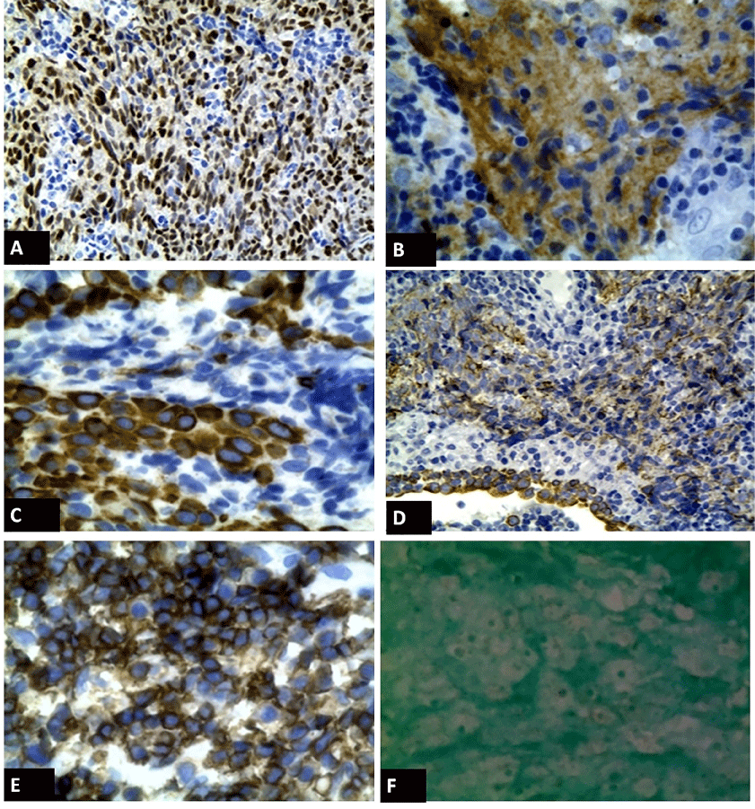
The patient underwent endoscopic nasopharyngeal biopsy and open lymph node biopsy. Due to the extreme vascularity of the tumor the patient required posterior tamponade and3 day hospitalization; was discharged with no evidence of epistaxis. The diagnosis of nasopharyngeal carcinoma was confirmed. Immunohistochemistry was performed with the following results (Figure 5): P63 + (Figure A), EGFR positive (Figure B), cytokeratin 5/6 positive (Figure C), cytokeratin AE1/AE3 positive (Figure D), CD45 negative (Figure E), EBER positive (Figure F). Radiotherapy was indicated with good results. The patient has a 13 month follow up with no evidence of disease.

    Figure 5:  Immunohistochemistry results: P63 + (Figure A), EGFR positive
(Figure B), cytokeratin 5/6 positive (Figure C), cytokeratin AE1/AE3 positive
(Figure D), CD45 negative (Figure E), EBER positive (Figure F).
Discussion
Nasopharyngeal carcinoma is a nonlymphomatous squamous cell carcinoma arising from the surface epithelium of the nasopharynx more common in south china and Taiwan [1-4]. Consumption of salted fish and Epstein Barr virus have been associated as causative factors. The highest presentation peaks are in the second and fourth to fifth decades.
Signs and symptoms of nasopharyngeal carcinoma are related to the primary tumor location and dissemination [5]. Because of its hidden location, disease could spread if not diagnose early [6]. The most common findings are: nasopharyngeal mass, neck mass and epistaxis. The presence of nasopharyngeal mass predicts nasopharyngeal carcinoma with a sensitivity of 90.7% and specificity of 28.4%; on the other hand, a neck mass has a lower sensitivity (66%) but higher specificity (80.9%) [4]. Chung-Chan et al. [4] calculated the sensibility and specificity of combining both the presence of a neck mass and a nasopharyngeal mass, finding 60% for sensibility and 89.6% for specificity, accuracy 65.6.
Regarding the otolaryngology manifestations of disease, the tumor frequently involves rosenmuller fossa and torus tubarius causing eustachian tube obstruction and subsequently OME. Although the otolaryngology community considers unilateral OME suggestive of a nasopharyngeal mass occupation, our patient had bilateral OME which demonstrates the dissemination of the disease. On the other hand, epistaxis is a non-specific symptom.
Cranial nerve involvement is rare, only n=11 (7.8%) of patients develop this symptom at the initial presentation of the disease [4] (Table 1). The most frequent affected cranial nerves are V and VI [4,5]. CN palsy is caused by skull base and cavernous sinus tumor invasion.
Table 1: Incidence of cranial nerve paralysis at presentation. Literature review.
Li et al. [5] reported that 75% of their patients with cranial nerve involvement had V, VI or XII palsy. Diplopia has been reported in another study [6] in 6.2% of patients at initial presentation. Furthermore Li et al. found a significant neurologic recovery after radiotherapy.
Lee et al. [3] studied 4768 patients; they found CN palsies from I to XII cranial nerves, although no percentages are described. Moreover, Mo et al. [7] reported 171 unilateral CN palsy and 7 bilateral palsy.
Symptoms of nasopharyngeal carcinoma are vague and nonspecific, especially if only neurotologic symptoms are evident at initial presentation. Furthermore, there is a lack of awareness in the medical community of the early signs of nasopharyngeal carcinoma [6].
Our patient presented with neck mass, diplopia, epistaxis, facial pain, OME, nasal obstruction and a nasopharyngeal mass evident on endoscopy. Paradoxically the patient only complained at the first visit of diplopia and the rest of symptoms where obtained by direct interrogation and physical exploration. Lee et al. [3] have described this discrepancy between symptoms and signs in nasopharyngeal carcinoma patients; otologic symptoms for example, being greater when specific audiological tests are done (20% vs. 62%). Also, frequent asymptomatic cervical node enlargement adds on the diagnostic difficulty of the disease. Furthermore greater survival is related to lower stages of the disease.
CN palsies, as in our case, can be underestimated by the medical faculty and delay diagnosis. We consider important to highlight the diverse and early clinical presentations of this disease. A high clinical suspicion should be raised when face with a CN palsy, especially V and VI cranial nerves, above all if nasal symptoms or a neck mass are present. Full diagnostic work up, including CT scan of paranasal sinuses and nasal endoscopy is recommended especially in an endemic region.
Conclusion
Patients with cranial nerve palsies and simultaneous nasal symptoms, such as epistaxis, nasal obstruction or the presence of a neck mass should be rule out for nasopharyngeal carcinoma. We recommend to perform a CT scan and nasal endoscopy, with appropriate referral to the otolaryngology department, especially in endemic areas.
References
- Cannon T, Zanation AM, Lai V, Weissler MC. Nasopharyngeal carcinoma in young patients: a systematic review of racial demographics. Laryngoscope. 2006; 116: 1021-1026.
- Su CY, Lui CC. Unilateral palate paralysis in patients with nasopharyngeal carcinoma: imaging and clinical correlations. Laryngoscope. 2001; 111: 645-649.
- Lee AW, Foo W, Law SC, Poon YF, Sze WM, O SK, et al. Nasopharyngeal carcinoma: presenting symptoms and duration before diagnosis. Hong Kong Med J. 1997; 3: 355-361.
- Chung-Chan H, Wen-Hung W, Yen-Chun L, Hsu-Huei W, Kam-Fai L. A large scale study of the association between biopsy results and clinical manifestations in patients with suspicion of nasopharyngeal carcinoma. Laryngoscope. 2012; 122: 1988-1993.
- Li JC, Mayr NA, Yuh WT, Wang JZ, Jiang GL. Cranial nerve involvement in nasopharyngeal carcinoma: response to radiotherapy and its clinical impact. Ann Otol Rhinol Laryngol. 2006; 115: 340-345.
- Adham M, Kurniawan AN, Muhtadi AI, Roezin A, Hermani B, Gondhowiardjo S, et al. Nasopharyngeal carcinoma in Indonesia: epidemiology, incidence, signs, and symptoms at presentation. Chin J Cancer. 2012; 31: 185-196.
- Mo HY, Sun R, Sun J, Zhang Q, Huang WJ, Li YX, et al. Prognostic value of pretreatment and recovery duration of cranial nerve palsy in nasopharyngeal carcinoma. Radiat Oncol. 2012; 7: 149.