Case Report
Austin J Otolaryngol. 2015; 2(8): 1058.
Three Cases of Dizziness of Central Lesion that Otolaryngologist can Experience in the Outpatient Clinic
Dong-Hee Lee*
Department of Otolaryngology-Head and Neck Surgery, Catholic University of Korea, Republic of Korea
*Corresponding author: Dong-Hee Lee, Department of Otolaryngology-Head and Neck Surgery, Uijeongbu St. Mary’s Hospital, The Catholic University of Korea, 271 Cheonbo Street, Uijeongbu City, Gyeonggi-Do, 11765, Republic of Korea
Received: July 14, 2015; Accepted: October 01, 2015; Published: October 03, 2015
Abstract
Diagnosis of dizziness of central origin remains a challenge, especially when a dizzy patient visits the outpatient clinic. Recently, we experienced 3 cases of central causes in the outpatient otolaryngology clinic. (1) A 37-yearold woman complaint floating dizziness lasting for 10 months. At the first attack of dizziness, she admitted in other hospital and got brain MRI, which did not reveal any pathologic finding. Temporal MRI showed an intracanalicular schwannoma of left ear. (2) A 70-year-old man with hypertension complaint lightheadedness, disequilibrium and tilting sensation, which developed suddenly two days ago. Brain MRI showed an extra-axial mass at left prepontine cistern, which compresses left pons. (3) A 52-year-old man with diabetes mellitus and hypertension complaint relapsing dizziness lasting for about a half hour 2-3 times a day. Its nature was lightheadedness and presyncope, which developed suddenly together with left tinnitus a half month ago. He complained that his vision was blurred during dizziness attack. Brain MRI showed left tortuous distal vertebral artery compressing medulla oblongata.
Keywords: Vertigo; Dizziness; Central vestibular disorders
Introduction
Central vertigo or dizziness is due to a disease originating from the central nervous system (CNS). In clinical practice, it often includes lesions of 8th cranial nerve root entry zone but lesions of 8th cranial nerve itself are often classified to peripheral vertigo. Central vertigo may be caused by hemorrhagic or ischemic insults to the cerebellum, the vestibular nuclei, and their connections within the brain stem. Other causes include CNS tumors, infection, trauma, and multiple sclerosis [1,2].
Over the years, one of the principal uses of vestibular function evaluations, both direct physical examination and laboratory studies, has been to differentiate between peripheral and central vestibular system disorders. In most cases well-defined abnormalities on pursuit tracking or with saccade testing are indicators of central vestibular system involvement. However, just as a significant caloric asymmetry would be taken as an indication of peripheral dysfunction, the abnormal central findings on vestibular laboratory testing need to fit with the symptom presentation to suggest that those findings relate to the patient’s presenting complaints.
Although there have been reported symptoms as well as nature of nystagmus that are more likely to be of peripheral origin compared to those of central origin, the identification of dizziness of central origin is still difficult for most clinicians. Recently, we encountered three cases of central vertigo at the outpatient clinic and shared our experiences.
Case Presentation
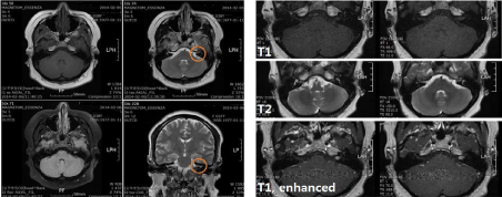
Case presentation 1: A 37-year-old woman without specific past medical history complaint floating dizziness lasting for 10 months. At the first attack of vertigo (10 months ago), she admitted in other general hospital and got brain MRI. Based on her own recall, clinicians of other hospital diagnosed that MRI was nonspecific for brain and managed her symptom conservatively. Just before visit to our outpatient clinic, she got admission and conservative management at other hospital because of recurrent attack of vertigo as well as ongoing dizziness. She denied any audiological symptom. On examination, she did not show obvious nystagmus or focal neurological sign. Pure tone audiogram (PTA) showed normal hearing of both ears. Videonystagmogram (VNG) demonstrated canal paresis (CP) of left 43% weakness and directional preponderance (DP) of left 14%. The SP/AP ratio of electrocochleogram was 0.207 in right and 0.292 left ear. Vestibular evoked myogenic potential (VEMP) was normal P1-N1 waves with asymmetry of 16%. While we reviewed brain MRI of other hospital, we found abnormal finding suspicious of acoustic neuroma. Temporal MRI confirmed an intracanalicular schwannoma of left ear.

    Figure 1:  A 37-year-old woman (case 1). Brain MRI taken at other hospital (left image) suspected acoustic neuroma (circle), which was missed. Temporal MRI
(right image) confirmed an intracanalicular schwannoma of left ear.

Figure 2: A 70-year-old man (case 2). Temporal MRI showed an extra-axial mass at left prepontine cistern, which compresses left pons.

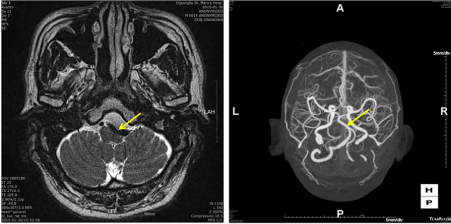
    Figure 3:  A 62-year-old man (case 3). Temporal MRI showed left tortuous distal vertebral artery compressing medulla oblongata. CT angiogram confirmed the
tortuosity of V4 segment of left vertebral artery, compressing brainstem.
Case presentation 2: A 70-year-old man with hypertension complaint lightheadedness, disequilibrium and tilting sensation, which developed suddenly two days ago and improved partially as time went by. He denied any audiological symptom. On neurootologic examination, he did not show obvious nystagmus but slight numbness was found on her left face. PTA showed down-slope sensorineural hearing loss of moderate degree in his both ears. VNG demonstrated CP of left 70% weakness and DP of right 44%. It also showed right beating nystagmus. Brain MRI showed an extra-axial mass at left prepontine cistern, which compresses left pons.
Case presentation 3: A 62-year-old man with diabetes mellitus and hypertension complaint relapsing dizziness lasting for about a half hour 2-3 times a day. Its nature was lightheadedness and presyncope, which developed suddenly together with left tinnitus a half month ago. He complaint that his vision was blurred during dizziness attack. Neuro-otologic examination failed to show any nystagmus or focal neurological sign. PTA showed high frequency sensorineural hearing loss of both ears. VNG demonstrated CP of left 4% weakness and DP of right 16%. It also showed right beating positional and positioning nystagmus. SP/AP ratio was 0.212 in right and 0.281 left ear. VEMP was normal P1-N1 waves with asymmetry of -35%. Brain MRI showed left tortuous distal vertebral artery compressing medulla oblongata.
Discussion
The clinician first should ascertain the nature of the patient’s vertigo or dizziness. Patients who have conditions known to cause central vertigo do not always complain strictly of vertigo. Vertigo implies an abnormal sensation of movement or rotation of the patient or his or her environment. Some patients with central disease may complain of disequilibrium, imbalance, or difficulty maintaining an upright posture. Other important historical factors include the presence of associated symptoms and their nature; the onset, duration, and positional dependence of symptoms; and medical history. If associated symptoms are present, they may suggest the nature of the underlying disease [3,4].
Central vertigo is still difficult to recognize in outpatient and emergency departments. This may be due to a low suspicion index or the complex presentations. In assessing the possibility of central vertigo related to cerebrovascular disease, inquire about important risk factors. The following are the risk factors associated with an increased incidence of cerebrovascular accident (CVA): hypertension, diabetes mellitus, atrial fibrillation, history of prior CVA, and advanced age [5]. Central vertigo often produces other neurologic symptoms, although this generalization has many exceptions. Detail neurological examination including 5th and lower cranial nerves was essential. Imaging of the posterior fossa is necessary if the clinician suspects a central lesion. MRI is the preferred modality to detect infarction, hemorrhage, tumor, and the white matter lesions of multiple sclerosis. If MRI is unavailable, CT scan with fine cuts through the posterior fossa may be used. Unfortunately, CT scan is limited by poorer resolution than MRI and bony artifact [6,7].
Case 1 emphasizes the importance of careful review of the imaging studies. Even though other medical doctor reviewed MRI, otologist should re-evaluate MRI in our viewpoint because neurologist or neurosurgeon, even diagnostic radiologist, can miss the lesion of internal auditory canal or inner ear. Case 2 teaches that we should take a history in more detail and perform thorough neurologic examination. Despite of definite VNG findings consistent with left vestibular weakness, symptom nature and facial numbness were clues to check MRI. Case 3 teaches that we should take a history in more detail. Despite of VNG findings suspicious of peripheral vestibular disease, symptom nature and accompanying blurring vision were clues to check MRI.
In conclusion, central vertigo should be suspected and brain imaging performed in the presence of neurologic symptoms, in older patients, or when several risk factors for cerebrovascular disease are present. Clinicians should keep in mind that the incidence of central causes for vertigo in patients presenting to an otorhinolaryngology is quite high. Diagnosis of central vertigo is very difficult and routine VNG examination is not sufficient for diagnosis. Therefore, in the presence of symptoms and negative tests, this should direct the physician to investigate further.
References
- Smouha E. Inner ear disorders. NeuroRehabilitation. 2013; 32: 455-462.
- Schneider JI, Olshaker JS. Vertigo, vertebrobasilar disease, and posterior circulation ischemic stroke. Emerg Med Clin North Am. 2012; 30: 681-693.
- Thompson TL, Amedee R. Vertigo: a review of common peripheral and central vestibular disorders. Ochsner J. 2009; 9: 20-26.
- Furman JM, Whitney SL. Central causes of dizziness. Phys Ther. 2000; 80: 179-187.
- Jauch EC, Saver JL, Adams HP, Jr., Bruno A, Connors JJ, Demaerschalk BM, et al. Guidelines for the early management of patients with acute ischemic stroke: a guideline for healthcare professionals from the American Heart Association/American Stroke Association. Stroke. 2013; 44: 870-947.
- Simmons Z, Biller J, Adams HP Jr, Dunn V, Jacoby CG. Cerebellar infarction: comparison of computed tomography and magnetic resonance imaging. Ann Neurol. 1986; 19: 291-293.
- Selesnick SH, Jackler RK, Pitts LW. The changing clinical presentation of acoustic tumors in the MRI era. Laryngoscope. 1993; 103: 431-436.