
Case Report
Austin J Otolaryngol. 2016; 3(1): 1071.
Positional Vertical Nystagmus in a Vestibular Migraine Patient
El-Badry MM*
Department of Otolaryngology, Minia University, Minia, Egypt
*Corresponding author: El-Badry MM, Department of Otolaryngology, Audio-Vestibular Unit, Minia University, Minia, Egypt
Received: May 09, 2016; Accepted: May 25, 2016; Published: May 27, 2016
Abstract
Vestibular Migraine (VM) is a very common cause of spontaneous recurrent vertigo in adults. Up to 70% of the VM patients have positional vertigo in the course of the disease. However, most of these patients have positional vertigo in combination with recurrent spontaneous vertigo, and only 1% present with positional vertigo as the sole vestibular symptom. In this repot, we present a case of VM patient, who no longer complained from spontaneous episodic vertigo and her presenting symptom was positional vertigo mimicking the presentation of Benign Paroxysmal Positional Vertigo (BPPV).
Keywords: Vestibular Migraine; Vertical Nystagmus
Case Presentation
Present history
Fifty one year- old housewife female presented with positional vertigo. She described the vertigo as severe sense of rotation of surroundings on lying on her back. Complain duration was 3 months. In the last month she abandoned lying flat in her back due to the severity of vertigo. She asked for medical advice and was told that she had Benign Paroxysmal Positional Vertigo (BPPV). Repositioning maneuvers were performed twice with one week apart. She refused to perform more repositioning maneuvers because she did notice any improvement; instead she felt dizzier during and after the maneuver.
Past history
She had history of recurrent spontaneous attacks of sense of rotation of surroundings, associated with vomiting and phonophobia. Durations of the attacks varied from hours to a whole day. The frequency of the attack was about one attack per month. Most attacks were severe enough to disable her from performing her daily activities. The spontaneous vertiginous attacks started 13 year ago and lasted for 10 years. During that period, she requested medical advices, had neurological and audio-vestibular evaluations, and took several medications; however, no definitive diagnosis was established. Luckily, the vertiginous attacks stopped in the last 3 years. Although the patient had history of typical migraine headache in her teenage and twenty age periods, the diagnosis of Vestibular Migraine (VM) was not made, probably because she had no attacks of migrainous headache in the period of vertiginous attacks and had no concurrent headache during the vertiginous attacks. She had no history of ear diseases. She had no history of hypertension, diabetes, other systemic disorders, or neurological disorder.
Examination
Neuro-otological and audio-vestibular evaluations were performed for the patient. She had no signs suggestive for neurological disorder. Audiometry revealed bilateral normal hearing sensitivity with 100% speech discrimination score in each ear. Immittancemetry revealed bilateral type A tympanograms with intact both ipsilateral and contralateral acoustic reflex, indicating normal middle ear functions. Vedio-nystagmographic examination revealed no spontaneous nystagmus, no gaze evoked nystagmus, and no posthead shake nystagmus. Saccadic eye movement was normal (i.e., normal saccadic accuracy, velocity and latency). However, smooth pursuit eye movement was abnormal and revealed catch-up saccades, and reduced gain in both directions (Figure 1). Dix-Hallpike test was negative for BPPV in both sides. Supine position with head center induced upbeat nystagmus with Slow Phase Velocity (SPV) of 33° (average SPV of 10 consecutive beats). Nystagmus had no latency, was persistent (i.e., nystagmus lasted as long as head remained in supine position), had similar intensity throughout the provoking position (i.e., had no specific peak or paroxysm), and disappeared by fixation. Figure 1 shows these nystagmus characters. Because both the catch-up saccades in smooth pursuit testing and the vertical positional nystagmus are signs of central vestibular lesions, MRI with contrast for petrous bone and brain were ordered. Results of the MRI revealed no abnormal findings.
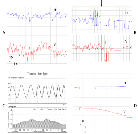
    Figure 1:  Video-nystagmographic recording of a vestibular migraine patient.
Panel A shows supine position with head center. Note the upbeat vertical nystagmus in the vertical eye movement. Panel B shows abolishment of nystagmus by
fixation (the arrow indicates the start of fixation).
Panel C shows catch up saccades in smooth pursuit testing.
Panel D shows disappearance of nystagmus after 2 weeks of Topamax treatment. Recording of horizontal eye movement (H) and vertical movement (V) are shown.
Diagnosis and management
Absence of neurological signs and normal MRI excluded Central Nervous System (CNS) disorder. The positional nystagmus did not have the main pathognomonic feature of BPPV, which is the peak or the paroxysm. Therefore, nystagmus does not fit the diagnosis of BPPV. On the other hand, the patient fulfills the criteria for VM according to International Classification of Headache Disorder (ICHD) committee of the International Headache Society (IHS) in its 2013 last edition (ICHD-3, 2013) [1]. These diagnostic criteria are: at least five episodes of moderate or severe intensity vestibular symptoms, current or past history of migraine without aura or migraine with aura, at least 50% of the episodes are associated with at least one of migrainous features (i.e., migrainous headache, photophobia, phonophobia, and visual aura) and the symptoms are not accounted by another vestibular disorder or another diagnosis listed in the international classification of headache disorder, 3rd version (ICHD-3, 2013). Applying these criteria show that our patients fulfills the diagnostic criteria of VM as she had past history of migraine, and had many episodes of spontaneous vertigo of severe degree associated with phonophobia. In addition, the symptom and signs are not accounted by other disorder.
Based on the diagnosis of VM, Topamax drug was described for the patient in a dose of 1 tablet (25mg) per day. Topamax medication is commonly described for the migrainous patients. It is an agonist for the GAPA inhibitory neurotransmitter. The drug was described for 2 weeks and the patient was re-examined. She had complete relief from the positional vertigo, and disappearance of the nystagmus. The patient was advised to continue taking the drug for 6 months in addition to other prophylactic measures for migraine. During this period, the patient was interviewed each 2 months and no symptoms recurred or nystagmus appeared.
Conclusion
Although VM is a very common cause of spontaneous recurrent vertigo in adults [2], it often missed as a cause for vertigo. In the majority of patients, VM manifest as recurrent attacks of spontaneous vertigo. Occasionally, VM presents with positional vertigo and positional nystagmus in the absence of spontaneous episodic vertiginous attacks or spontaneous nystagmus [3]. Our case belongs to this category of VM patients. Thorough history taking, nystagmus characters that differentiate VM nystagmus from that of the BPPV, and exclusion CNS diseases will help in the diagnosis. Anti-migraine medications are considered in such case. Symptom relief and disappearance of nystagmus with medications establish the diagnosis.
References
- Headache classification committee of International Headache Society (IHS). The international classification of headache disorders, 3rd edition (beta version). Cephalagia. 2013; 33: 629-808.
- Strupp M, Versino M, Brandt T. Vestibular migraine. In: Handbook of Clinical Neurology, Vol. 97 (3rd series) Headache, Nappi G, Moskowitz (Eds), Elsevier B.V. 2011; 755-771.
- Dieterich M, Brandt T. Episodic vertigo related to migraine (90 cases: vestibular migraine?) J Neurol. 1999; 246: 883-892.