
Research Article
Austin J Otolaryngol. 2016; 3(3): 1081.
Comparison between Millard’s Rotational Advancement Flap and Tennison-Randall Flap Techniques for Surgical Correction of Unilateral Cleft Lip Deformity
Gadre P*, Borle R, Rudagi BM, Bhola N and Yadav A
Department of Oral and Maxillofacial Surgery, Sharad Pawar Dental College, Sawangi Meghe, Wardha, Maharashtra, India
*Corresponding author: Pushkar Gadre, Department of Oral and Maxillofacial Surgery, Consultant Gadre Clinic, 2132, Vijaynagar Colony, 204 ‘Trimurti’ Apartments, Above Hotel Masemari, off Tilak Road, Behind Girja Juice Center, Maharashtra, India
Received: July 16, 2016; Accepted: September 28, 2016; Published: September 30, 2016
Abstract
A congenital cleft lip deformity has significant physical and psychological. Successful repair of cleft lip deformity is a challenging as well as rewarding task. Though localized to a small anatomic area, the face it demands more attention and priorities. It is a three-dimensional anomaly involving hard tissue that changes in the fourth dimension with growth and function. The treatment goals of correction are early tension-free correction to attain an early tension free closure and have mobile and balanced lip. Many techniques have been used since eons for the correction of the unilateral cleft lip deformity and each has its own merits and demerits.
The present study was carried out in the Department of Oral and Maxillofacial Surgery, Sharad Pawar Dental College and Hospital and in Acharya Vinobha Bhave Rural Hospital, (AVBRH) Sawangi, Wardha. Surgeries were performed on consecutive patients with unilateral cleft lip deformity. 60 unilateral cleft lip patients were randomly assigned to two groups, each compromising of thirty patients (Millard’s -Group M and Tennison- Randall- Group T).
All the patients were evaluated preoperatively, on 7th postoperative day and on one month follow up. Comparison between the two techniques was made keeping in mind the aesthetic and functional aspects of the repair.
Keywords: Unilateral Cleft Lip (UCL); Millard’s rotational advancement flap; Tennison-Randall flap; Lip Notching; Scar; Alar base symmetry
Introduction
The comprehensive management of cleft lip and palate has received significant attention in the surgical literature over the last half century. It is the most common congenital facial malformation. It has a significant developmental, physical, and psychological impact on the affected individuals and their families. Treatment of the deformity presents a constant challenge and hence, a plethora of treatment philosophies have been propounded. Each philosophy has its ardent advocates, as well as equally emphatic opponents [1]. Successful correction of the deformity is one of the most professionally satisfying experiences for a surgeon.
Compared with the non-cleft individuals, the three groups of superficial facial muscles (i.e., the nasolabial, bilabial, and labiomental) are all displaced inferiorly. The orbicularis oris muscle finds a new and abnormal insertion on the left side and a partially distorted insertion on the non-cleft side. The Cupid’s bow on the left side and the white skin roll on either side are also distorted [2].
The treatment goals for cleft lip defects are early correction of the cleft, with primary correction to a tension-free, mobile, and balanced lip. The repair of any cleft lip deformity should not only be accounted for a mere closure but, also a functional anatomical repair of the underlying hard and soft tissues [2].
One of the most popular methods for Unilateral Cleft Lip [UCL] repair is the original or modified rotational advancement technique of Dr Millard which was given in 1955. Through the years, he added various refinements to his initial technique. Many authors have reported its modifications [3].
Tennison and Marcks (1950-1960) and colleagues introduced triangular flap which created a Z-plasty at lower part of lip. Subsequently, Randall used the same design as Tennison but, reduced size of triangular flap [4].
Aim and Objective
To compare the outcomes of two different surgical techniques namely Millard’s Rotational Advancement Flap Technique and Tennison- Randall Flap Technique or Triangular Flap Technique for correction of UCL defect in terms of aesthetic and function and to find out a better suited technique amongst the two.
Materials and Methods
The prospective clinical randomized clinical trial was carried out in The Department of Oral and Maxillofacial Surgery, Sharad Pawar Dental College and Hospital and in Acharya Vinobha Bhave Rural Hospital, (AVBRH) Sawangi, Wardha between August 2010 to May 2012. Approval for the present study was obtained from our institution’s Experimental Medical Research and Practicing Center Ethical Committee. Informed consent was obtained from all patients who were enrolled in the study.
Sixty unilateral cleft lip patients were included in present comparative study from the patients reporting to our department. The UCL patients were randomly assigned to be treated by one of the technique.
Group M: Patients treated using Millard’s Rotational Advancement Flap- 30 patients.
Group T: Patients treated using Tennison-Randall Flap (Triangular Flap technique) - 30 patients.
The inclusion criteria were – (a) Non syndromic patients, (b) UCL patients with complete or partial cleft, (c) Age ranging from 6 months to 60 years, (d) Either of the sexes and (e) ASA I and ASA II category. Patients with Orofacial cleft, Bilateral cleft lip, and requiring secondary lip revision with ASA III and ASA IV were excluded from the study.
All the patients were hospitalized two days prior to surgery to facilitate investigations and complete pre-surgical and preanesthetic evaluation. The patients were counseled by the cleft team comprising of surgeon, orthodontist, anesthetist and psychiatrist. They underwent a mandatory pre-surgical dental checkup and orthodontic treatment whenever necessary. All patients received the same preanasthetic medication which included anxiolytics, laxative, antacids and antibiotics.
Operative technique
The patients were operated by a team of senior consultants, but their surgical differences were minimized in the present study by following a standard protocol of surgical procedure. A standard aseptic principle and optimum degree of sterilization of instruments followed in all the surgeries.
Standard Triangular Flap Technique (Group T) (Figure 1) Millard’s Rotational Advancement Flap Technique (Group M) (Figure 2) and was performed for 30 patients each. For the patients below the age of 4 yrs and having associated cleft palate, anterior palatal repair was carried out in single stage. This was done in order to avoid anterior palatal fistula occurrence during the later palatal repair.
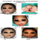
Figure 1: Tennison- Randall technique.
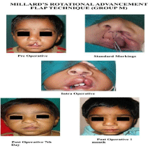
Figure 2: Millard’s rotational advancement technique.
Lip closure was performed was carried out in layers composing of muscle and subcutaneous suturing using 4-0 Vicryl suture respectively and 6-0 ethilon sutures were placed in the vermilion and the mucosa of the lip completing the closure. The nostril sill was closed with ethilon sutures. The alar cartilage on the left side was repositioned independently of the overlying alar skin by placing a through-andthrough suture tied over a bolster for duration of 1 week.
Antibiotics, analgesics and antacids were administered to the patients via suitable route till 7th postoperative day.
Suture removal was done on 7th postoperative day, followed by discharge and instructions on wound care. Subsequently, the patients were followed on outdoor basis on the 30th post operative day.
In this prospective cohort study, analysis of two types of techniques for evaluating the residual facial deformity in patients with repaired unilateral cleft lip with preoperative and post operative findings was be based upon following criteria’s regarding:
- White roll match
- Scar quality (i.e. satisfactory, hypertrophic or stretched)
- Cupids bow symmetry
- Lip length
- Lip height
- Notching
- Alar base symmetry
We used Vernier caliper for lip length and height measurements as used by Court B. Cutting and Joseph H. Dayan [5].
The points for the measurements were
1. Lip height=The measurements from highest point of cupid’s bow to the base of columella on cleft and non cleft side, respectively (Figure 3).
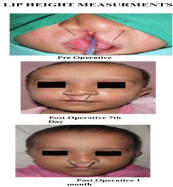
Figure 3: Lip Height.
2. Total lip length=Sum of measurements from corner of mouth to the fading of white roll on cleft side and from corner of mouth to the fading of white roll on non cleft side (Figure 4).
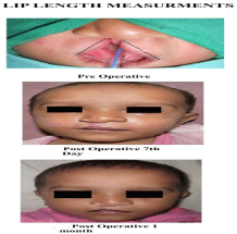
Figure 4: Lip Length.
Data was collected preoperatively, on 7th and 30th postoperative days. Complete analysis was done by same observer for more consistent results. The analytic observations were based on demographic data of patients (age, sex, side of cleft lip involved and the type of defect), the aesthetic values like white roll match, scar quality, alar base symmetry, Cupid’s bow symmetry, notching. The functional analysis was done with respect to lip length and lip height of upper lip (Figure 5, 6).
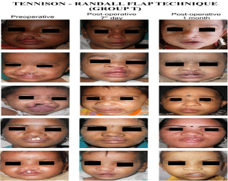
Figure 5: Group T.
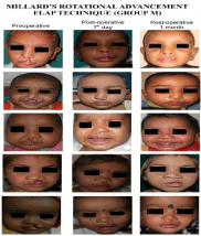
Figure 6: Group M.
Statistical analysis
The study variables like white roll match, cupids bow symmetry, scar quality, notching and alar base symmetry were analyzed using parametric test like ‘Fisher’s Exact Test’ test and non parametric test like ‘Unpaired t Test’ and ‘Paired t Test’ test for testing significant difference between pre and post surgical observations.
Results
Of the 60 patients enrolled in the study, Group M had mean age of 12 months with 17 patients between age group of 1 to 5 years. Similarly, UCL patients in Group T were mostly less than 1 year of age (i.e. 16 patients) with 11 months as their mean age.
The total male patients were 39 to females adding up-to 21. Equal male and female distribution was present within the two groups. Cleft lip of left side was prominently observed 19 in Group M and 24 in Group T respectively adding to 43 patients out of 60.
40 patients had complete unilateral cleft lip and 20 had incomplete unilateral cleft lip. The patients having complete cleft lip anomaly in Group M were 16 and that in Group T were 24. While the number of patients involving incomplete unilateral cleft lip deformity were 14 and 6 in Group M and T respectively.
The comparison with respect to the presence of white roll match on 7th postoperative day and on follow up presented similar results (Table 1). The total number of patients having white rolls matched postoperatively and on follow up were found out to be 53 patients to that of white roll match absent was 7 patients. In general, more patients in Group T showed presence of white roll matched.
    Post Op. Day
    WHITE ROLL MATCH
    Group M
    Group T
    Fisher’s Exact Test
      p value


    Day-7
    Present
    25

    28
    0.424 NS


    Absent
    5
    2


    Day-30
    Present
    25
    28
    0.424 NS


    Absent
    5
    2
Table 1: Postoperative and on follow-up white roll match in each group.
On 7th postoperative day, the patients with satisfactory scar quality in the Group T (27 patients) were more than in Group M (19 patients). ‘P’ value came out to 0.03 and was significant (Table 2). But, an improvement in the scar quality was seen in Group M on follow up. Though insignificant, the patients in Group M had symmetrical alar base in all three analysis days than those in Group T (Table 3).
    Post Op. Day
    SCAR QUALITY
    Group M
    Group T
    Comparison of Satisfactory Proportions.
      Fisher’s Exact Test
      p value


    Day-7
    Satisfactory
    19
    27
    0.03 Sig


    Stretch
    8
    2


    H
    3
    1


    Day-30
    Satisfactory
    21
    27
    0.104 NS


    Stretch
    5
    2


    H
    4
    1
Table 2: Postoperative and on follow-up scar quality in each group.
    Analytic Day
    ALAR BASE SYMMETRY
    Group M
    Group T
    Fisher’s Exact Test
      p value


    Day - 0
    Symmetry
    10
    6
    0.382 NS


    Asymmetry
    20
    24


    Day -7
    Symmetry
    19
    15
    0.435 NS


    Asymmetry
    11
    15


    Day -30
    Symmetry
    20
    15
    0.295 NS


    Asymmetry
    10
    15
Table 3: Alar Base symmetry in each group.
With respect to Cupid’s bow symmetry, the total number of patients with symmetrical Cupid’s bow were 50 overall on 7th postoperative day. This value reduced to 46 patients on one month follow up. Group M had less number of patients with symmetrical Cupid’s bow than those in Group T (Table 4). Group T patients had less incidences of lip notching than those in group M. Also presence of notching in group M increased from 2 to 6 on follow up to that on 7th postoperative day. Similarly 1 patient presented with lip notching on follow up in Group T (Table 5).
    Post Op. Day
    CUPIDS BOW SYMMETRY
    Group M
    Group T
    Fisher’s Exact Test
      p value


    Day -7
    Symmetry
    23
    27
    0.299 NS


    Asymmetry
    7
    3


    Day -30
    Symmetry
    21
    25
    0.360 NS


    Asymmetry
    9
    5
Table 4: Cupid’s Bow symmetry postoperatively and on follow-up in each group.
    Post Op. Day
    Notching
    Group M
    Group T
    Fisher’s Exact Test
      p value


    Day -7
    Absent
    28
    30
    0.492 NS


    Present
    2
    0


    Day -30
    Absent
    24
    29
    0.103 NS


    Present
    6
    1
Table 5: Notching postoperatively and on follow up in each group.
The total lip length, in both the groups remained approximately same on 7th postoperative and one month follow up day. The lip length reduced more in Group T postoperatively than that to preoperative analysis (Table 6). The cleft side lip height increased postoperatively in both groups. But, no significant change in lip height was observed on the analytic days (Table 7).
    Analytic Day
    LIP LENGTH (mm)
    Group M
    Group T
    Unpaired t
    p value


    Day - 0
    Mean
    43.20
    40.73
    1.409
    0.164 NS


    Sd
    7.508
    5.965


    Day -7
    Mean
    42.07
    40.03
    1.179
    0.243 NS


    Sd
    7.446
    5.810


    Day -30
    Mean
    42.07
    40.10
    1.139
    0.259 NS


    Sd
    7.446
    5.827
Table 6: Comparison of lip length in each group.
    Analytic Day
    LIP HEIGHT (Cleft side) (mm)
    Group M
    Group T
    Unpaired
      t
    p value


    Day - 0
    Mean
    8.70
    7.90
    1.466
    0.148 NS


    Sd
    2.307
    1.90


    Day -7
    Mean
    9.90
    9.13
    1.301
    0.198 NS


    Sd
    2.51
    2.03


    Day -30
    Mean
    9.87
    9.13
    1.239
    0.220 NS


    Sd
    2.53
    2.03
Table 7: Comparison of lip height in each group.
Discussion
The goals of UCL repair include the creation of an intact upper lip with appropriate vertical length and symmetry, repair of the underlying muscular structures to achieve normal function, and the management of the associated nasal deformity. The Tennison – Randall and Millard’s rotational advancement flap technique remains the most accepted techniques. With the need of time and situation, certain modifications in both techniques are made and combinations of both have been utilized.
In the present study, the presence of white roll match on 7th postoperative day and on one month follow up did not vary. Rajesh S. Powar et al [7] suggested that the white roll matching is important for balanced repair of cleft lip. A comparison between three incisions (Millard incision, Pfeifer wave line incision, or Afroze incisions) by Srinivas Gosla Reddy et al [2] concluded the Millard’s incision presented with least white roll matched.
The postoperative scar appearance in 27 and 19 patients of Group T and M respectively was satisfactory. However, difference was noticed on 1 month follow up, the satisfactory scar quality was seen in 21 and 27 patients of group M and T, respectively. This suggests that the scar remained constant in Group T on postoperative 7th day and on follow up of one month .But, the incisional scars settled satisfactorily in Group M patients on follow up. The observations of present study match those of Barbel Holtmann and R. Chris Wray [6] who compared Triangular and Rotation- Advancement flaps for correction of UCL repair concluded that the frequency of hypertropic scar following rotation- advancement repair was more than triangular flap. Similarly, N.A. Chowdri et al [8] stated that, more patients (i.e. 6.5%) treated by the rotation-advancement flap developed hypertrophic scar in comparison to those treated with the triangular flap.
The alar base symmetry showed improvement postoperatively with twice the number of patients presenting with symmetrical alar base in the Group M. However, in the Tennisson group, the number of patients having symmetrical alar base remained constant postoperatively. These results match the study conducted by Barbel Holtmann and R. Chris Wray [6] and Chowdri et al [8] where in, they found out that overall appearance of lip and nose postoperatively in Millard’s and Tennison –Randall techniques was same. These findings might be due to the better access to the alar cartilage and base gained by use of Millard’s rotational advancement technique.
The studies by Linas Zaleckas et al [9] and Tomohiro Yamada et al [10] where in, they observed better symmetry of the Cupid’s bow been restored by use of Tennison’s technique, contradicts the findings reported in the present study.
Overall, lip notching was less in group T. The patients presenting with lip notching on follow up can be attributed to the contracture of the incisional scar. Taik Jong Lee et al [11] indicated that a repaired unilateral cleft retained the vertical and horizontal dimensions determined at the time of the initial repair. Also, the comparison between Fisher technique and rotational advancement flap technique by Donita Dyalram- Silverberg et al [12] concluded that the Fisher technique shows virtually no vermillion border notching and was better than rotational advancement technique.
The comparison between the preoperative to 7th postoperative day and preoperative day to one month follow within the groups M and T, respectively presented with a decrease in total lip length postoperatively. In contrast, the comparison between postoperative days 7th and 30th within the two groups gave a non significant result. Thus showing that, not much change had occured on one month follow up in the lip length. Srinivas Gosla Reddy et al [8] concluded that Afroze incision is superior with regard to lip length and always gave superior results compared to the Millard or Pfeifer incision. H. Xing et al [13] on comparing preoperative and 1-year followup measurements of the modified Millard’s lip repair showed no change in linear correlation between the preoperative and 1-year postoperative lip length.
Overall, postoperatively the lip height increased on the left side. The findings are consistent with those of N.A. Chowdri et al [8] who concluded that the cleft lip height measurements were having no influence on outcomes of comparison of the two techniques.
Thus, the present study suggests that both Millard’s rotational advancement and Tennison –Randall technique gave similar kind of results with respect to white roll match, alar base symmetry, Cupid’s bow symmetry and the lip length. However, the incisional scars on 7th postoperative day in patients treated with Tennison- Randall technique was significantly better than those patients treated using Millard’s rotational advancement flap technique. Also, though insignificant, the lip notching in number of patients was less in patients treated with Triangular technique. Postoperatively, cleft side lip height improved in group M patients. The saying ‘Cut as you go.’ stands true. During the surgical procedure, the use of Millard’s technique was found to be simpler to perform than the triangular flap technique as also, the access to alar cartilage was possible with the Millard’s procedure. In contrast, the Tennison-Randall technique was found to be mathematically precise.
Thus, the choice of technique for surgical correction of UCL should be based on evidence that shows the best functional and aesthetic outcomes. Certain preoperative anatomical features may lead the surgeon to choose one particular incision pattern in preference to another, but in this study it was found that both the techniques of repair can be used satisfactorily for correction of unilateral cleft lip deformity. The findings of this study support the view that these two methods of cleft lip repair have their own advantages and disadvantages. It is desirable that the operator be familiar with several of the excellent methods of lip repair, their modifications, so that the best procedure for each case can be selected.
A long term follow up of patients along with dynamic lip movements is required for better analysis and comparison of the two techniques of unilateral cleft lip repair.
Our results might be altered with cleft size, severity of defect and age at which surgical intervention was carried out.
References
- Sykes JM, Tollefson TT. Management of cleft lip deformity. Facial Plastic Clinics of North America. 2005; 13: 157-167.
- Reddy GS, Reddy RR, Bronkhorst EM, Prasad R, Jagtman AMK, Berge S. Comparison of Three Incisions to Repair Complete Unilateral Cleft Lip. Plastic and Reconstructive Surgery. 2010; 125: 1208-1216.
- Koh KS, Hong JP. Unilateral complete cleft lip repair: orthotopic positioning of skin flaps. BJPS. 2005; 58: 147–152.
- Lazarus DD, Hudson DA, Fleming AN, Fernandes D. Repair of unilateral cleft lip: Comparison of five techniques. Annals of Plastic Surgery. 1998; 41: 587- 594.
- Cutting CB, Dayan JH. Lip Height and Lip Width after Extended Mohler Unilateral Cleft Lip Repair. Plastic and Reconstructive Surgery. 2002; 111: 17-23.
- Barbel H, Wray RC. Randomized comparison of triangular and rotation advancement unilateral cleft lip repairs. Plast. & Reconstr Surgery. 1983; 71: 172-179.
- Powar RS, Patil SM, Kleinman ME. A geometricallysound technique of vermilion repair in unilateral cleft lip. Journal of Plastic, Reconstructive & Aesthetic Surgery. 2007; 60: 422-425.
- Chowdri NA, Darzi MA, Ashraf MM. A comparative study of surgical results with rotation advancement and triangular flap techniques in unilateral cleft lip. British Journal of Plastic Surgery. 1990; 43: 551-556.
- Zaleckas L, Linkeviciene L, Olekas J, Kutra N. The Comparison of Different Surgical Techniques Used for Repair of Complete Unilateral Cleft Lip. Medicina (Kaunas). 2011; 47: 85-90.
- Yamada T, Mori Y, Minami K, Mishima K, Sugahara T. Three-dimensional facial morphology, following primary cleft lip repair using the triangular flap with or without rotation advancement. Journal of Craniomaxillofacial Surgery. 2002; 30: 337–342.
- Lee TJ. Upper Lip Measurements at the Time of Surgery and Follow- Up after Modified Rotation-Advancement Flap Repair in Unilateral Cleft Lip Patients. Plastic & Reconstructive Surgery. 1999; 104: 911-915.
- Dyalram-Silverberg D, Jamali M, Hoffman D, Lazow SK, Berger JR. Comparison of the Rotational Advancement Flap and an Anatomical Subunit Approximation Technique for Closure of Unilateral Cleft Lip - poster in American Association of Oral and Maxillofacial Surgery. 2006; 80-81.
- Xing H, Bing S, Kamdar M, Yang L, Qian Z, Sheng L, Yan W. Changes in lip 1 year after modified Millard repair. Int Journal of Oral and Maxillofacial Surgery. 2008; 37: 117–122.