Case Presentation
Austin J Otolaryngol. 2017; 4(2): 1092.
Epidermal Choristoma Presenting as an Enlarging Tongue Mass
Lindquist NR¹, Firan M² and Mukerji SS¹*
¹Department of Otolaryngology – Head and Neck Surgery, Baylor College of Medicine, USA
²Department of Pathology and Immunology, Baylor College of Medicine, USA
*Corresponding author: Shraddha Siddharth Mukerji, Department of Otolaryngology, Texas Children’s Hospital, Clinical Care Tower, Houston, USA
Received: June 23, 2017; Accepted: July 14, 2017; Published: July 21, 2017
Abstract
Epidermal choristoma is an extremely rare entity characterized by histologically normal-appearing skin with associated hair follicles, sebaceous glands, and adnexa in an abnormal location. When localized to the oral cavity, this lesion presents most-commonly as an asymptomatic hyper pigmented macule or patch on the lingual dorsum of a male patient. Herein, we present an unusual case of an epidermal choristoma presenting as an acutely infected mass of the deep anterior tongue in a teenage female.
Keywords: Epidermal choristoma; Follicular choristoma; Congenital tongue mass
Introduction
Choristomas are a developmental abnormality characterized by the proliferation of histologically normal tissue in an ectopic location. Choristomas of the head and neck have been noted in the tongue [1], floor of mouth [2], nasopharynx [3], pharynx [4], hypopharynx [5], and submandibular [6] regions and are clinically important lesions as they may present as a cause of airway obstruction or feeding/ swallowing difficulties, especially in the pediatric population [7]. In the oral cavity, heterotopic tissue consistent with choristoma has been reported with otherwise histologically-normal salivary gland, cartilaginous, osseous, thyroid, sebaceous, glial, gastric mucosa, and epidermal tissues although cartilaginous, osseous, and lingual thyroid choristomas are relatively more common [7]. “Epidermal choristoma” or “cutaneous choristoma” is defined by the presence of stratified squamous epithelium (epidermis) with associated adnexal structures including sebaceous glands, apocrine glands, and hair follicles. Epidermal choristoma is a very rare lesion of the oral cavity, with only five reported examples in the literature to date. All reported cases have occurred in males, with the majority presenting as a hyper pigmented macule or plaque (80%), most commonly of the dorsal tongue [8]. Follicular choristoma appear to be a related lesion defined by pigmented epidermis, sebaceous glands, sweat glands, and mature hair follicles with the additional finding of keratin-containing cysts. To date, there are two cases of follicular choristoma reported in the English language literature. Herein, we report a unique example of an epidermal choristoma presenting as an infected tongue mass in a female teenager.
Case Presentation
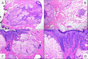
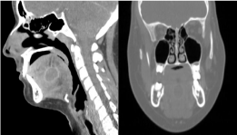
A 14-year-old female with no significant medical history presented to the emergency room with two days of tongue swelling, painful swallowing, fever, and difficulty speaking. The patient complained of some difficulty in breathing. Physical exam revealed moderate swelling and tenderness of the dorsum of the tongue with a 1cm right paramedian mass just anterior to the circumvallate papillae. Of note, there was no pigmentation changes or ulceration reported. The patient did not have stridor. Laboratory studies showed mild leukocytosis. Computerized Tomography (CT) scan with contrast demonstrated a rim-enhancing hypodense mass in the deep midline tongue measuring 2.2 x 1.6 x 1.8 cm with mild bilateral suprahyoid jugular chain lymphadenopathy (Figure 1). The patient was started on a course of intravenous clindamycin and dexamethasone for continued swelling of the tongue and concern for airway compromise. Upon clinical improvement the patient was discharged with oral clindamycin, steroid taper, and close follow-up for definitive treatment. Interval surgical excision of a 1.0 x 1.0 x 0.2 cm tan, rubbery, soft mass was performed in the operating room with nasotracheal intubation and general anesthesia using sharp dissection and electrocautery. No cystic structure or keratinaceous debris was identified grossly. Histopathologic analysis demonstrated keratinizing squamous epithelium with acanthosis and hypergranulosis associated with abundant sebaceous glands, apocrine glands rare hair follicles, and mature adipose tissue overlying a segment of excised lingual skeletal muscle. No melanin pigment was identified histologically (Figure 2). The patient has been symptom free and without any evidence of recurrence at the one month follow-up period.

    Figure 1:  Sagittal and coronal views of contrast-enhanced CT images
demonstrate a hypo-intense, rim-enhancing, low-attenuation mass in the
midline deep tongue superior to the geniohyoid-genioglossus complex and
measuring 2.2 x 1.6 x 1.8 cm (anteroposterior by transverse by craniocaudal).

    Figure 2:  Histopathological photomicrographs. A,B: Low power view (4X)
showing squamous keratinizing epithelium with sebaceous glands and hair
follicles overlying mature adipose tissue with minor salivary glands and
skeletal muscle. C: Medium power view (10X) showing acanthosis and
hypergranulosis of the squamous epithelium as well as sebaceous glands,
apocrine glands and hair follicle. D: Medium power (20X) view showing
absence of melanin pigment.
Discussion
The presence of ectopic epidermal, sebaceous, or adnexal tissue in the oral cavity is not uncommon. However, the association of such structures with hair follicles is rare [9,10]. In 1973 Arwill et al. termed “follicular choristoma” or “folliculoma” for a previously unreported lesion of hair follicles with sebaceous glands, melanocytes, and keratin-containing cysts in the alveolar gingiva of a nine-year-old female [11]. Similarly, Azorin et al. coined the term “epidermal choristoma” in 2005 to describe a pigmented macule exhibiting hyperorthokeratosis, hypergranulosis, melanin pigmentation, sebaceous glands, hair follicles, and apocrine glands in the tongue of a neonate [12]. Originally, this term was intended to describe a lesion meeting the criteria both for a melanotic macule as well as sebaceous glands in an ectopic location: the tongue.
After extensive literature search, there are only five other cases of epidermal choristoma reported in the English language literature (Table 1) [8,11,13-15]. Oral cavity epidermal choristomas commonly present in male patients as an asymptomatic brown or black-pigmented macule on the dorsum of the tongue (60%) with the majority carrying a presumptive clinical diagnosis of melanotic macule.8 Four out of the five cases previously reported have presented as an asymptomatic hyperpigmented macule or plaque. The remaining case presented as an elastic, soft polypoid mass with normal-appearing oral mucosa on clinical exam but melanin pigmentation clearly evident on histologic review [14]. None of the previously described cases showed evidence of local inflammatory reaction. Our case has several unique and unreported features that raise questions as to the ‘classic’ variant of epidermal and follicular choristomas. This is the first report of an epidermal choristoma in a female patient without any evidence of tongue pigmentation, as confirmed by histopathology. She was clearly symptomatic at the time of presentation and required oral antibiotics, steroids and airway monitoring. In addition, the radiological evaluation at presentation was that of a rim-enhancing hypodense mass concerning for infected foregut duplication cyst, dermoid cyst, or atypical thyroglossal duct cyst. No cystic structure was identified after excision raising the possibility that the initial presentation was due to enlargement of the sebaceous and/or apocrine glands that were missing the appropriate draining system normally seen in the skin. The patient responded well to initial conservative management followed by definitive surgical excision.
    Author
    Sex
    Age
    Location
    Size (mm)
    Presentation
    Clinical Diagnosis
    Diagnosis


    Arwill et al. (1973)
    Female
    9 years
    Mandibular alveolar gingiva
    Not specified
    Tumor-like nodular projection
    Benign tumor
    Follicular choristoma


    Azorin et al. (1973)
    Male
    1 month
    Dorsal tongue
    2 – 6
    Three brown macules
    Melanotic macule
    Epidermal choristoma


    Chi et al. (2010)
    Male
    32 years
    Buccal mucosa
    11 x 8
    Brown-and-white plaque
    Biting trauma vs. melanoma
    Epidermal choristoma


    Chi et al. (2010)
    Male
    56 years
    Dorsal tongue
    3
    Brown macule
    Melanotic macule
    Epidermal choristoma


    Curto-Barredo et al. (2015)
    Male
    1 month
    Dorsal tongue
    4 x 3
    Pigmented macule
    Melanotic macule
    Epidermal choristoma


    Sood et al. (2000)
    Male
    46 years
    Anterior floor of mouth
    10 x 10 x 5
    Hair growth
    Sublingual dermoid cyst
    Follicular choristoma


    Yoshioka et al. (2012)
    Male
    2 months
    Maxillary alveolar gingiva
    10 x 6 x 5
    Polypoid mass
    Congential epulis
    Epidermal choristoma


    Current case
    Female
    14 years
    Deep dorsal tongue
    10 x 10 x 2
    Infected cyst, fever, painful swallowing
    Foregut duplication cyst vs. dermoid cyst
    Epidermal choristoma
Table 1: Reports of epidermal and follicular choristomas.
The development of epidermal and follicular choristoma is not well-understood. Sood et al. proposed that epidermal and follicular choristoma stem from relics of ectodermal and dermal adnexa as the result of aberrant embryogenesis or the marsupialization of previously undetected dermoid cysts [15]. There is the possibility that pluripotent epithelium in the oral cavity may differentiate toward skin elements rather than normal oral mucosa [13]. Acquired lesions co-localizing hair follicles and sebaceous glands in the oral cavity have also been noted from traumatic implantation of hair and the invagination of a cutaneous sinus tracts [13,16].
The differential diagnosis for a cystic mass in the pediatric oral cavity is broad but should include dermoid cyst, foregut duplication cyst, lymphatic or venous malformation, mucocele, atypical thyroglossal duct cyst, as well as oral cavity choristoma. A significant portion of pediatric choristomas in the tongue/floor of mouth present as symptomatic or asymptomatic cysts demonstrating respiratory, gastric, and squamous epithelium on pathologic analysis [1]. When symptomatic, this age group may present with signs of infection, difficulty feeding/swallowing, and partial airway obstruction. In terms of radiographic characterization, Magnetic Resonance Imaging (MRI) provides better soft-tissue definition without ionizing radiation, although CT is often more practical in terms of scheduling as well as the potential to avoid sedation for neonates and infants. In most cases, the treatment of choice is complete surgical excision with routine post-operative follow-up to confirm wound healing and absence of recurrence. Long term monitoring is generally not required, with no recurrences reported to date.
Conclusion
In summary, epidermal choristomas are exceedingly rare and possibly under-recognized lesions of the oral cavity, previously described as hyperpigmented macules or plaques of the dorsal tongue seen in male patients. Sebaceous, follicular, and epidermal choristomas, especially when located on the tongue, may represent a spectrum of the same lesion and they should be part of the differential diagnosis for cystic masses in the oral cavity. This is the first reported case of an epidermal choristoma in a female presenting with acute infection and concern for airway obstruction, emphasizing a significant complication for these lesions. Such lesions are best treated by complete surgical excision.
References
- Chai RL, Ozolek JA, Branstetter BF, Mehta DK, Simons JP. Congenital choristomas of the oral cavity in children. The Laryngoscope. 2011; 121: 2100-2106.
- Pentenero M, Marino R, Familiari U, Gandolfo S. Choristoma involving the floor of the mouth and the anterior tongue: a case of teratoid cyst with gastric and respiratory epithelia. J Oral Maxillofac Surg. 2013; 71: 1706-1711.
- Liao SL, Lien R, Hsueh C, Chung MT. Nasopharyngeal choristoma: case report and review of the literature. J Otolaryngol. 2003; 32: 68-70.
- Chang H, Ahn Y, Lim YS, Hah JH. Gastric choristoma of the oropharynx. Clin Exp Otorhinolaryngol. 2009; 2: 103-105.
- Van Abel KM, Carlson ML, Narendra S, Cofer SA, Thompson DM. Hypopharyngeal gastric choristoma in an infant resulting in airway obstruction. Archives of otolaryngology--head & neck surgery. 2011; 137: 947-951.
- Johann AC, Garcia BG, Nacif TR, de Freitas JB, do Carmo MA, Mesquita RA. Submandibular osseous choristoma. J Craniomaxillofac Surg. 2006; 34: 57-59.
- Chou LS, Hansen LS, Daniels TE. Choristomas of the oral cavity: a review. Oral surgery, oral medicine, and oral pathology. 1991; 72: 584-593.
- Batsakis JG, el-Naggar AK. Sebaceous lesions of salivary glands and oral cavity. Ann Otol Rhinol Laryngol. 1990; 99: 416-418.
- Batsakis JG, el-Naggar AK, Hicks MJ. Epithelial choristomas and teratomas of the tongue. Ann Otol Rhinol Laryngol. 1993; 102: 567-569.
- Arwill T, Heyden G, Ramstedt A. Follicular choristoma of the gingiva: a peculiar lesion. Oral surgery, oral medicine, and oral pathology. 1973; 35: 89-92.
- Azorin D, Enriquez de Salamanca J, de Prada I, Colmenero I, Gonzalez Mediero I. Congenital melanotic macules and Sebaceous Choristoma arising on the tongue of a newborn: epidermal choristoma? J Cutan Pathol. 2005; 32: 251-253.
- Chi AC, Mapes IL, Javed T, Neville BW. Epidermal choristoma of the oral cavity: report of 2 cases of an extremely rare entity. J Oral Maxillofac Surg. 2010; 68: 451-455.
- Yoshioka I, Marutsuka K, Igawa Ket, Jyunko Nagata, Maho Yoshida, Takashi Baba, et al. Epidermal choristoma arising on the midline gingiva as a congenital epulis: a case report. J Craniomaxillofac Surg. 2012; 40: 812-814.
- Sood V. An oral ‘follicular’ choristoma presenting in the anterior floor of the mouth. Dent Update. 2000; 27: 231-233.
- Mitchell DA. A bizarre facial sinus. Dent Update. 1994; 21: 303-304.