
Case Presentation
Austin J Otolaryngol. 2017; 4(2): 1093.
Cerebrospinal Fluid Nasal Fistula of the Lateral Sphenoid Recess, Associated with Idiopathic Intracranial Hypertension and Primary Empty Sella Syndrome
Vargas-Aguayo AM* and Sanchez-Castro GF
Department of Otorhinolaryngology, Centro Médico ABC, Mexico
*Corresponding author: Alejandro Martín Vargas Aguayo, Department of Otorhinolaryngology, Centro Médico ABC, Mexico
Received: June 19, 2017; Accepted: August 09, 2017; Published: August 16, 2017
Abstract
Current treatment of nasal Cerebrospinal Fluid (CSF) leaks, are preferable realized by endoscopic approach, and several techniques describe a high success rate when it comes to defect closure. However when associated with high intracranial pressure, the success rate decreases importantly without the establishment of a complete treatment. Up to 45% of the spontaneouslyappearing fistulas can be associated to an elevation in intracranial pressure. A prompt and adequate recognize of the clinical and radiological findings, such as meningoceles, empty sella syndrome, etc., represent a great impact on the prognosis. Complementary measures include long term medical treatment to decrease the production of CSF and weight loss. A 40 years old female patient with high body mass index, presented with symptoms of nasal CSF leak, diagnosis was made with biochemical parameters and radiological studies to determinate a bony defect on left sphenoidal lateral recess wall. The leak was successfully sealed in a multilayer fashion with complementary treatment.
Keywords: Cerebrospinal fluid leak; Spontaneous; Transanal endoscopic surgical procedures; Idiopathic intracranial hypertension
Introduction
The importance of recognizing the etiology of a disease, directly affects the prognosis of the patient. At present, when it comes to Cerebrospinal Fluid (CSF) nasal fistulas, there has been an ideological change concerning its cause. The current consensus establishes that many CSF nasal fistulas that appear “spontaneously” are associated with idiopathic intracranial hypertension and with empty sella syndrome, which comes up on imaging. The handling of this disease must be directed towards reestablishing normal physiology, in order to have a higher success rate. We will now present a case with that has the above mentioned characteristics, but with difficult endoscopic access to close the fistula (lateral sphenoid recess).
Clinical Case
40-year-old female patient, with spontaneous onset of rhinorrhea, accompanied by an intermittent pulsing headache that had been going on for 4 months and that had increased some 3 weeks before, after the patient took a 7-hour flight. The rinorrhea was constant, but was more evident when the Valsalva maneuvers were performed.
Physical exploration revealed slightly elevated arterial pressure, 130/90mm of Hg, a body weight of 84kg, a height of 1.67cm, with a body mass index of 30.1kg/m2. The nasal endoscopy identified a very moist nasal mucosa in the left nostril. Noticeable leak when the head was inclined, turbinates appeared to be normal. Both tympanic membranes showed no reaction to light, the oropharynx showed evidence of post-nasal hyaline discharge.
The rhinorrhea liquid was collected to make a cytochemical that was compatible with CSF, since Beta-2 Transferrin was not available.
In the computerized tomography of the nose and paranasal sinuses, a highly pneumatized sphenoid was observed with what appeared to be something with the density of soft tissue on it; a bone defect in the left lateral sphenoid recess, around the optical carotid recess, was also identified (Figures 1, 2). The T2 magnetic resonance of the skull gave evidence of a hyperintense signal coming from the shpenoid’s interior, strongly suggesting the presence of cerebrospinal fluid (Figure 3). Empty sella syndrome, which showed arachnoid herniation through the sellar diaphragm, also showed up in the T1 sequence, compressing the pituitary gland in the characteristic image of the anchor shape (Figure 4).
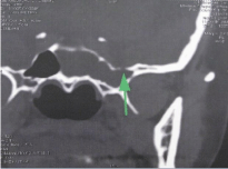
    Figure 1:  Computerized tomography, in coronal section. The bone defect
is noted in the posterosuperior portion of the lateral recess of the sphenoid.
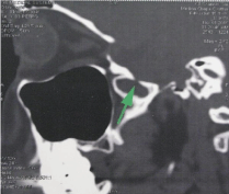
    Figure 2:  Computerized tomography, in the sagittal section. The bony defect
is noted in the posterosuperior portion of the lateral recess of the sphenoid.
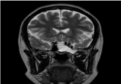
    Figure 3:  Magnetic resonance, T2 sequence, coronal section. Presence of
cerebrospinal fluid with a hypertense signal inside the left sphenoid sinus.
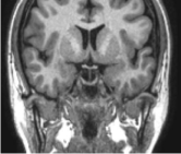
    Figure 4:  Magnetic resonance imaging, T1 sequence, coronal section.
Characteristic image of the “anchor sign”, which denotes arachnoid herniation
through the diaphragm seal.
With the clinical and radiographic data, a diagnosis of cerebrospinal fluid nasal fistula was given, located in the left lateral sphenoid recess, associated with idiopathic intracranial hypertension and empty sella syndrome. Cisternotomography was not considered necessary, since the site of the leak was identified.
A planned surgical intervention was carried out to close the fistula, utilizing an endoscopic approach. Fluorescein was administered at 10% (0.2ml diluted in 10ml of LCR), through the placement of a subarachnoid catheter, which operated as a temporary shunt for 4 days.
During surgery, an ample transnasal sphenoidotomy was carried out, becoming more ample with a posterior ethmoidectomy. A very pneumatized sphenoid was observed, as well as the presence of abundant CFS with fluorescein, coming from a meningocele located in the left lateral recess of the sphenoid, with a bone defect of approximately 5mm (Figures 5, 6). The tough lateral location of the bone defect made the use of instruments on the site difficult, for which reason it was decided use the “overlay” method to close it in layers, the dural regeneration matrix, autologous abdominal fat and fibrin glue (Figures 7, 8). The Valsalva maneuvers confirmed the absence of a leak and a temporary seal of absorbing material was applied.
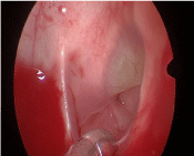
    Figure 5:  Intraoperative image, through the left sphenoid sinus, the bone
defect and menigocele are observed with fluorescein against light.
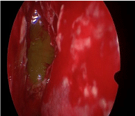
    Figure 6:  Intraoperative image. Bone defect posterior to the reduction of
menigocele.
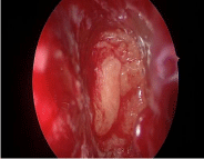
    Figure 7:  Transoperative image. Seal with autologous abdominal fat, with no
evidence of leakage of cerebrospinal fluid.
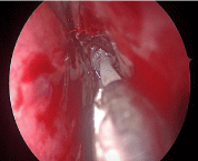
    Figure 8:  Transoperative image. Fibrin glue being applied over the dural
regeneration matrix and abdominal fat.
The patient had bed rest for 4 days, consumption of the arachnoid catheter was monitored, head elevated at 30º, laxatives. Administration of acetazolamide began immediately after surgery, as well as control of serum electrolytes. After removing the catheter, the patient began mobilization and could leave the hospital on the fifth postoperative day.
To this day, there have been no signs of recurrence of symptoms in the follow-up that has been done 1 year after surgery, but the patient continues using acetazolamide, since headaches flare up again when the medicine is discontinued.
Discussion
Spontaneously-appearing fistulas are those which occur despite an absence of a triggering event and up to 45% of them can be due to an elevation in intracranial pressure. Adequately recognizing the clinical and visual characteristics of intracranial pressure has a huge impact on planning and treatment, and therefore, on the prognosis of recurrence [1,2].
In which types of patients with spontaneous CFS nasal fistulas should primary empty sella syndrome, associated with idiopathic intracranial hypertension, be suspected. In female patients, middleaged, with a BMI above 30 and with accompanying characteristic symptoms such as intermittent headaches, pulsatile tinnitus or blurry vision. The anchor sign, characteristic of empty sella syndrome, should be intentionally sought out in the T1 sequence of the magnetic resonance [2].
Other radiographic findings associated with idiopathic intracranial hypertension are, arachnoid pits in 63% of patients,3 multiple bone defects in 31%,4 the formation of meningoceles in 50% - 100%, empty sella syndrome in 85% of the patients, bone thinning in the cranial base, dilatation in the nerve sheaths, etc [3-6].
Patients have an elevated CFS opening pressure at the time the lumbar puncture is performed. This elevation in the intracranial CFS has been associated with the possibility of greater failure in surgical closure (up to 25%) [2-7]. At the moment of the lumbar punction, the pressure of our patient’s cerebrospinal fluid was to be taken to be able to apply the fluorescein, but the device failed.
When a fistula is associated with idiopathic intracranial hypertension, the treatment of these should include complementary measures such as carbonic anhydrase inhibitors (acetazolamide); another important measure to implement is weight loss. Obesity represents another independent risk factor in the development of a CFS nasal fistula, raising the risk by 1.61, for each 5kg/m2 over a body mass index of 25kg/m2 [8-10]; CFS shunt, either by lumbar drainage or less frequently at present a ventrilculostomy; most of these shunts are maintained temporarily, however, some indications for permanent (lumboperitoneal or ventricular-motor) include high risk of failure, hydrocephalus, history of surgical failure or symptoms of intracranial hypertension that are not medically controllable.
There are many existing techniques for fistula closure. The “underlay” technique of placing a mesh in various layers has proven to be the most effective. In this case, the orientation and location of the defect and its proximity to vascular and nervous structures make it difficult and increase the risk of iatrogenic, for which reason the overlay technique was employed. Although the transpteygoid approach presents an alternative to improve the exposure of the defect, it is not exempt from possible comorbidities related to bleeding and never damage, without providing a surgical advantage, in this case. It was decided that the fistula would be sealed with a dural regeneration matrix, obliteration with fat and fibrin glue, since this is described as a valid technique for fistula closure [8].
Long-term follow-up for these patients is mandatory. All of the comorbidities that could allow for the reopening of the fistula must be prevented.
Conclusion
When treating patients with a spontaneous CFS nasal fistula, it must always be considered that this type of fistula is associated with idiopathic intracranial pressure. The intentional search for clinical data and imaging clues that could point to empty sella syndrome is mandatory. Quick and effective surgical treatment must be accompanied by measures which reduce the risk of relapse. The surgeon must know what the alternatives for surgical fistula closure are and must be prepared to resolve any scenario that doesn’t offer optimal conditions. The approach of making a permanent CFS shunt should be effectively communicated to the patient.
References
- Carrau RL, Snyderman CH, Kassam AB. The Management of Cerebrospinal Fluid Leaks in Patients at Risk for High-Pressure Hydrocephalus. The Laryngoscope. 2005; 115: 205-212.
- Chaaban MR, Illing E, Riley KO, Woodworth BA. Spontaneous cerebrospinal fluid leak repair: A five-year prospective evaluation. The Laryngoscope. 2014; 124: 70-75.
- Shetty PG, Shroff MM, Fatterpekar GM, Sahani DV, Kirtane MV. A retrospective analysis of spontaneous sphenoid sinus fistula: MR and CT findings. Am J Neuroradiol. 2000; 21: 337-342.
- Schlosser RJ, Bolger WE. Nasal cerebrospinal fluid leaks: critical review and surgical considerations. The Laryngoscope. 2004; 114: 255-265.
- Corbett JJ, Thompson HS. The rational management of idiopathic intracranial hypertension. Arch Neurol. 1989; 46: 1049-1051.
- Hubbard JL, McDonald TJ, Pearson BW, Laws ER Jr. Spontaneous cerebrospinal fluid rhinorrhea: evolving concepts in diagnosis and surgical management based on the Mayo Clinic experience from 1970 through 1981. Neurosurgery. 1985; 16: 314-321.
- Shetty PG, Shroff MM, Fatterpekar GM, Sahani DV, Kirtane MV. A retrospective analysis of spontaneous sphenoid sinus fistula: MR and CT findings. American journal of neuroradiology. 2000; 21: 337-342.
- Lee TJ, Huang CC, Chuang CC, Huang SF. Transnasal endoscopic repair of cerebrospinal fluid rhinorrhea and skull base defect: ten-year experience. The Laryngoscope. 2004; 114: 1475-1481.
- Chaaban MR, Illing E, Riley KO, Woodworth BA. Acetazolamide for high intracranial pressure cerebrospinal fluid leaks. In International forum of allergy & rhinology. 2013; 3: 718-721.
- Dlouhy BJ, Madhavan K, Clinger JD, Reddy A, Dawson JD, O'Brien EK, et al. Elevated body mass index and risk of postoperative CSF leak following transsphenoidal surgery. J Neurosurg. 2012; 116: 1311-1317.