
Case Report
Austin J Otolaryngol. 2017; 4(2): 1096.
Myositis Ossificians Traumatica of the Left Pterygoid Muscles
Department of Oral and Maxillofacial Surgery, Howard University Hospital, Washington, USA
Natoya Reid, Department of Oral and Maxillofacial Surgery, Howard University Hospital, Washington, District of Columbia, USA
*Corresponding author: Natoya Reid, Department of Oral and Maxillofacial Surgery, Howard University Hospital, Washington, District of Columbia, USA
Received: November 28, 2017; Accepted: December 20, 2017; Published: December 29, 2017
Abstract
Myositis Ossificans (MO) is a rare benign ossifying lesion that is self-limiting. It affects the soft tissue and may develop in the muscle, fat, nerves or tendons. Myositis ossificans can be categorized into nonhereditary and hereditary types which are distinct entities. There are very few reports of Myositis Ossificans in the masticatory muscles. The treatment is usually complex and the relapse and recurrence rates are high. This case study discusses the successful management of a patient who developed Myositis Ossificans after a gunshot wound injury. The patient had relapse after initial surgical intervention however, he has remained disease free after the second surgical intervention and implementation of aggressive and early physiotherapy. We review the diagnosis, clinical manifestations, possible causes and recommendations for prevention as described in the literature.
Case Presentation
A 37-year-old male patient with no significant past medical history underwent closed reduction with Intermaxillary fixation after sustaining a gunshot wound to the face in 2002. Two years later he had developed chronic trismus. He had undergone a left coronoidectomy to improve mouth opening at another facility. The procedure did not result in improvement of his trismus. After reviewing the patient history, physical exam, laboratory and radiographic studies, it was concluded that the patient suffered from Myositis Ossificans Traumatica involving the left Pterygoid muscles as a result of a gunshot wound suffered in 2002. On presentation, his Maximal Incisal Opening (MIO) was 15mm. Subsequently, the patient was taken to the operating room and an osteotomy of the lesion through a transoral approach and a brisement procedure were performed to mobilize the mandible. The range of motion improved immediately after the surgery, the patient had an MIO of 30mm.
Shortly after the procedure, the patient was incarcerated and was lost to follow up. Physiotherapy could not be successfully executed. The patient later returned to our clinic after his release from the correctional facility in 2012. On presentation his MIO was 5mm. At that time, he was unable to perform protrusive or excursive movements. Computerized tomography (CT) scanning revealed an ossification in the left medial and lateral pterygoid muscles (Figure 1). Three-dimensional CT showed the position and shape of the ossification and the dimension of fusion of the pterygoid muscles to the pterygoid plates (Figure 2).
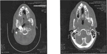
    Figure 1:  Left, Axial view showing initial presentation; Right, Axial view
showing the patient after relapse in 2012.
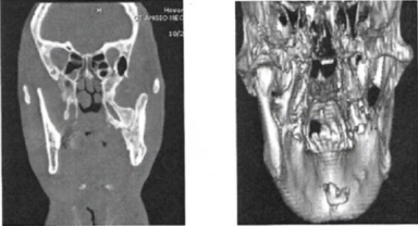
    Figure 2:  Left, coronal view showing heterotopic bone in the region of the
left Pterygoid plate and extends to the medial of the ascending ramus; Right,
three dimensional image showing the heterotopic bone formation.
All laboratory results including calcium, phosphorus, alkaline phosphatase, and parathyroid hormone levels were within normal limits. In October 2012, the patient was taken back to the operating room for an osteotomy of the left mandibular pterygoid heterotrophic bone, brisement procedure and reconstruction under general anesthesia.
The patient was brought to the operating room and an awake fiber optic-assisted nasotracheal intubation was performed. The performed osteotomy included the lateral aspect of the ramus, the body of the mandible including the entire Myositis Ossificans mass. A gap of 4cm was left and an interpositional abdominal fat graft (Figure 3) was placed between the medial aspect of the ramus and the pterygoid plates. A part of the osteotomized bone that was removed from the body and ascending ramus was replaced and held in position with a reconstruction plate (Figure 4). The patient started physiotherapy on day 5 postoperatively and remained in physiotherapy for 4 months. Figure 5 illustrates the post-operative changes on the panoramic view and Figure 6 illustrates the before and after surgery computed tomography images after his second surgery. The patient continues to be monitored on a frequent recall basis to assess for relapse or recurrence. He was last seen in June 2017 and has shown no evidence of relapse. He continues to have a full range of motion (Figure 7).
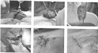
    Figure 3:  Abdominal Fat harvest was obtained using a transverse incision
with subcutaneous dissection making sure we were over the abdominal
muscular fascia.
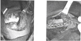
    Figure 4:  A part of the osteotomized bone that was removed from the body
and ascending ramus was replaced and held in position with a reconstruction
plate.
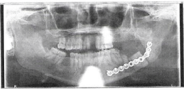
    Figure 5:  Post-Operative orthopanthomograph showing well adapted
reconstruction plate with 4 screws on the distal segment, one in the autograft
and 3 in the proximal segment.
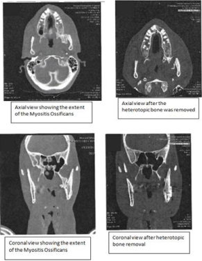
Figure 6: Axial view, Coronal view.
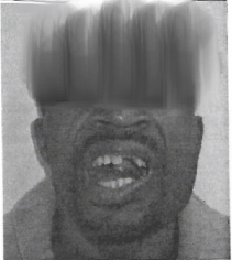
    Figure 7:  The patient continues to be monitored on a frequent recall basis to
assess for relapse or recurrence.
Discussion
The etiology of MOT is not completely understood. According to [1], the pathophysiology of Myositis Ossificans Traumatica is the implantation of active periosteum into a muscle seen in penetrating injuries. Another proposed mechanism is the overproduction of bone morphogenic protein-4 in response to injury [2], originally reported the Zone Phenomenon as typical histologic findings of Myositis Ossificans Traumatica. The zone phenomenon is composed of the inner zone consisting of undifferentiated cells with hemorrhagic and necrotic muscular tissue; the middle zone consists of osteoblasts and immature osteoid formation, and the outer zone consists of mature bone with collagenous fibrous stroma.
There are two types of Myositis Ossificans. The first type is known as Myositis Ossificans Progressiva (MOP) and is also referred to as Fibrodysplasia Ossificans Progressiva. It is inherited with an autosomal dominant pattern in which ossification typically occurs without trauma. The growth or progression of this condition follows a predictable pattern. The second, and more common of the two which is the case being reported, is Nonhereditary Myositis Ossificans also known Myositis Ossificans Traumatica (MOT), myositis ossificans circumscripta, Fibrodysplasia ossificans circumscripta or localized myositis ossificans. It involves calcifications at the site of injured muscle, most commonly in the arms or in the quadriceps of the thighs. Although, as cited in literature, in as many as 25 percent of cases, a history of trauma cannot be elicited and there is often little, if any, inflammation. Consequently, some authors have chosen to refer to the phenomenon as heterotopic ossification or pseudomalignant osseous tumor of soft issue. Unlike MOP, MOT is managed surgically, although some patients are refractory to treatment.
Patients often present with symptoms of trismus, pain and facial swelling. Trismus is the most common symptom. Diagnosis of MOT is usually both clinical and radiographic. The typical radiographic finding is a well-circumscribed, high-attenuating periphery, with a low-attenuating central portion. This was described by [3], as the pathognomonic feature of MOT. However, there have been varied radiographic presentations. Whenever the ossifying soft-tissue mass is detected it can be non-specific and malignancies must be ruled out.
Effective treatment is usually excision of the lesion and this is often done in conjunction with the brisement technique for early mobilization of the mandible. In the management of MOT, forced opening of the jaw with the application of brisement force has been an effective modality. In this procedure, the jaw is forced open by means of a mouth gag and mobilized as much as possible by forceful manipulation. Forced dilatation (brisement) is performed to ensure an incisal opening under anesthesia of > 50mm. The widest opening possible is produced at surgery to help ensure a stable incisal opening beyond 40mm on completion of physical therapy and rehabilitation.
Myositis Ossificans Traumatica as opposed to Myositis Ossificans Progressiva is reactive and also in contrast responds to surgical excision. The recurrence rate has been found to be high and as a result numerous adjunctive treatments have been proposed to improve success rate which have still proven futile in many cases. This poses a significant challenge to managing clinicians. Adjunctive treatments such as bisphosphonates, nonsteroidal anti-inflammatory agents, radiation and physiotherapy have been used to prevent recurrence after surgery. A few authors have reported the use of fat from the abdomen or buccal fat pad as interpositional grafts after excision of the lesion to improve post-operative success rate. In the case reported, we utilized fat graft from the abdomen as the interpositional material, as suggested by [4]. The graft is intended to prevent heterotrophic calcifications.
Physiotherapy is a critical component in the management of Myositis Ossificans. As with our patient, patients have relapses despite surgical treatment. The difference in the outcome at the second surgical intervention is attributed to the intense physiotherapy postoperatively. It is also important that due to the relapse potential that the patient has postoperative follow up [5-9].
We conclude that, in patients who present with trismus, Myositis Ossificans should always be a consideration, although it is a rare entity. The recurrence rate can be high, which is attributable to a poorly understood etiopathogenesis and optimal treatment approach. However, successful outcomes can be achieved. The case report demonstrates that surgical excision and aggressive physical therapy can provide a long-term and stable result. Further research is still needed on the specific causes and pathophysiology of Myositis Ossificans Traumatica which will result in more predictable treatment of the condition.
References
- Marx, Robert E and Stern, Diane. Oral and Maxillofacial Pathology: A Rationale for Diagnosis and Treatment, 2nd 2012.
- Ackerman LV. Extra-osseous Localized Non-Neoplastic Bone and Cartilage Formation (So-called Myositis Ossificans): clinical and pathological confusion with malignant neoplasms. J Bone Joint Surg Am. 1958; 40: 279-298.
- Boffano P, Zavattero E, Bosco G, Berrone S. Myositis ossificans of the left medial pterygoid muscle: case report and review of the literature of myositis ossificans of masticatory muscles. Craniomaxillofacial Trauma Reconstr. 2014; 7: 43-50.
- Thangavelu A, Vaidhyanathan A, Narendar R. Myositis ossificans traumatica of the medial pterygoid. Int J Oral Maxillofac Surg. 2011; 40: 545-558.
- Ellis E, Walker RV. Treatment of Malocclusion and TMJ Dysfunction Secondary to Condylar Fractures. Craniomaxillofacial Trauma & Reconstruction. 2009; 2: 1-18.
- Fonseca, Raymond J, Marciani, Robert D, Turvey, Timothy A. Oral and Maxillofacial Surgery (3rd Edition). 2009; 2: 900.
- Ghali GE. Temporomandibular disorders. Milaro M, editor. In: Peterson’s Principles of Oral and Maxillofacial Surgery. 2nd, Vol. 2. People's medical publishing house-USA: B.C. Decker Inc. 2004; 933-1033.
- Joshi UM, Patil SG, Shah K, Allurkar S. Brisement force in fibrous ankylosis: A technique revisited. Indian J Dent Res. 2016; 27: 661-663.
- Torres AC, Nardis RA, da Silva, Savioli C. Myositis ossificans traumatica of the medial pterygoid muscle following a third molar extraction A.M. International Journal of Oral & Maxillofacial Surgery. 2015; 44: 488-490.