Case Presentation
Austin J Otolaryngol. 2018; 5(1): 1098.
A Case of an Uncommon Giant Concha Bullosa Associated with Nasal Obstruction and Headache
Altindal AS¹* and Ünal N²
¹Department of Otolaryngology, Kutahya Tavsanli State Hospital, Kutahya, Turkey
²Department of Patholology, Kutahya Tavsanli State Hospital, Kutahya, Turkey
*Corresponding author: Aysegul Sule Altindal, Department of Otolaryngology, Kutahya Tavsanli State Hospital, Kutahya, Turkey
Received: March 28, 2018; Accepted: April 13, 2018; Published: April 20, 2018
Introduction
The middle turbinate in endoscopic sinus surgery (ESS) is an important anatomical landmark for surgeons and builds the medial wall of the ethmoid sinuses. It has important functions such as moisturizing the nasal cavity, lubrication of the upper airways, air flow or heat regulation, olfaction and air filtration [1].
Different types of middle turbinates, including pneumatized, paradoxally curved, bifurcate, trifurcate, secondary and accessory, have been described [2-4] but Concha bullosa (CB) also known as the middle turbinate pneumatizasyon, is the most frequently encountered anatomical variation of the nose and paranasal sinuses in which one or both of the nasal turbinates are pneumatized [5,6]. In the literature various studies have reported an incidence that varies from 14 to 53% [7]. The reason for this pneumatization uncertain. Although it is typically remains asymptomatic, it should be considered in patients presenting with nasal obstruction, headache, and impaired smell function, rhinosinusitis or pyocele [2,8,9].
Clinical examination and endoscopy are diagnostic together with paranasal sinus computerized tomography (CT). Because of the extensive use of coronal paranasal sinus CT and ESS increased the importance for determining pathological changes in the paranasal sinuses and anatomic variations such as CB [10,11].
We present here an unusual case of giant CB presenting as a nasal mass blocking the cavity which leaded to nasal obstruction and headache. Mucosal contact between the nasal walls caused headache probably.
Case Presentation
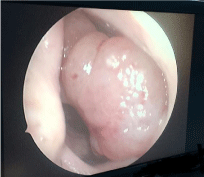
A 47 year old male presented to the outpatient unit of our department with complaints of right side nasal obstruction over the last few years associated with frequent attacks of headache. He did not have a history of hospital admission, examination for these complaints, epistaxis, trauma, previous nasal surgery or constitutional symptoms. Anterior rhinoscopik and endoscopic examination revealed a giant mass which filled the right nasal cavity almost completely, reaching the nasal floor, compressing the right lower concha, originating from the lateral middle turbinate and pushing the septum to the contralateral side. The lesion had a smooth, regular and pale surface, showed no pulsation, and looked similar to inverted papilloma (Figure 1). The color and size of the mass did not change with the valsalva maneuver.

    Figure 1:  Endoscopic examination, giant nasal mass filling right nasal cavity
and almost about to exceed the vestibule.
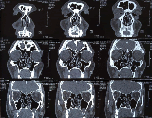
The paranasal sinus CT showed a mass in the right nasal cavity that originated from middle turbinate, almost completely filled the right nasal passage, pressed into the right inferior turbinate and pushed the septum to the left. The mass was identified as a giant CB. Also one retention cyst inside the left maxillary sinus was found on CT scan (Figure 2).

    Figure 2:  Coronal section Paranasal sinus CT, demonstrating the giant
concha bullosa. It pressed the right inferior turbinate and pushed the septum
to the left side.
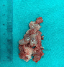
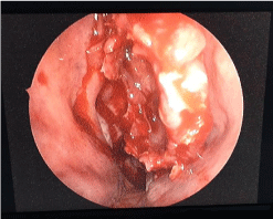
ESS was offered to the patient, including septoplasty plus conchoplasty and he underwent under general anesthesia. On the operation, a vertical incision was made on the midline of the anterior wall of the mass and lateral and medial lamellae of the CB were separated and the lateral lamella was then cut out (Figures 3 and 4). Finally, septoplasty and conchaplasty were performed. Histopathological examination of the excised material from the CB revealed similar to nasal mucosa tissue. The patient was discharged from the hospital postoperatively on the next day and he started to use saline nasal spray and nasal steroid for one month. His symptoms reduced postoperatively and endoscopic examination after two weeks showed that clear nasal passage. Nasal breathing and headache complaints ceased during 2 weeks postoperatively. 12 months follow was uneventful.

Figure 3: Giant CB material after complete excision.

Figure 4: Intra operative image after CB excision.
Discussion
CB is one of the most common anatomical variant of middle turbinate [7]. Although the etiologic factor of CB is not certain yet, ‘ex vacua’ theory is the possible mechanism which associated with air flow patterns of the nasal passage. According to this theory, air flow decreases on the side of the septal deviation while it increases on the opposite side. So, conchal aeration on the opposite side of the deviation also increases [12,13]. Also a strong association between unilateral or dominant CB and contralateral nasal septal deviation reported in the literature [14]. In present case, CB and septal deviation was seen together possibly due to reduced airflow in the deviated side and increased airflow in the opposite side.
Bolger et al. classified them into three types according to the site of pneumatization; Lamellar concha is characterized by vertical lamellar pneumatization, Bulbous type defined by pneumatization of inferior segment and Extensive type defined as pneumatization of both components [15]. Although the relation between the size of CB and complaints of patients is debated, while the extensive type is symptomatic, lamellar and bullous type is usually asymptomatic and discovered incidentally[16]. In the presence of huge concha bullosa, contact region between lateral nasal wall and septal mucosa may cause headache or facial pain [17]. It may cause nasal obstruction, sinusitis, postnasal rain age or olfactory disorders according to size of pneumatization. But the authors still in polemic that whether CB may cause sinus pathologies due to mass effect. In our case, an extensive type CB was contacting to both lateral nasal wall and ethmoid bullae and this possibly caused the patient’s headache. But endoscopic and radiological examinations indicated no chronic infection sign in the neighboring sinuses.
Consideration of patient’s family history, physical examination, endoscopic examination, radiological imaging and, if necessary, a biopsy should be performed to maintain the success of differential diagnosis. Radiologically CT scan has become an essential part of the preoperative examination for patients undergoing ESS. CT is very helpful for visualizing the nasal-paranasal structures and pathologies, but if any suspect of soft tissue or orbital disease occur, magnetic resonance imaging (MRI) may be necessary. CBs could be differentiated from benign nasal polyps, papillomas, tumors and the other nasal masses with following these steps.
While asymptomatic CB does not need special treatment, surgical approach is the definitive treatment of symptomatic CB which cause obstruction of the ostiomeatal complex, paranasal sinuses diseases and airway obstruction. These must treated by performing ESS. The most commonly used surgical technique and effective procedure is resection of the lateral lamella of the middle concha [18]. Also elimination of the contact surfaces with surgical treatment has the potential to decrease pain [19]. In our case report, the lateral segments of right bullous conchae were excised endoscopically, and septoplasty plus conchaplasty was performed. His headache significantly decreased and nasal breathing was significantly improved within 2 weeks postoperatively.
Conclusion
We suggest that if a patient has a CB with nasal obstruction and headache, resection of the lateral wall of the CB and ESS will be needed to properly treat this condition.
References
- Lee HY, Kim CH, Kim JY, et al. Surgical anatomy of the middle turbinate. Clin Anat. 2006; 19: 493-496.
- Braun H, Stammberger H. Pneumatization of turbinates. Laryngoscope. 2003; 113: 668-672.
- Eweiss A, Khatwa MM, Zeitoun H. Trifurcate middle turbinate: An unusual anatomical variation. Rhinology. 2008; 46: 246-248.
- Lin YL, Lin YS, Su WF, et al. A secondary middle turbinate co-existing with an accessory middle turbinate: an unusual combination of two anatomic variations. Acta Otolaryngol. 2006; 126: 429-431.
- Unlü HH, Akyar S, Caylan R, Nalça Y. Concha bullosa. J Otolaryngol. 1994; 23: 23-27.
- Lee JS, Ko IJ, Kang HD, Lee HS. Massive concha bullosa with secondary maxillary sinusitis. Clinical and experimental otorhinolaryngology. 2008; 1: 221-223.
- Bahadir O, Imamoglu M, Bektas D. Massive concha bullosa pyocele with orbital extention. Auris Nasus Larynx. 2006; 33: 195–198.
- Moideen SP, Mohan M, Arun G. Giant Concha Bullosa with Secondary Maxillary Sinusitis. Annals of International Medical and Dental Research. 2017; 3: 1-2.
- Öner F, Tatar A. A Rare Case of Acute Nasal Obstruction with a Giant Middle Turbinate Pyocele: Case Report. Turkish J Rhinology. 2016; 5: 12-15.
- Kaplanoglu H, Kaplanoglu V, Dilli A, Toprak U, Hekimoglu B. An Analysis of the Anatomic Variations of the Paranasal Sinuses and Ethmoid Roof Using Computed Tomography. Eurasian J Med. 2013; 45: 115-125.
- Clark ST, Babin RW, Salazar J. The incidence of concha bullosa and its relationship to chronic sinonasal disease. Am J Rhinol. 1989; 3: 11-12.
- Uygur K, Tuz M, Dogru H. The correlation between septal deviation and concha bullosa. Otolaryngol Head Neck Surg. 2003; 129: 33-36.
- Ural A, Uslu SS, Ileri F, Atilla MH, Özbilen S, Köybasioglu A. Dev konka bülloza. KBB ve BBC Dergisi. 2002; 10: 89-92.
- Stallman JS, Lobo JN, Som PM. The incidence of concha bullosa and its relationship to nasal septal deviation and paranasal sinus disease. AJNR Am J Neuroradiol. 2004; 25: 1613-1618.
- Bolger WE, Butzin CA, Parsons DS. Paranasal sinus bony anatomic variations and mucosal abnormalities: CT analysis for endoscopic sinus surgery. Laryngoscope. 1991; 101: 56–64.
- Ozturan O, Degirmenci N. Pneumo-concha dilatans: Middle concha growing in anterior and lateral directions. J Craniofac Surg. 2013; 24: 444-446.
- Shihada R, Luntz M. A concha bullosa mucopyocele manifesting as migraine headaches: A case report and literature review. Ear Nose Throat J. 2012; 91: 16-18.
- Caughey RJ, Jameson MJ, Gross CW, Han JK. Anatomic risk factors for sinus disease: fact or fiction? Am J Rhinol. 2005; 19: 334-339.
- Huang HH, Lee TJ, Huang CC, et al. Non-sinusitis related rhinogenous headache: A ten year experience. Am J Otolaryngol. 2008; 29: 326-332.