Case Presentation
Austin J Otolaryngol. 2018; 5(2): 1102.
Obstructive Sphenoid Mucocele after Orbital Decompression for Graves’ Ophthalmopathy: A Case Report
Choi AM*, Brown RJ and Chen PG
Department of Otolaryngology, The University of Texas Health at San Antonio, USA
*Corresponding author: Choi AM, Department of Otolaryngology, The University of Texas Health at San Antonio, USA
Received: April 30, 2018; Accepted: May 22, 2018; Published: May 29, 2018
Abstract
Orbital decompression is a method of surgical intervention used in Graves’ ophthalmopathy. The surgery involves reconstruction of the orbital walls to allow for herniation of orbital contents into the paranasal sinuses and orbital pressure relief. Sinonasal complications relating to this surgery are uncommon but can occur resulting in obstructive sinusitis and mucoceles of the maxillary and frontal sinuses. The sphenoid sinus is usually not involved with this procedure. We report a case of sphenoid sinus obstruction and mucocele formation as a result of orbital decompression for Graves’ ophthalmopathy.
Keywords: Endoscopic Orbital Decompression; Endoscopic Sinus Surgery; Graves’ Ophthalmopathy; Sinonasal Complications; Sphenoid Sinusitis; Sphenoid Mucocele Formation
Introduction
Endoscopic orbital decompression is regularly performed for Graves’ ophthalmopathy and the associated orbital complications including exposure keratitis, diplopia, optic neuropathy, blindness, and proptosis [1]. Endoscopic decompression typically involves removal of the bony medial and inferior orbital walls and opening of the periorbita, which relieves orbital pressure by allowing orbital contents to prolapse into the paranasal sinuses. Sinonasal complications resulting from orbital decompression are uncommon; nonetheless, it is important to take steps intraopertively to prevent obstruction of sinus ostia with resultant frontal and maxillary obstructive sinusitis and mucoceles [2,3]. For example, the lamina papyracea is left in situ at the level of the frontal ostium to prevent herniated fat from obstructing the outflow tract. Additionally, a wide maxillary antrostomy is performed [2,3]. Less commonly are specific measures made to prevent sphenoid sinus obstruction. We report a case of sphenoid sinus obstruction and mucocele formation after endoscopic orbital decompression for Graves’ ophthalmopathy.
Case Presentation
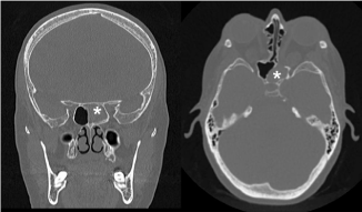
A 68-year-old female with a history of Graves’ ophthalmopathy was referred for left chronic sphenoid sinusitis after revision bilateral endoscopic and external orbital decompression surgeries. The patient denied a prior history of sinusitis, but subsequent to her most recent revisional endoscopic bilateral orbital decompression, she developed symptoms of left-sided nasal congestion, facial pressure, and rhinorrhea. The symptoms were refractory to one year of medical management including nasal saline irrigation, topical nasal steroids, and antibiotics. Computed tomography (CT) scan of the sinuses revealed complete opacification of the left sphenoid sinus and partial opacification of the left posterior ethmoid sinus (Figure 1). All other sinuses were clear.

    Figure 1:  Computed tomography (CT) scan of the sinuses revealing complete
opacification of the left sphenoid sinus and partial opacification of the left
posterior ethmoid sinus. * = left sphenoid sinus.
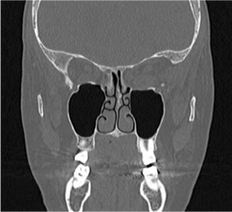
Rigid nasal endoscopy revealed left septal deviation and evidence of bilateral medial orbital decompression with mucosalized orbital fat, more significant on the left and obstructing the left sphenoid ostium and posterior ethmoid sinus (Figure 2). There was significant scarring and mild mucosal edema on the left.

    Figure 2:  CT scan indicating left septal deviation and bilateral medial orbital
decompression with mucosalized orbital fat obstructing the left sphenoid
ostium and posterior ethmoid sinus.
Given the refractory nature of the patient’s sinus disease and the mechanical nature of sinus obstruction, the decision was made to proceed with endoscopic sinus surgery to relieve the sphenoid obstruction and clear the mucocele.

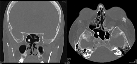
    Figure 3:  CT scan of widely patent right sphenoid ostia with an opened
intersinus septum, and a completely resolved left sphenoid mucocele. R =
right sphenoid sinus, L = left sphenoid sinus.
Due to the prior surgeries, the left lamina papyracea and lateral sphenoid wall were absent, with additional narrowing of the sinus passage due to the prolapsed orbital contents. This anatomical configuration placed the medial rectus muscle at significant risk for damage during revision surgery. To protect the left medial rectus muscle while also adequately exposing and opening the sphenoid sinus, a transseptal approach was used. Maintaining a corridor under the septal mucosa, the left sphenoid sinus was identified and opened widely, including removal of the floor and portion of the rostrum.

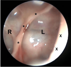
    Figure 4:  Endoscopic view with 30-degree endoscope looking through
the right sphenoid osteotomy into the now healthy left sphenoid sinus. R
= right sphenoid sinus, L = left sphenoid sinus, X = medial edge of right
sphenoidotomy, * = remnant sphenoid intersinus septum.
To further facilitate drainage from the left sphenoid, a portion of the sphenoid intersinus septum was removed to allow drainage from the left into the right sphenoid. Additionally, a small posterior septectomy was performed. The final sphenoid neo-ostium spanned the distance between the superior turbinates. A dissolvable mometasone-eluting stent was placed in the neo-ostium of the sphenoid sinuses. The patient tolerated the surgery well and was discharged from the hospital the same day.
At the 1-week postoperative follow-up visit, the patient reported significant improvement in nasal congestion and facial pressure. Rigid nasal endoscopy revealed a patent sphenoid sinus and posterior septectomy with expected crusting and edema. The patient was kept on a regimen of nasal saline irrigations and topical steroids. Seven months after surgery the patient reported resolution of all sinus symptoms. Nasal endoscopy showed widely patent right sphenoid ostia with healthy sphenoid mucosa. The left sphenoid ostium was stenotic, but the left sinus could be easily visualized from the right side through the opened intersinus septum (Figure 3). Postoperative CT scan of the sinuses showed complete resolution of the left sphenoid sinus mucocele (Figure 4).
Discussion
Graves’ orbitopathy (or ophthalmopathy) is an autoimmune ocular disease occurring in patients with hyperthyroidism secondary to Graves’ disease. The pathophysiology of Graves’ orbitopathy involves glycosaminoglycan deposition and adipocyte accumulation in the orbit as well as fibrosis and edema of the extra ocular muscles. This is thought to occur secondary to orbital fibroblast activation by auto antibodies. Graves’ orbitopathy is more common in women, but severe forms affect men and women nearly equally [4]. The most common clinical manifestations of Graves’ orbitopathy include proptosis and periorbital edema. Other signs and symptoms include epiphora, retro-orbital pain, diplopia, blurry vision, and loss of vision [5]. Treatment begins with addressing the underlying issue of autoimmune hyperthyroidism either through medication or surgery. Medications that have shown efficacy against Graves’ orbitopathy include selenium, glucocorticoids, and rituximab. These medications provide anti-inflammatory, immunosuppressive, and antiautoantibody properties, respectively, which may improve symptoms of Graves’ orbitopathy [6-9]. However, no treatment has been shown to successfully reverse the pathologic orbital changes.
Indications for orbital decompression include failure of medical management, the threatened loss of vision, optic neuropathy, exposure keratopathy, diplopia, and cosmetic reasons [1,10]. The surgery involves a combination of removal of the medial, inferior, and/or lateral orbital walls through an endoscopic, external, or combined approach. The goal of surgery is to increase orbital volume and reduce orbital pressure by allowing orbital contents to herniate into the paranasal sinuses.
The overall incidence of complications following orbital decompression is 9.3% [11]. The most common complications include worsening of pre-existing diplopia and new-onset diplopia occurring in 15 to 63% of patients [1]. Less common side effects include postoperative sinusitis (0.42%), mucocele formation (0.33%), postoperative hemorrhage, cerebrospinal fluid leak, and facial numbness [2,12]. Previously reported sinonasal complications of orbital decompression have involved the frontal, maxillary, and ethmoid sinuses [2,3]. A recent review of surgical complications of orbital decompression for Graves’ orbitopathy by Sellari-Franceschini showed that the occurrence of sinusitis and mucocele formation is below 1% in most large series [2]. Chronic sinusitis and mucocele formation after orbital decompression occur due to sinus outflow tract obstruction by herniated orbital contents. Maxillary sinusitis can be corrected or prevented by creating a large inferiorly-oriented maxillary antrostomy (“mega-antrostomy”), allowing outflow around the obstructive orbital contents. A transaxillary frontal sinusotomy with stent placement has been described for management of frontal sinusitis after orbital decompression. Measures to prevent frontal sinus obstruction include leaving the superior portion of the lamina papyracea near the frontal recess intact and resecting the middle turbinate [3].
To our knowledge, this is the first reported case of sphenoid sinusitis as a complication of orbital decompression. The fact that the sphenoid sinus is not usually affected after orbital decompression can potentially be explained by the location of the sphenoid ostium medial to the superior turbinate and away from the orbit. An intact superior turbinate shields the sphenoid ostium from obstruction by herniated fat, so it is arguable that the turbinate should be left intact whenever possible. Unfortunately, in severe cases of ophthalmopathy and exophthalmos, the greater degree of decompression may not allow leaving the superior turbinate possible. Careful review of the preoperative CT scan of the sinuses in this case reveals that the left superior turbinate was partially resected and situated more medially when compared to the right-factors that may have diminished the protective effect of the superior turbinate. Additionally, multiple prior decompression surgeries may have led to scarring around the sphenoid ostium, which could further increase the risk of sinus obstruction. Long-term follow up to assess for sphenoid sinusitis and mucocele formation should be considered in patients undergoing decompression.
Conclusion
Obstructive sinusitis after endoscopic orbital decompression is uncommon and typically involves the frontal and maxillary sinuses. This is the first report of obstructive sphenoid sinusitis and mucocele after orbital decompression surgery. A transseptal approach with removal of the sphenoid intersinus septum was performed to address the obstructed sinus.
References
- Ting JY, Sindwani R. Endoscopic orbital decompression. Operative Techniques in Otolaryngology. 2014; 25: 213-217.
- Sellari-Franceschini S, Dallan I, Bajraktari A, Fiacchini G, Casani AP, Nardi M, et al. Surgical complications in orbital decompression for Graves’ orbitopathy. Acta Otorhinolaryngo logicaItalica. 2016; 36: 265-274.
- Antisdel JL, Gumber D, Holmes J, Sindwani R. Management of sinonasal complications after endoscopic orbital decompression for Graves’ orbitopathy. The Laryngoscope. 2013; 123: 2094-2098.
- Maheshwari R, Weis E. Thyroid associated orbitopathy. Indian Journal of Ophthalmology. 2012; 60: 87-93.
- Bahn RS. Graves’ ophthalmopathy. New England Journal of Medicine. 2010; 362: 726-738.
- Rayman MP. The Importance of Selenium to human health. Lancet. 2000; 356: 233-241.
- Wiersinga WM. Advances in treatment of active, moderate-to-severe Graves’ ophthalmopathy. Lancet Diabetes Endocrinology. 2017; 5: 134-142.
- Salvi M, Campi I. Medical Treatment of Graves’ Orbitopathy. Hormone and Metabolic Research. 2015; 47: 779-788.
- ElFassi D, Banga JP, Gilbert JA, Padoa C, Hegedüs L, Nielsen CH. Treatment of Graves’ disease with rituximab specifically reduces the production of thyroid stimulating autoantibodies. Clinical Immunology. 2009; 130: 252-258.
- Lyons CJ, Rootman J. Orbital decompression for disfiguring exophthalmos in thyroid orbitopathy. Ophthalmology. 1994; 101: 223-230.
- Leong SC, Karkos PD, Macewen CJ, White PS. A systematic review of outcomes following surgical decompression for dysthyroid orbitopathy. Laryngoscope. 2009; 119: 1106-1115.
- Rootman D. Orbital decompression for thyroid eye disease. Survey of Ophthalmology. 2018; 63: 86-104.