Case Report
Austin J Otolaryngol. 2020; 7(1): 1108.
Lacrimal Gland Choristoma in the Setting of Acute on Chronic Sinusitis: A Rare Case Report
Holmes CP* and Sharma AR
Department of Otolaryngology ENT Head and Neck Surgery, University of Saskatchewan, Canada
*Corresponding author: Holmes CP, College of Medicine, University of Saskatchewan, Canada
Received: March 30, 2020; Accepted: April 29, 2020; Published: May 06, 2020
Abstract
Objective: The current study presents the first reported case of an incidental lacrimal gland choristoma in the setting of acute on chronic sinusitis with presumed orbital involvement.
Background: Choristomas are benign masses comprised of histologically normal tissue located at abnormal sites likely due to aberrant implantation of embryonic cells. Only a handful of cases of lacrimal gland choristomas have been reported in the literature.
Clinical Case: A 12-year-old boy presents with orbital proptosis in the context of acute on chronic sinusitis. Attempted intra-operative drainage incidentally revealed an orbital tumor.
Conclusion: In the context of presumed orbital complications of acute on chronic sinusitis, orbital tumors must be considered. All sinus cases involving the orbit require an ophthalmology consult to ensure appropriate diagnostic and surgical management for these patients.
Keywords: Sinusitis; Orbital Complications of Sinusitis; FESS; Incidental Orbital Tumor; Lacrimal Gland Choristoma
Abbreviations
CT: Contrast-Enhanced Computed Tomography; LGC: Lacrimal Gland Choristoma
Introduction
The orbit is the most commonly involved structure in the complications of sinusitis [1]. Approximately 3% of sinusitis patients suffer orbital complications [2]. The pediatric population is more prone to orbital complications likely due to higher rates of sinusitis, upper respiratory tract infections and the age-dependent development of the frontal and sphenoid sinuses [1]. In children, the first manifestations of sinusitis may be orbital in nature [2]. Evaluation of a patient with sinusitis presenting with orbital findings includes a thorough history including recent upper respiratory infections, duration and progression of symptoms, recent trauma, swimming, ear infection, dental surgery or infection and other systemic illnesses [1]. Generally, the patient will present with fever and findings consistent with acute sinusitis. Examination requires multidisciplinary input from the otolaryngologist and the ophthalmologist [2]. Common signs and symptoms include orbital edema, proptosis, pain, and fever [2]. More advanced cases may present with gaze restriction and changes in visual acuity [2]. The Chandler classification system is useful for staging orbital complications, in order of increasing severity these include: preseptal cellulitis; orbital cellulitis; subperiosteal abscess; orbital abscess and cavernous sinus septic thrombosis [2]. Contrast- Enhanced Computed Tomography (CT) scanning is considered the gold standard imaging modality when evaluating for post septal involvement and aids in surgical planning [1,2].

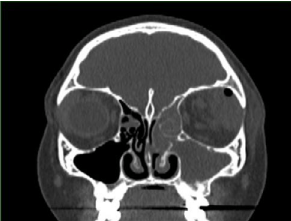
    Figure 1: A coronal CT scan of the head depicting potential periorbital
abscess and/or lesion of the left upper lateral portion of the eyelid.
Patients presenting with sinusitis and presumed orbital complications should be admitted to hospital and started on broad-spectrum intravenous antibiotics [2]. Surgical drainage is recommended when there is CT evidence of abscess formation, worsening visual acuity or blindness, rapid progression of orbital signs and symptoms or lack of improvement despite aggressive medical therapy [1]. Regardless of the method, underlying sinus pathology and abscesses must be addressed as at least 10% of untreated cases result in blindness [2]. While sinusitis is a common cause of orbital infection, it is important to rule out other possible causes of ophthalmological symptoms. The differential diagnosis can include orbital pseudotumor (pediatrics), orbital rhabdomyosarcoma, ethmoid mucocele, allergic fungal rhinosinusitis, iatrogenic injury and neoplasm [2]. Choristomas are benign masses of histologically normal tissue located at abnormal sites [3]. Choristomas consisting of more than one type of tissue are considered complex. Choristomas containing ectopic lacrimal glands have been reported in the orbit, outer eye, conjunctiva, cornea, sclera or in the intraocular tissue [4]. The following case is the first reported incident of an incidental Lacrimal Gland Choristoma (LGC) in the setting of acute on chronic sinusitis with presumed orbital involvement.
Case Report
Demographics
A 12-year-old male recently immigrated from France to Canada of African descent.
Medical History: Chronic rhinosinusitis patient who previously underwent Functional Endoscopic Sinus Surgery (FESS) in Europe. Otherwise healthy with no allergies and up-to-date vaccinations.
Signs and Symptoms
The patient was admitted 8 days prior to surgery for retro-orbital eye collection. The patient presented with purulent nasal discharge, throbbing facial pain, intermittent episodes of spiking fevers, swelling of left upper and lower eyelids and some difficulty opening his eyelids. CT scan revealed evidence of potential periorbital abscess and/or lesion of the left upper lateral portion of the eyelid.
Treatment and Test results: The patient was treated with a course of IV ceftriaxone, moxifloxacin, and vancomycin. The patient then underwent image-guided FESS, septoplasty and submucosal resection of turbinates, left maxillary antrostomy, left anteriorposterior ethmoidectomy, left frontal sinusotomy and antral lavage 8 days after admission. The left maxillary sinus had a significant amount of pus which was drained out and sent for culture and sensitivity. Intra-operative drainage incidentally revealed an orbital tumor. A left superior orbital mass resembling lymphoma was submitted to the pathology department. Histopathological analysis of excisional biopsy revealed a lacrimal gland choristoma with focal acute inflammation. More specifically, the tumor tissue showed mainly acini glands resembling lacrimal glands with focal reactive changes subsequent to acute inflammation. Some decompression of the eye was noted after the procedure. There were no intraoperative complications. The patient was handed over to ophthalmology for the management of eye pathology.
Discussion and Conclusion
Lacrimal Gland Choristomas (LGC) are rare congenital lesions thought to be caused by sequestration of parts of the lacrimal anlagen [5]. Macroscopically these specimens resemble normal lacrimal gland tissue [5]. Microscopic examination generally reveals normal lacrimal gland tissue, commonly accompanied by moderate nonspecific inflammatory cell infiltration [5]. Alyahya et al. in their study reviewed 61 ectopic lacrimal gland specimens collected over 50 years. 62% of these specimens were considered complex choristomas, while the remainder contained only ectopic lacrimal gland tissue [6]. In our case, the histopathological analysis revealed only ectopic lacrimal gland tissue. Although symptoms of LGC’s can manifest at any age, they most commonly present during the first 3 decades of life, the reason for their often-delayed presentation is unknown [5]. Generally, the complications of an LGC are inflammatory in nature [5]. However, vascular lesions, pleomorphic adenomas and very rarely carcinomas have developed in ectopic LGCs [5]. Our patient presented with suspected orbital involvement presumably attributable to an inflammatory process from acute on chronic sinusitis. Incidentally, a LGC was found that was likely contributing to his orbital symptoms. This case emphasizes that in the context of presumed orbital complications of sinusitis, orbital tumors must be considered. All sinus cases involving the orbit require ophthalmology consult to ensure appropriate diagnostic and surgical management for these patients.
References
- Giannoni CM, Weinberger DG, Ferguson BJ, Ryan MW. Complications of Rhinosinusitis editors. Head & Neck Surgery- Otolaryngology Lippincot Williams & Wilkins. 2006; 2: 493-504.
- Goldstein BJ, Goldenberg D, Goldstein BJ, Goldenberg D. Orbital Complications of Sinusitis editors. Handbook of Otolaryngology Thieme. 2018; 228-230.
- Zwaan J, Karcioglu ZA. Orbital Tumors editor. In Benign Pediatric Tumors Springer. 2015; 337-348.
- Ranganathan D, Lenhart P, Hubbard G, Grossniklaus H. Lacrimal gland choristoma in a preterm infant, presenting with spontaneous hyphema and increased intraocular pressure. Journal of Perinatology. 2010; 30: 757-759.
- Verdijk RM, Grossniklaus H , Heegaard S. The Orbit, Including the Lacrimal Gland and Lacrimal Drainage System In Eye Pathology. 2015; 547-731.
- Alyahya G, Bangsgaard R, Prause J, Heegaard S. Occurrence of lacrimal gland tissue outside the lacrimal fossa. comparison of clinical and histopathological findings Acta Ophthalmologica Scandinavica. 2005; 83: 100-103.