
Review Article
Austin J Otolaryngol. 2020; 7(2): 1116.
Chondrocutaneous Rotation Flap for Helical Repair
Skaria AM*
Department of Dermatology, University of Bern, Switzerland
*Corresponding author: Skaria AM, Department of Dermatology, University of Bern, Switzerland
Received: November 12, 2020; Accepted: December 08, 2020; Published: December 15, 2020
Abstract
Purpose: Helical rim repair after tumor surgery can prove to be challenging. In literature, a variety of reconstruction methods are recommended for these cases. The Antia-Buch technique, an advancement flap, is the workhorse flap in this indication alternatively to retro-auricular interpolation flaps, as twotimes procedures. We adopted a reconstruction method applying a chondrocutaneous rotation flap for helical rim repair which does not require second stage corrections and does not alter the length of the ear.
Methods: We reviewed the medical records and photographs of 27 patients which had a chondro-cutaneous rotation flap reconstruction for helical rim defects after Mohs surgery and a follow up for at least 1 year.
Results: All patients showed good to excellent results, without notching or cupping of the ear.
Conclusion: The chondro-cutaneous rotation flap is a valuable single staged alternative to the classic approach of the Antia-Buch advancement flap due to the nature of flap harvesting, recovery and success rates. The chondrocutaneous rotation flap has several advantages towards the approach of Antia- Buch which will be discussed in this article.
Introduction
The helical rim is a frequent site for skin cancers [1]. Reconstruction of postsurgical defects, may prove to be challenging due to the special anatomy. The risk, to create an uneven rim, with a notch or ear cupping is high for mainly two reasons; 1. The cartilage is rigid and creates tension on the skin 2. the skin of the ear is thin with limited elasticity which can barely compensate this tension for example in direct closure [2].
The mention of the chondro-cutaneous advancement flap for the reconstruction of the helical rim by Antia-Buch in 1967 got the standard method of helical rim repair [3]. Various modified helical rim advancement flaps where documented in literature, respecting the helical sulcus as incision line [4,5]. We present 27 cases of chondrocutaneous rotation flaps. As it replaced completely the advancement flap of Antia-Buch in our daily practice, we think it is justified to publish a review of our well documented cases of the last 8 years.
Anatomy
The ear, is a complex three-dimensional structure with welldefined anatomical landmarks and exposed contours. Visually, the ear is defined by the outer form of the helix and the ear lobe. The form of the helix is given by the cartilaginous framework which takes 2/3 of the upper part of the ear. The cartilaginous framework forms the scaphoid fossa, which goes over to the helical margin and finally vanishes about one to two cm above the ear lobe at the vertical height of the antitragus. This is a landmark, where the helical rim is without cartilage support and the ear lobe begins.
The ear lobe shows great variability between individuals. It might insert directly at the mandibular angle to form a straight line until the beginning of the helical sulcus and is then called, an attached ear lobe. When there is no direct attachment, it is called a free or detached ear lobe.
The vascularization of the ear is in a random pattern, and takes its origin from branches of the posterior auricular artery, the occipital, the superficial temporal and superior auricular artery [6]. A dense network of anastomosis between the superior and inferior branch of the superficial temporal artery guarantees the vascular supply. Shokrollahi et al demonstrated a fine and rich vascular network that exists throughout the postauricular fascia with multiple anastomoses [6]. They called it Intrinsic Postauricular Fascia (IPF) and defined it as a distinct anatomical entity. Concordant with Park et al [7]. They noted in their assessment of the vascularity of the pinna that among the rich vascular network it was often not possible to ascertain which components of the network arose from the posterior auricular artery and which from ascending branches of the superficial temporal artery and occipital artery. [6,7]
They conclude, that on the basis of their findings in relation to vascularity and macro-anatomy, the IPF could reasonably contribute to reconstruction of defects of the helix. Because of the dense vascular network, its elastic properties, and codominant blood supply the IPF has the potential for both reconstructive flaps in the local vicinity, as well as acting as a free graft.
Technique
The flap is harvested from the caudal part of the full-thickness defect, including the whole rim with the sandwiched cartilage (Figure 1). The flap is designed in a triangular shape. From the most anterior caudal point of the defect (Figure 1B) on the stem of the antihelix a line is drawn, traversing the scapha to reach the helical margin between helical rim and antihelix to finally join the ear lobe. (C in Figure 1b), When the incision goes only to the helical tail we might produce slight curbing of the helical rim (Figure 6 a-d). From this most caudal point of the flap, a line is drawn upwards on the posterior side of the helix until 1 cm below the defect (D in Figure 1b), which corresponds to beginning of the pedicle. From here the line goes cranial oblique until the postauricular sulcus to delimitate the pedicle of the rotation flap (E in Figure 1b). The incision starts from the epidermis and dermis of the anterior skin of the helix, through the cartilage (Figure 1b, 1c). The incision ends at the ear lobe. The incision goes not deeper than the retro-auricular perichondrium layer on the cartilage (Figure 1c,1d). The pedicle of the chondro-cutaneous rotation flap is the Intrinsic Postauricular Fascia (IPF) and postauricular skin, therefore we have to be careful not to harm this layer (Figure 1c and 1d). The flap is then harvested from the caudal part of the designed flap involving the whole rim until the posterior retro-auricular incision line joins the incision line of the pedicle. (CD in Figure 1b, 1c, 3d, 4b) From here on the postauricular skin and IPF is separated by blunt dissection from the surface of the postauricular concha cartilage in the perichondrium layer until there is enough mobility. (Figure 1d, 3d, 4b) Finally, we incise the skin and postauricular fascia (IPF) from caudal to cranial in a posterior anterior direction. (Figure 1c, d, 3d, 4b)
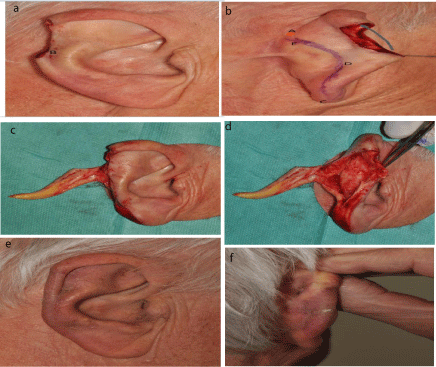
    Figure 1: a) 79 years old patient with an infiltrative BCC on the helix after
Mohs surgery.b) design of the flap retroauricular, point A (orange) is the
rotation axis of the flap (B,C,D and E are the extremities of the flap).
c) d) the flap harvested and reclined, remark the fascia and thickness of
the pedicle. e) f) result after 6 months, observe the standing cone nearly
resorbed.
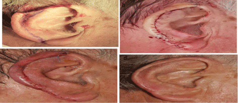
    Figure 2: a) 74-years old patient after mohs surgery for micronodular BCC on
the right ear. b) the rotation flap in place. c) suture removal D7 after surgery,
no sign of ischemia, sutures on the top will be removed 2 days later d) result
after one year.
The pedicle has to be oblique from caudal to cranial which allows later tension free rotation of the flap (Figure 1c, d, e and 3d, 4e). The longer the pedicle, the easier is the rotation movement around the axis A (Figure 1a,b,3c,4e) but blood supply may get more critical. The flap has to be placed without tension to the pedicle to guarantee good vascularization and to avoid to create a notch (Figure 6 a-c). In some rare cases, the operator can reduce the radius of rotation by shaving the scapha to decrease the tension on the pedicle. The cartilage is adapted with Thyco® braided nylon 5.0 Sutures and small round cardiopoint needle® (CV-331) (11mm), this kind of sutures are used in heart walve surgery. The skin is adapted with Polypropylene 6.0 sutures after having undermined the rest of the postauricular skin and IPF caudal to the pedicle. It can rarely happen, that after rotation of the flap a small skin defect might be left which can be left to second intention healing on the postauricular side. A stabilizing bandage, is put and kept for 24 hours. For one month, the patient is advised to be careful not to fold the ear.
The patient is advertised that cartilage surgery of the ear is often accompanied with slight pain on the ear for about 6 weeks.
Results
We have collected 27 cases of helical repairs by chondrocutaneous rotation flaps. All these cases have been documented and followed up for at least 1 year clinically and by photographs. Out of the 27 patients, 23 where men and 4 where women. The age range was from 48 to 101 years with a mean age of 72 years. The size of the defect ranges between 10x12 mm up to 30x16 mm (Table 1).
![]()
    Patient
    AgeY
    sex
    left/ right
    Size mmxmm
    Diagnosis
    Complication, results
    Figure


    RH
    82
    m
    r
    18 x 15
    CSCC
    No complaint, good result


    FC
    65
    m
    r
    20x25
    mock
    No complaint, good result


    JC
    71
    m
    r
    20x17
    iBCC
    No complaint, excellent result


    JD
    67
    m
    l
    20x12
    mBCC
    No complaint, excellent result


    JD
    48
    m
    l
    17x18
    BCC
    No complaint, good result


    JF
    85
    m
    r
    21x15
    CSCC
    Suture infected, good result
    Fig 3


    VR
    64
    m
    l
    28x15
    mBCC
    No complaint, good result


    HL
    74
    m
    r
    30x15
    mnBCC
    No complaint, excellent result
    Fig 2


    RH
    82
    m
    l
    18x15
    CSCC
    No complaint, good result


    AJ
    77
    m
    l
    20x15
    CSCC
    No complaint, good result


    MK
    79
    m
    r
    25x20
    iBCC
    No complaint, excellent result
    Fig 1


    RK
    73
    m
    l
    25x15
    MM
    No complaint, good result


    YC
    81
    f
    r
    18x17
    CSCC
    No complaint, excellent result


    CK
    55
    f
    r
    15x10
    MM
    Minor notch, correct result
    Fig 6


    DL
    85
    f
    l
    20x20
    mBCC
    No complaint, excellent result


    MT
    79
    f
    l
    24x16
    BCC
    No complaint, good result,    slight curb
    Fig 5


    AT
    91
    m
    l
    20x11
    BCC
    No complaint, excellent result


    CM
    79
    m
    l
    18x17
    CSCC
    No complaint, good result


    MP
    101
    m
    l
    19x16
    CM
    Suture infected, good result


    MR
    52
    m
    l
    30x15
    iBCC
    No complaint, excellent result


    VR
    64
    m
    l
    28x45
    BCC
    No complaint, excellent result


    BS
    67
    m
    r
    24x18
    CM
    No complaint, good result


    KS
    77
    m
    r
    12x10
    BCC and cSCC
    No complaint, excellent result


    JS
    70
    m
    r
    20x18
    BCC
    No complaint, good result


    JT
    85
    m
    r
    24x12
    BCC
    No complaint, good result
    Fig 4


    AZ
    81
    m
    l
    25x16
    cSCC
    No complaint, excellent result


    EH
    82
    m
    r
    20x20
    mBCC
    No complaint, excellent result
Table 1: Summary of 27 patients with rotation flaps.
In all cases the alignment of the rim without dehiscence was maintained over the one year period. One patient (Figure 6 a-d) showed a minor notch, but this was already the case when suturing and not a secondary dehiscence as can be seen in (Figure 6 a-d). The esthetic results, where good to excellent in all cases. None of the patients was disturbed by the esthetic outcome, nor did any patient demand for correction. In two patients, one of the cartilage nylon sutures got infected and could not be treated by local and systemic antibiotics, wherefore the suture had to be removed with no secondary problems after 6 months (Figure 3 a- f).
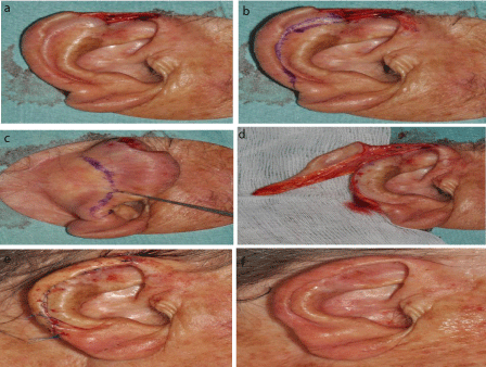
    Figure 3: a) 85-years old patient after mohs surgery for an infiltrative
squamous cell carcinoma on the apex of the helix, patient with dermaporotic
skin. b) c) The defect extends more on the helix as case 2. In consequence
the flap is broader.d) observe the thickness of the pedicle with the IF in a
patient, with important actinic damage and atrophy of the skin.e) f) flap in
place after surgery and result 2 years later.
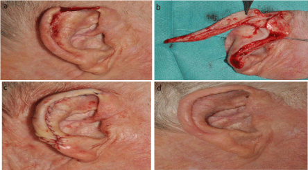
    Figure 4: a) 85 year old male patient with a BCC excision by Mohs surgery on
the apex of the right ear b) observe the thick pediclein this 85 year old patient
c) after surgery and d)3 month later.
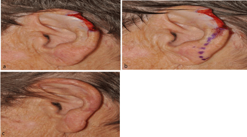
    Figure 5: a) a 79 year old female patient with a BCC excision by Mohs
surgery on the left ear, the 24 mm long defect is in the middle third of the
helix b) the flap design which is unfortunately not going to far on the ear lobe
which produces a slight curb between ear lobe and helix c)d) immediately
after surgery and 2 month later.
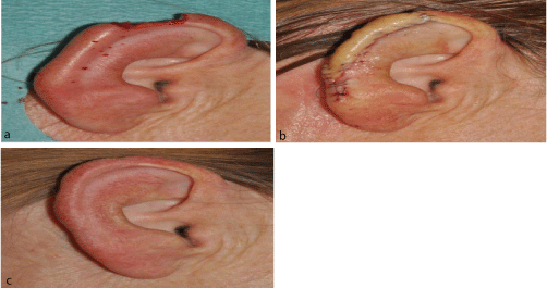
    Figure 6: a) 55 years old patient with excision for melanoma on the right ear.
b) flap in place, but a creation of a notch because of limited adaption of the
underlying cartilage c) suture removal D7 without sign of ischemia.
Discussion
Of all NMSC, 8.8% are involving the ear, the helix is the most common site [1]. Different techniques have been proposed for reconstruction of helical defects. For small defects, wedge excision or star excision have been proposed with the risk of cupping or notching of the ear [3]. Retro-auricular advancement flaps, banner flaps, transposition flaps require cartilage grafting and may be a multistage procedure [2-4]. The chondro-cutaneous advancement flap of Antia-Buch is a major step in ear reconstruction [2-4]. It allows to reconstruct full-thickness helical rim defects by an advancement flap harvesting tissue from the ear lobe. Limitation of this flap is first, that the patient has to have an ear lobe and secondly the size of the ear lobe may diminish. These two points are mentioned by Butler as a major drawback of the advancement flap criticizing the loss of vertical height and distortion of the lobe contour in this kind of reconstruction [2]. An advancement flap on the helical rim always creates tension as the flap movement is perpendicular to the scar with the risk to create a notch, this point is also outlined by Butlerm [2]. Finally, the advancement flap of Antia-Buch is less convenient for defects of the spine of the helix and the apex of the ear.
For all these reasons, we do prefer the chondro-cutaneous rotation flap. The rotation movement around the circumferential radius of the helix allows tension free closure, without notching and/ or cupping of the helix. Further, flap harvesting does not depend on the size of the ear lobe and may be applied to any form of the ear. The whole helical rim and helical tail are rotated into the defect. In consequence, it allows to maintain the form of the ear with the only drawback of slight narrowing of the horizontal diameter of the ear. This distinguishes it clearly from an advancement flap with all the disadvantages mentioned by Butler [2]. Diminishing the horizontal diameter of the ear is less disfiguring than loss or reduction of the ear lobe and decreased vertical size. Direct comparison of the horizontal diameter from both ears is visually impossible in daily life.
A problem might be cartilage necrosis, described in free grafts but until now we have not encountered this complication in this compound flap reconstruction [5]. This might be due to the fact that random pattern vascularization of the ear is very dense, as shown by Shokrollahi et al, and cartilage is sandwiched in the skin of the ear rim and may not be comparable to free cartilage grafts [6].
We did not see secondary notching of the scar, Butler who has mentioned this reaction in advancement flaps of the ear suggested that this might be prevented by Z-plasty [2]. This seems in our eyes not sufficient as the problem lies in the very slow healing of cartilage and the tension of the spreading underlying cartilage in advancement flaps. We use a non-resorbable Tycon® suture with a CV-needle for cartilage adaption. Tycon® sutures are used in heart valve surgery and there are three points to favor this kind of sutures; 1. The suture is braided and therefore avoids cutting the cartilage 2. The PX needle is a round needle and avoids splitting of the cartilage in contrary to a cutting needle 3. The non-resorbable suture keeps the slow regenerating cartilage stabilized for a long period, to allow better healing.
All our patients showed a good to excellent result. None of the patients showed flap necrosis, major infections, notching or scar dehiscence.
Conclusion
This flap is not mentioned in textbooks in reference to the reconstruction of helical defects. It shows four main advantages towards the workhorse flap of Antia-Buch. First it does not shorten the ear, second it can be practiced in patients with physiological attached ear lobes or attached ear lobes after face lifts, third, as it is a rotation flap there is nearly no tension on the scar avoiding notches, and finally, this flap can easily be performed for defects of the proximal apex of the helix in contrary to the Antia-Buch technique.
We think this flap is a valuable alternative to the reconstruction of Antia-Buch and it should make part of the panoplia of ear reconstruction techniques.
References
- Ragi JM, Patel D, Masud A, Rao BK. Nonmelanoma skin cancer of the Ear: Frequency, Patient’s knowledge, and Photoprotection Practices. Dermatol Surg 2010; 36: 1232-1239.
- Butler CE. Reconstruction of marginal ear defects with modified chondrocutaneous helical rim advancement flap. Plast and Reconstr Surg 2003; 111: 2009-2013.
- Antia NH, BuchVI. Chondrocutaneous advancement flap for the marginal defect of the ear. Plast Reconstr Surg 1967; 39: 472-7.
- Menick FJ. Reconstruction of the ear after tumor excision. Clin Plast Surg 1990; 17: 405-15.
- Burget GC, Menick FJ. Aesthetic reconstruction of the nose 2nd Edition 1993, Mosby.
- Shokrollahi K, Taylor JP, Le Roux CM, Ashton MW, Rozen WM, Jones NS, et al. The postauricular fascia: classification, anatomy, and potential surgical applications. Ann Plast Surg. 2014; 73: 92-7.
- Park C, Lee TJ, Shin KS, et al. A single-stage two-flap method of total ear reconstruction. Plast Reconstr Surg. 1991; 88: 404-412.