Clinical Image
Austin J Otolaryngol. 2021; 8(1): 1120.
Suspicious Hard Palate Tumor Revealing HIV Infection
Allouch I*, Belhaj N, Benkhraba N, Bencheikh R, Benbouzid MA and Essakalli L
Department of ENT, The Rabat Specialty Hospital, Rabat Salé University Hospital, Moracco
*Corresponding author: Allouch Ihssane, Department of ENT, The Rabat Specialty Hospital, Rabat Salé University Hospital, Moracco
Received: April 19, 2021; Accepted: May 25, 2021; Published: June 01, 2021
Clinical Image
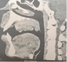
This is a 31-year-old patient with no notable pathological history, who presents an ulcerative-budding mass of the hard palate increasing rapidly and bleeding easily on contact, without palpable cervical lymphadenopathy or other associated signs, the injected face CT scan objectified the presence of a tissue lesion process of the hard palate, lateralized to the left, lysing the alveolar bone, bulging into the oral cavity and extending to the soft palate and soft gingival parts.

Figure 1: Clinical image showing an ulcerative mass of the hard palate.

Figure 2: CT scan showing a tumor of the palate with bone lysis.
The patient underwent a biopsy of the tumor and an anatomopathological study was performed showing an ulcerated spindle cell proliferation leading to a kaposia sarcoma, the immunohistochemistry confirmed the diagnosis by a positive antiCD34 and anti HHV-8 antibodies. HIV (human immunodeficiency virus) serology and its viral load subsequently returned positive.