
Case Series
Austin J Otolaryngol. 2023; 9(1): 1129.
Endoscopic Septoplasty vs Conventional Septoplasty: A Comparitive Study
Rukma Bhandary¹; Rohan Shetty²*
¹Associate Professor, Department of Ent, Ajims, Mangalore, India
²Post Graduate, Department of Ent, Ajims, Mangalore
*Corresponding author: Rohan Shetty Department of ENT, AJIMS, Mangalore, #36, 1st Main, 4th Cross, Chinanna Layout, Kavalbyrasandra, R.T.Nagar, Bangalore North, Bangalore, Karnataka - 560032, India Email: Rohshetty1095@gmail.Com
Received: September 20, 2023 Accepted: October 30, 2023 Published: November 06, 2023
Abstract
Introduction: Nasal obstruction due to deviated nasal septum is a common problem encountered by most otolaryngologists. A variety of surgical procedures have been tried in the treatment of the same.
Objective: This study was conducted to evaluate the outcomes and complications of endoscopic and conventional septoplasty and to compare both.
Materials and Methods: This is a prospective, randomized study done in tertiary health center, Mangalore in which 60 patients with symptomatic deviated nasal septum were included in the study. Twenty two of them underwent conventional septoplasty and the remaining thirty eight underwent endoscopic septoplasty.
Results: Endoscopic septoplasty had better outcome with respect to complications and proved as an easier method to correct posterior deviations and isolated spurs. Post operative symptoms were also better in the group which underwent endoscopic septoplasty.
Conclusion: Endoscopic septoplasty allows accurate, conservative repair of obstructive nasal septum deviations, with fewer complications and better functional results compared to conventional septoplasty.
Keywords: Conventional septoplasty; Deviated nasal septum; Endoscopic septoplasty; Septoplasty
Introduction
Nasal obstruction is one of the most common complaints that a otorhinolaryngologist faces in the day to day practice [1]. Deviated nasal septum being the most common cause for the nasal obstruction [1]. It not only causes breathing difficulties but also results in improper aeration of paranasal sinuses predisposing to sinusitis and also results in drying of mucosa leading to crusting and epistaxis [1]. Various surgeries have been proposed for the correction of deviated nasal septum [1]. It has undergone several modifications since its inception [1]. Initially submucous resection of septum was done which was a radical surgery and was associated with lot of complications [1]. Later septoplasty was developed as it had advantages of minimal resection of septum and less complications [1].
With the introduction of endoscope into the field of otolaryngology, there were efforts to use it for the correction of deviated nasal septum targeting the surgical procedure in removing only the deviated portion, spur and maxillary crest [1]. It is more effective with minimal manipulation [1]. And also had the advantage of diagnosing and treating the abnormalities of the lateral wall of the nose at the same sitting [1]. Hence the present study was taken up to compare the two techniques i.e. conventional and endoscopic septoplasty [1]. Preoperative symptom analysis, techniques of surgeries, post operative analysis and complications are presented in this study [1].
Objectives
1. To compare the results post operative of conventional septoplasty with those of endoscopic septoplasty, in cases of septal deviation and septal spurs [1]
2. To compare the improvement in symptoms based on history taken pre and post operation.
3. To compare the complications post operatively of conventional septoplasty with those of endoscopic septoplasty.
4. Informed Consent- Obtained
Methodology
The present study was carried out in the Department of Otorhinolaryngology at a tertiary sector [1]. All patients attending the Out Patient Department of Otorhinolaryngology with symptomatic deviated nasal septum were included in the study [1]. Patients with age less than 10 years, allergic rhinitis, vasomotor rhinitis and with acute infection were excluded [1]. Data was collected by selecting the patients with symptomatic deviated nasal septum willing for surgery [1]. They were divided into two groups; one group undergoing conventional septoplasty and the other endoscopic septoplasty by random selection. 1Sixty patients were included in the study. Twenty two of them underwent the conventional septoplasty and rest of 38 patients underwent endoscopic septoplasty.
Under aseptic precautions ,parts painted and draped.A 0 degree rigid nasal endoscope (4 mm), held in the left hand and nasal cavity was visualized [1]. Anterior nasal packing was done with adrenaline soaked packs. Infiltration given to the septum with lignocaine and adrenaline. An incision was made 2 mm posterior to the caudal end of the septum on the side of the deviation (hemitransfixation) [1]. The initial mucoperichondrial flap was elevated using Freer’s elevator [1]. All the above procedures were done under visualisation with the 0 degree endoscope. Further elevation was done using 0 degree rigid nasal endoscope (4 mm), held in the left hand, keeping the tip of the endoscope between the mucoperichondrial flap and the septal cartilage and suction tip in the right hand, this allowed for better visualisation while elevating [1]. The right hand was used for instrumentation [1]. Flap elevation was done in the correct plane to minimize the bleeding [1], this was achieved by constantly using the suction tip while elevating. In cases with caudal dislocation or anterior buckling of the cartilage, this part was corrected last after correcting the rest of the septum [1]. A spur without any other obvious septal deformity, was resected after incision and exposure made anterior to the spur [1]. In this case the incision and elevation along with dislocation of the spur was made with the help of freer’s elevator .The dislocated part was now removed with the help of lux forceps. IVALON merocel nasal packs were then placed in both nasal cavities with the help of endoscope after significant haemostasis was achieved. Post-operatively patients were put on antibiotics at least for a week, along with analgesics and decongestants [1]. Nasal packs were removed 24 h after the surgery [1].
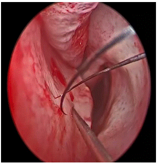
Figure 1: Infiltration to Septum.
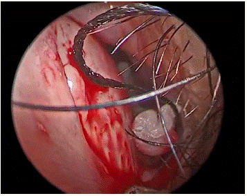
Figure 2: Incision on Side of Deviation.
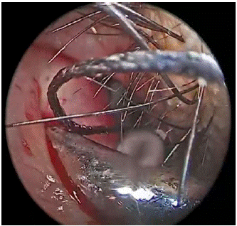
Figure 3: Elevation of Flap With.
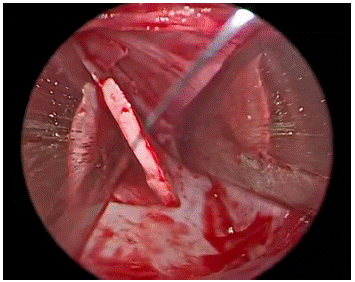
Figure 4: Exposure of Cartilage.
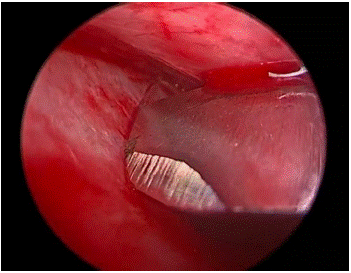
Figure 5: Cartilage Incision.
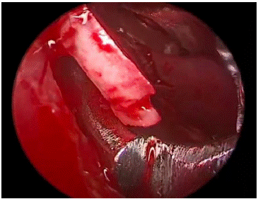
Figure 6: Elevation of Cartilage.
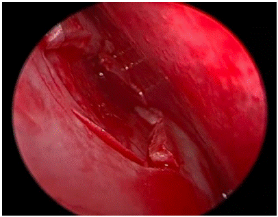
Figure 7: Cartilage Excised.
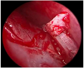
Figure 8: Elevation of Spur.
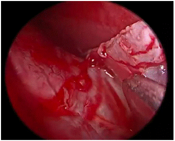
Figure 9: Excision of Spur.
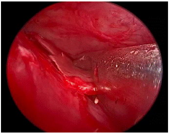
Figure 10: Blakesley to Remove the Remaining Cartilage and Bone.
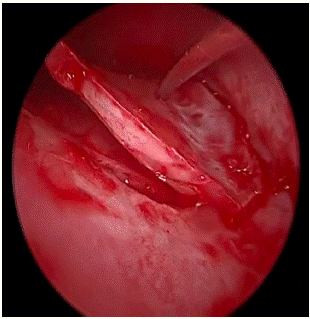
Figure 11: Freers to Remove Remaining Cartilage.
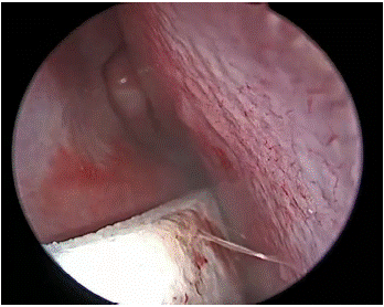
Figure 12: Merocel Nasal Pack Placed.
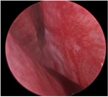
Figure 13: Isolated Spur Noted.
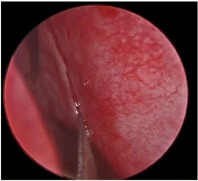
Figure 14: Incision Anterior to Spur with Freer’s.
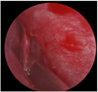
Figure 15: Elevation of Flap.
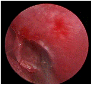
Figure 16: Spur With Bleksley.
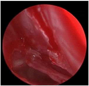
Figure 17: Status Post Spur Excision.
Results
Type of Surgical Intervention Of the total 60 cases in our study, 22 patients underwent conventional septoplasty and 38 patients underwent endoscopic septoplasty [1].
Postoperative Symptomatology - Post operatively the patients were reviewed on 3rd, 7th, 30th day [1]. During each visit, patients were asked about benefits from their symptoms [1]. Out of 52 patients with nasal obstruction, 46 of the 52 patients were relieved of the symptom of which 14 of the patients belonged to conventional and 32 patients belonged to endoscopic septoplasty group.
Nasal discharge persisted in 4 patients of conventional group as well as endoscopic group. Headache persisted in only 2 patients of endoscopic group as compared 4 in conventional group. Epistaxis was relieved in patients belonging to conventional septoplasty group and endoscopic septoplasty group.
Complications
The intraoperative and postoperative complications were as follows [1]. In this study, three patients in conventional septoplasty group had intraoperative hemorrhage and only one patient in the endoscopic septoplasty group developed intraoperative hemorrhage [1]. Mucosal tear occurred in three patients belonging to conventional septoplasty group and 3 patients belonging to endoscopic septoplasty group [1]. Two patients belonging to conventional septoplasty had synechae formation in between septum and inferior turbinate and only one patient belonging to endoscopic septoplasty had this [1]. Two patients belonging to conventional septoplasty and one patient belonging to endoscopic septoplasty group had residual deviation [1]. None of the patients belonging to conventional septoplasty had septal perforation and none of the patient belonging to endoscopic septoplasty had this either.
Discussion
Endoscopy reduced operating time in septoplasty [2]. In learning curve studies, operating time is classically used as primary assessment criterion, as it corresponds to the technical ease with which a procedure is performed along the learning curve [2]. Subjectively, Anatomic results seem better with endoscopy , providing significantly better anatomic correction of septal deviation [2].
This may be due to:
• Better intraoperative visualization of anatomy, diagnostic endoscopy giving direct and precise visualization of septal deformity there is also the possibility of checking for residual deformity at end of procedure, with complementary correction if need be [2];
• Less mucosal damage, thanks to direct visualization of the flap during detachment [2].
Endoscopy is considered to be useful in patients with previous septal cartilage resection, limiting flap dissection and adapting cartilage resection and thus reducing the risk of complications and especially of septal perforation [2]. The conventional technique is performed under direct visualization, with a limited view of the operative field, making it difficult to determine the relations between the nasal septum and the lateral structures of the nose, especially in case of posterior deviation [2].
Endoscopy induces fewer postoperative complications and Less mucosal damage [2]. Better anatomic visualization during flap dissection and detachment may also reduce the rate of complications [2]. This anatomic advantage of endoscopy also resulted in better functional results as reported [2].
Hospital stay was shorter with endoscopy, as the MEROCEL pack placed could be removed in 24 hrs. Shorter hospital stay although of great interest is no longer a fundamental issue now that endoscopic septoplasty is performed as day surgery [2].
There are connections between septoplasty and the rest of nasal surgery. 2Instruments are the same [2]. When septal surgery is required ahead of endonasal surgery, endoscopy facilitates the passage from one step to the other, thus shortening surgery time [1]. Some endonasal procedures require prior septoplasty simply to give access to the sinus [2].
The endoscope is connected up to a video camera, making the technique easier to teach and thus helping junior surgeons to learn it [2]. The narrow operative field in conventional septoplasty means that only the operator can monitor performance: for junior surgeons, it is difficult to acquire the technique, understand the procedure and be supervised by a senior surgeon when operating alone [2].
Conclusion
Evolution of endoscopic septoplasty is of great importance in septal surgery [1]. It helps in dealing with posterior deviations, high deviations and isolated spurs [1]. It gives better and precise vision of the anatomy of nasal cavity and thus helps in proper planning of the surgery [1]. In our study although the objective assessment showed insignificant difference in the functional outcome of both, the complications significantly occurred in the conventional septoplasty group [1]. The subjective assessment of symptoms was insignificant [1]. The following are the technical advantages of endoscopic septoplasty [1]. Endoscopic septoplasty is performed with minimal incision and minimal manipulation [1]. This resulted in minimal damage to the tissues, minimal removal of septum in cases where the deviated part of septum only needed to be removed and hence precise reconstruction [1]. So the stability of the septum is not compromised, mucosal tears are avoided and hence synechae formation [1].
Under endoscopic guidance we could identify the bleeding points and reduce the incidence of haemorrhage [1].
In cases of isolated spurs it is easier to avoid mucosal tears as the vision is better in endoscopic technique unlike the conventional septoplasty where the region inferior and posterior to the spur is relatively invisible leading to mucosal tears and excessive manipulation of tissues leading to synechae formation [1]. Contact points can be precisely addressed due to better visualistation [1]. Our study concluded that it was easier to correct posterior deviation, high deviation and isolated spurs with endoscopic septoplasty [1].
References
- Sathyaki DC, Geetha C, Munishwara GB, Mohan M, Manjuanth K. A comparative study of endoscopic septoplasty versus conventional septoplasty. Indian J Otolaryngol Head Neck Surg. 2014; 66: 155-61.
- Champagne C, Ballivet de Régloix S, Genestier L, Crambert A, Maurin O, Pons Y. Endoscopic vs. conventional septoplasty: a review of the literature. Eur Ann Orl Head Neck Dis. 2016; 133: 43-6.
- Gleeson M, Scott-Brown W. Scott-Brown’s otorhinolaryngology, head and neck surgery. 7th ed. London: Hodder. Arnold. 2008; 1574.
- Maran AGD. Septoplasty. J Laryngol Otol. 1974; 88: 393-405.
- Cantrell H. Limited septoplasty for endoscopic sinus surgery. Otolaryngol head neck Surg. 1997; 116: 274-7.
- Sautter NB, Smith TL. Endoscopic septoplasty. Otolaryngol Clin North Am. 2009; 42: 253-60.
- Lanza DC, Rosin DF, Kennedy DW. Endoscopic septal spur resection. Am J Rhinol. 1993; 7: 213-6.
- Muhammad IA, Rahman N-U. Complications of the surgery for deviated nasal septum. J Coll Phys Surg Pak. 2003; 13: 565-8.
- Nayak DR, Balakrishnan R, Murthy KD. An endoscopic approach to the deviated nasal septum-a preliminary study. J Laryngol Otol. 1998; 112: 934-9.