Letter to the Editor
Organoid nevi of the eccrine apparatus are very rare, with a variety of clinical and histological presentations. Most are described as classic eccrine nevi, including Mucinous Eccrine Nevi (MEN) and Eccrine Angiomatous Hamartomas (EAH) which are all characterized by an increase in mature eccrine ducts and/or glands with additional components [1,2]. We report two additional cases of MEN with typical histopathological features and review the pertinent literature.
The first patient is a 47-year old male who presented with a solitary 8 mm lesion on the right thumb, for at least a few years duration described as raised and rough with no associated pain or tenderness. The clinical impression was a wart or a chronically traumatized nevus and an excision was performed.
The second patient is a 28 year-old male who presented with a solitary lesion of a couple years duration on the right index finger described as a non-tender, ballotable mass. The clinical impression was a mucous cyst and an excision was performed.
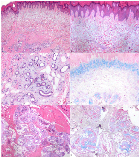
Both patients denied prior history of trauma or surgery in the area. The histopathological findings of both excisions were consistent with MEN (Figure 1). No further treatment was given and both patients failed to return for follow up.

    Figure 1: Mucinous Eccrine Nevus. Case 1: A-C) Low and medium power
views (H&E stain, 20X, 40X, 100X) showing a verrucous epidermis with
increased eccrine glands within the deep dermis with prominent mucin
deposition within the superficial dermis and around the eccrine glands. D)
Alcian blue stain highlighting the mucin (20X). Case 2: E) Medium-power view
showing prominent eccrine glands with surrounding mucin (H&E, 40X). F)
Alcian blue stain highlighting the peri-eccrine mucin (40X).
MEN was first described by Romer and Taira in 1994 [2]. The characteristic features consist of a proliferation of benign eccrine glands in a mucinous stroma. Only 11 cases have been previously reported in the literature (Table 1), and now total 13 with the addition of our 2 cases [1-11].
The pathogenesis of MEN is unclear. Llombart et al. [3] suggests it arises from a defect in embryogenesis, but is less favored as cases have been reported in adulthood. Sweat gland proliferations have been observed in healing wounds, suggesting that cases arising during adulthood may be caused by trauma or surgery [2]. Espana and colleagues [5] hypothesized MEN may be the result of migration of genetically identical clones during fetal development, following the lines of Blaschko, becoming evident during adulthood only after significant trauma which would stimulate fibroblasts to synthesize mucin. Only one case presented after surgery, and was thought to be related to the exceptional bilateral distribution found in that particular case [7].
Most cases present as unilateral, solitary lesions on the lower extremities (Table 1). Only two cases had bilateral presentations which were in a patient with chronic granulomatous disease [11] and after an anterior resection of the rectum for stage I rectal cancer [7]. Espana and colleagues [5] reported a unilateral case with multiple lesions following the lines of Blaschko.
Case
Age (y)/
Location
Onset(mo)
Laterality/#
Presentation
Size (cm)
Clinical Dx
Hx of Trauma (T) or Hyperhidrosis (H)
Treatment
Sex
47/F
R LE
24
U/1
Erythematous, tender nodule
1
Erythema nodosum
No
---
2/F
Buttock
21
U/2
Firm brown irregular nodules
1
Mastocytoma
No
None
3[4]
46/M
L 4th toe
1
U/1
Erythematous, tender, firm, swollen patch
2.5
Cellulitis
No
None
32/F
L buttock, leg
240
U/
Brown irregular firm nodules along lines of Blaschko
1.5
---
Yes (H)
---
multiple
5[6]
18/F
L foot
204
U/1
Hyperpigmented, firm, tender triangle-shaped lesion
4
Pigmented nevus
Yes (H)
Plastic surgery
6[7]
57/M
Inner thighs
36
B/
Brown irregular, firm nodules
3
---
Yes (T-rectal surgery)
IL steroids
multiple
7[8]
4 mo/F
R forearm
1
U/
Brown irregular, firm nodules
1.5
Mastocytoma, nevus of sweat gland or smooth muscle
No
Topical steroids
multiple
8[9]
5 mo/F
L wrist
5
U/1
Brown to flesh colored plaque
3
---
Yes (H)
---
9[10]
47/M
L LE
72
U/1
Purple-red tender, firm mass
2
Angiolymphoid hyperplasia with eosinophilia
Yes (H)
None
10[11]
N/M
R knee, ankle, heel, foot, toes & L ankle, elbow
---
B/
Red-brown, firm, hyperkeratotic plaques
8
---
No
None
multiple
11[1]
3 mo/M
Low back
3
U/1
Erythematous firm oval plaque
2.1
Connective tissue nevus, epidermal nevus, mastocytoma
No
---
12
47/M
R thumb
36
U/1
Raised, rough lesion
0.8
Wart, traumatized nevus
No
---
(Current case 1)
13 (Current case 2)
28/M
R index finger
24
U/1
Non-tender, ballotable mass
---
Mucous cyst
No
---
Cm: Centimeter; Dx: Diagnosis; F: Female; Hx: History; IL: Intralesional; L: Left; LE: Lower extremity; M: Male; mo: months; N: Newborn; R: Right; U: Unilateral
Table 1: Summary of MEN cases reported in the English literature (n=13).
MEN occurs in both sexes equally (M=7; F=6) and usually presents before puberty, with 3 cases noticed at birth. Typically, they are red-brown nodules, plaques, or well-defined masses, with or without hyperhidrosis and range in size from 0.8-8 (mean=2.53) cm.
Histopathological findings are similar in all cases. The deep dermis often shows a proliferation of morphologically normal eccrine glands with both increased number and diameter. Mucin deposition is confirmed by Alcian Blue staining in the stroma surrounding the eccrine glands in the deep dermis and less often in the superficial dermis [8].
Since MEN is a benign entity, additional treatment is not necessary, except for esthetic purposes and bothersome symptoms [3,4]. Most cases did not undergo additional treatment. Plastic surgery was performed in one case for hypertrophy and intralesional and topical steroids were administered in two cases for persistent symptoms [6-8].
In summary, MEN is a very rare variant of eccrine nevus with a variety of clinical presentations but specific histopathological findings. The etiology of MEN remains unclear and additional cases will provide more insight into the pathogenesis of this rare entity.
References
- Tempark T, Shwayder T. Mucinous eccrine naevus: case report and review of the literature. Clin Exp Dermatol. 2013; 38: 1-4.
- Romer JC, Taira JW. Mucinous eccrine nevus. Cutis. 1994; 53: 259-261.
- Llombart B, Molina I, Monteagudo C, Ramón D, Martín JM, Sanchez R, et al. Mucinous eccrine nevus: an unusual lesion in a child. Pediatr Dermatol. 2003; 20: 137-139.
- Park HS, Lee UH, Choi JC, Chun DK. Mucinous eccrine nevus. J Dermatol. 2004; 31: 687-689.
- España A, Marquina M, Idoate MA. Extensive mucinous eccrine naevus following the lines of Blaschko: a new type of eccrine naevus. Br J Dermatol. 2006; 154: 1004-1006.
- Man XY, Cai SQ, Zhang AH, Zheng M. Mucinous eccrine naevus presenting with hyperhidrosis: a case report. Acta Derm Venereol. 2006; 86: 554-555.
- Lee WJ, Chang SE, Lee MW, Choi JH, Moon KC, Koh JK, et al. Bilateral mucinous eccrine nevus in an adult. J Dermatol. 2008; 35: 552-554.
- Yoshizawa J, Hozumi Y, Katagiri Y, Kawaguchi M, Shimanuki M, Suzuki T. Mucinous eccrine naevus. J Eur Acad Dermatol Venereol. 2009; 23: 348-349.
- Chen CW, Tsai TF, Chen YF, Hung CM. Congenital mucinous eccrine nevus in a 5-month-old girl with frequent intertriginous dermatitis. Pediatric dermatology. 2008; 25: 573-574.
- Chen J, Sun JF, Zeng XS, Liu Y, Jiang YQ, Li AM, et al. Mucinous eccrine nevus: a case report and literature review. Am J Dermatopathol. 2009; 31: 387-390.
- Gross SS, Fridlington E, Stone MS. Congenital mucinous eccrine nevi in an infant with chronic granulomatous disease. Arch Dermatol. 2012; 148: 140- 142.
