
Rapid Communication
Austin J Pathol Lab Med. 2018; 5(1): 1021.
Ameliorative Effect of Selenium against Endosulfan Induced Toxicity in Sertoli Cells of Mice
Kumar B1, Sinha A1, Sahay R1, Lal N3, Kumar R2, Ali M2 and Kumar A2*
1Department of Zoology, Patna University, India
2Mahavir Cancer Institute & Research Centre, India
3Department of Zoology, Dr.Syama Prasad Mukherjee University, India
*Corresponding author: Sachin Sinha, Department of Oral Pathology and Microbiology, Daswani Dental College, Kota, India
Received: May 25, 2018; Accepted: July 27, 2018; Published: August 03, 2018
Abstract
Indiscriminate use of pesticides in the recent times is widely used by the farmers for the better yield of their crops. Although, the yield of the crops has increased many folds but many health related diseases has arised in the population. Endosulfan is an organochlorine pesticide which is widely used by the farmers of India, but in recent times it has led to cause various health related problems in the population. Therefore, the present research work on animal deciphers the ameliorative effect of selenium in Endosulfan induced reproductive toxicity in male mice.
Endosulfan at the dose of 3mg/Kg body weight was administered orally to male mice for 5 weeks. Thereafter, Selenium in the form Sodium seleniteat the dose of 10µg/Kg body weight was administered for 4 weeks to observe the ameliorative effect of it on sertoli cells of male mice. The study reveals that after the administration of Endosulfan, there was significant degeneration in the in the sertoli cells of mice at the cellular as well as sub cellular level. But, after administration of selenium, there was significant restoration in the sertoli cells at the cellular as well as sub cellular levels denotes that selenium possesses restorative property. Thus, it acts as one of the best antidote against endosulfan induced cellular toxicity.
Keywords: Endosulfan; Selenium; Histopathology; Electron microscopy
Introduction
Agrochemicals pollute the environment primarily because of their wasteful application and due to the fact that crops use them inefficiently. Fertilizer use efficiency is revealed to be only 30-35%, the remaining proportion reaches the groundwater resources causing the pollution. Indiscriminate use of agrochemicals under conventional agriculture, not only cause severe health hazards for human beings but also has numerous other side effects on the environment including destruction of the biodiversity. Endosulfan and organochlorine, is widely used to kill pests of cereals, cotton, oilseeds, tea, coffee, fruits, potato, and vegetables. According to US Environmental Protection Agency [1]. it belongs to highly hazardous category. Moreover, it is easily absorbed by the stomach, lungs and skin and exposure through any route can be hazardous. The commercially produced endosulfan contains two isomers α- endosulfan and β- endosulfan. Both of the forms are known to be genotoxic to human gonads [2,3].
Pesticide safety is classified by the World Health Organisation (WHO) according to the results of LD50 tests, which document the amount of a chemical required to kill 50% of a population of laboratory rats. Under this system, endosulfan is currently classified as Class II – moderately hazardous to human health. However, the United States’ Environmental Protection Agency (EPA) rates endosulfan as Category Ib – highly hazardous [4]. LD50 data for endosulfan are in conformity with some published results indicating that the chemical should be in the WHO’s Class Ib, according to the organisation’s own criteria. Evidence of the threats to human health posed by endosulfan are abundant, and the chemical has been banned outright or severely restricted in a number of countries as a result. Independent of LD50 results, these threats warrant the immediate upgrading of endosulfan to WHO Class Ib [5]. Endosulfan has been associated with estrogenic activity both in vivo and in vitro [6]. Exposure to this pesticide causes hormonal imbalance leading to impaired testicular cells function [7].
Selenium is an essential macro element required in body during biochemical and physiological processes including the biosynthesis of coenzyme Q, regulation of ion fluxes across membranes, maintenance of the integrity of keratins, stimulation of antibody synthesis, and activation of glutathione peroxidase [8]. Selenium basically interact with xenobiotics at several sites like during absorption and excretion, transport of metals in the body, binding to target proteins, metabolism and sequestration of toxic metals, and oxidative stress [9]. Besides this, they may also serve as required prosthetic groups in active sites or as co-enzymes for indispensable metallo-enzymes. Various studies have shown that antioxidant nutrients protect cells against deleterious effects of environmental agents [10]. Selenium has received considerable attention as an essential micronutrient for both animal and human beings. Therefore, in the present study selenium has been used to observe its ameliorative effect against the pesticide endosulfan.
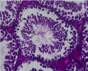
Figure 1: Microphotograph section of Control testis of mice showing seminiferous tubules with normal architecture of stages of Spermatogenesis (S), Sertoli Cells (SC) and Interstial Cells (IS). (X 500).
    Histopathological Study:
Figure 1: Microphotograph section of Control testis of mice showing
seminiferous tubules with normal architecture of stages of Spermatogenesis
(S), Sertoli Cells (SC) and Interstial Cells (IS). (X 500).
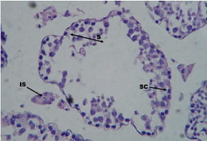
    Figure 2: Microphotograph section of testis of mice with Endosulfan 5
weeks treated showing abnormal architecture of seminiferous tubules with
degenerated stages of Spermatogenesis (S), Sertoli Cells (SC) and Interstial
Cells (IS). (X 500).
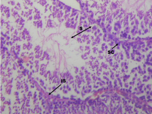
    Figure 3: Microphotograph section of testis of mice with Selenium treated
on Endosulfan 5 weeks pretreated group showing significant restoration in
seminiferous with normal architecture of stages of Spermatogenesis (S),
Sertoli Cells (SC) and Interstial Cells (IS). (X 500).
Materials and Methods
Animals
Swiss albino mice were bred at the mice room of Prof.A.Nath, Department of Zoology, Patna University, Patna, Bihar, India. The animals had free access to water and feed pellets (mixed formulated feed prepared by the laboratory itself). The age of mice for the experiment was 12 weeks old. The average body weight of experimental mice was 30 ± 2g.
Test chemical
Pesticide Endosulfan, manufactured by Excel India Pvt. Ltd., Mumbai with EC 35% was utilized for the experiment.
Preparation of selenium dose
In the present study, Selenium in the form of Sodium Selenite was procured from Himedia Laboratories, India and aqueous dose of 10µg/Kg body weight was prepared for oral administration after estimation of LD50 value (80µg/Kg body).
Study groups & sampling
The animals were grouped into three groups, group I, control n=6, received distilled water as drinking water while the group II, Endosulfan treated group (n=18) received Endosulfan 3mg/kg b.w daily by gavage method for 5 weeks. After this, the (n=12) animals (from the group II) were administered Sodium selenite at the dose of 10µg/Kg body weight /day upon the Endosulfan treated group for 4 weeks. All the animals were sacrificed after the entire experimentation. A midsaggital incision was made and testicular tissue from all the mice were removed and fixed in 10% neutral formalin for histopathological study. For Transmission Electronmicroscopy the tissues were fixed in 2.5% gluteraldehyde in 0.1M sodium phosphate buffer (pH 7.2) for 24 hours. After rinsing with phosphate buffer, tissues were postfixed with 2% osmium tetraoxide in sodium phosphate buffer. Dehydration was accomplished by gradual ethanol series and tissues were embedded in epoxy resin. Semi thin sections were stained with toluidine blue and examined with a light microscope (Olympus, LXi,Tokyo, Japan). Ultrathin sections (800 nm) were stained with uranyl acetate and lead citrate. Sections were thenviewed and photographed with Morgagini - 268 D TEM (SEI Co.) at SAIF-EM facility Unit (Sophisticated Analytical Instruments Facility) at All India Institute of Medical Sciences (AIIMS), New Delhi, India. For the histopathological study, the haemotoxylin-eosin stained slides were prepared and the sections were viewed under light microscope.
Results
Histopathological findings
The microphotograph of the control shows normal architecture of seminiferous tubules with distinct sertoli cells. Different stages of spermatogenesis are quiet distinct with the normal spermatozoas (Figure 1). The Endosulfan 5 weeks treated group showed high degree of degeneration as the seminiferous tubules showed lost stages of spermatogenesis. The sertolicells were rudimentary in condition with no spermatozoa. The integrity of the membrane of seminiferous tubules is completely lost along with the interstitial cells (Figure 2). The selenium treated group showed very significant restoration in the seminiferous tubules. The stages of spermatogenesis is quiet prominent with the spermatozoas (Figure 3). The restoration in about 90% of animals was observed as compare to 100% degenerated Endosulfan treated groups.
Transmission electron microscopic findings
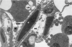
Transmission Electron Micrographs of control testis of mice showed sertoli cells with double membrane of nucleus with normal chromatin material. Mitochondria as well as the ribosomes were distinct while mitochondrial cristae and lipid droplets were clearly visible with normal endoplasmic reticulum (Figure 4). The endosulfan 5 weeks treated testis sertoli cells showed very high degree of degeneration in them. The nuclear membrane and the chromatin materials were in degenerated condition. Severe vacuolisation in the cytoplasm with damaged mitochondria were observed. Lipid droplets were highly increased in the cytoplasmic region while rudimentary plasma membrane were observed (Figure 5). Endosulfan 5 weeks followed by Selenium 4 weeks administered testis showed significant restoration in the sertoli cells. The nuclear membrane along with the chromatin material have restored at much extent. The spermatid is quiet in normal architecture (Figure 6).
Figure 4: Transmission Electron Micrographs of Control testis of mice showing normal architecture of Sertoli Cells (sc) with double membrane of Nucleus (NM), Chromatin material (C) and Lipid Droplets (LD). Mitochondria (M) with cristae are clearly visible. (X 22,000).
    Transmission Electron Microscopic Study:
Figure 4: Transmission Electron Micrographs of Control testis of mice
showing normal architecture of Sertoli Cells (sc) with double membrane of
Nucleus (NM), Chromatin material (C) and Lipid Droplets (LD). Mitochondria
(M) with cristae are clearly visible. (X 22,000).
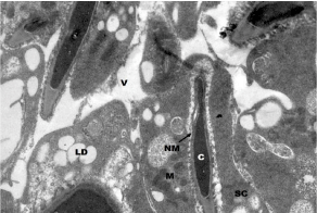
    Figure 5: Transmission Electron Micrographs of testis of mice with
Endosulfan 5 weeks treated showing Sertoli Cells (SC) in highly degenerated
condition. The Nuclear Membrane (NM) and the Chromatin materials (C) are
in degenerated condition. Severe Vacuolisation (V) the cytoplasm is clearly
with damaged mitochondria. (X 22,000).
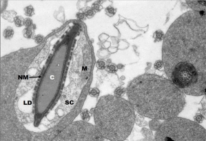
    Figure 6: Transmission Electron Micrographs of testis of mice with Selenium
treated on Endosulfan 5 week's pretreated group showing restoration in the
Sertoli Cells (SC). The Nuclear Membrane (NM) along with the Chromatin
material (C) have restored at much extent. The spermatid is quiet in normal
architecture along with Mitochondria (M) with Lipid Droplets (LD). (X 22,000).
Discussion
Pesticides in the recent times have caused lots of health hazards in the population. It produces oxidative stress and hormonal imbalance [11]. Endosulfan has caused lots of health hazards like hepatic, renal and reproductive disorders in the animal models [12]. It also causes decrease in the semen quality, increase in testicular and prostate cancer and increase in defects in male sex organs [13,14].
The sertoli cell is the principal structural element of the seminiferous epithelium, providing physical support and an environment conducive to germ cell development and maturation. Sertoli cells consist of cytoskeleton which is composed of major three elements: actin, vimentin, and tubulin. Actin makes up the microfilament network, and tubulin comprises the microtubule cytoskeleton. An extensive intermediate filament cytoskeleton composed primarily of the type III family member vimentin is present in the sertoli cells.
But, in the present study, Endosulfan has caused high degree of degeneration in the testicular cells especially in the seminiferous tubules as there is complete loss of spermatogenetic stages in mice. The sertoli cells at the other side are almost lost or in damaged condition denotes the complete failure of the formation of spermatozoa. The cytoskeleton of the sertoli cells are in the ruminant condition in Endosulfan treated groups. Similar, study at biochemical level have been well documented [15-18].
Selenium in the present study has played vital role in the restoration of the normal architecture of the sertoli cells. The spermatogenetic stages has revived at much extent after the administration of selenium. The significant amelioration in the architecture of the sertoli cells with the spermatids is the novel finding of the study denotes that selenium rebinds the actin, vimentin and tubulins of the cytoskeleton.
Various studies have proven the efficacy of the selenium as the ameliorative agent, as it is known to be very good antioxidant. The massive restoration in the sertoli cells observed in the present study proves its ameliorative effect. It is essential for the activity of glutathione peroxidase, an enzyme that protects against reactive oxygen species and subsequent cell membrane damage and is an integral part of more than about 30 known proteins [19,20]. Furthermore, it is one of the vital and the only trace element to be specified in the genetic code, as selenocysteine, which when incorporated into selenoproteins, protects tissues and membranes from oxidative stress and controls cell redox status [21,22]. The functions of many of the twenty five human selenoproteins are as yet unknown, although they generally participate in antioxidant and anabolic processes [23]. In a recent systematic review and meta-analysis of antioxidant supplements for the prevention of gastrointestinal cancers has assessed the evidence for an effect of selenium. It acts as an antioxidant and helps protect the body against the damaging effects of free radicals [24,25].
Conclusion
Therefore, from the present study it can be concluded that selenium helps in the recovery from the damage caused by endosulfan. It possesses ameliorative effect against endosulfan induced toxicity.
References
- EPA (Environmental Protection Agency). "Endosulfan Updated Risk Assessments; Notice of Availability, and Solicitation of Usage Information". Federal Register. 2007; 72: 64624-64626.
- Helle RA, Marie VA, Thomas HR, Marianne GI, Eva CBJ. "Effects of currently used pesticides in assays for estrogenicity, androgenicity, and aromatase activity in vitro". Toxicology and Applied Pharmacology. 2002; 179: 1-12.
- Agency of Toxic Substances and Disease Registry. Toxicological Profile for Endosulfan. 2000.
- World Health Organization. The WHO Recommended Classification of Pesticides by Hazard. 2005.
- EPA, Reregistration Eligibility Decision for Endosulfan. 2002.
- Bretveld RW, Thomas CM, Scheepers PT, Zielhuis GA, Roeleveld N. Pesticide exposure: the hormonal function of the female reproductive system disrupted? Reproductive Biology and Endocrinology. 2006; 4: 30.
- Safe SH. Environmental and dietary estrogens and human health: is there a problem? Environ Health Perspect. 1995; 103: 346-351.
- Fordyce F. "Selenium Geochemistry and Health". AMBIO: A Journal of the Human Environment. 2007; 36: 94-97.
- Miller S, Walker SW, Arthur JR, Nicol F, Pickard K. Selenite protects human endothelial cells from oxidative damage and induces thioredoxin reductase. Clin Sci. 2001; 100: 543-550.
- El-Demerdash FM. Antioxidant effect of vitamin E and selenium on lipid peroxidation, enzyme activities and biochemical parameters in rats exposed to aluminium. J Trace Elem Med Biol. 2004; 18: 113-122.
- Ahmad MM, Ahmad MM, Saleha S. Effects of endosulfan and chlorpyrifos on the reproductive organs and sex hormones of neonatal rats. Pakistan Journal of Zoology. 1993; 25: 11-14.
- Sinha P, Verma P, Kumar A, Nath A. Testicular atrophy in mice Mus musculus under sublethal doses of endosulfan. J Ecophysiol & Occup Hlth. 2004; 4: 191-196.
- Hileman B, Washington EN. Environmental estrogens linked to reproductive abnormalities. Cancer C & EN. 1994; 72: 19-23.
- Singh JK, Ali M, Kumar R, Nath A, Kumar A. Possible occurrence of apoptosis in Endosulfan induced testicular toxicity in mice. J Ecophysiol Occup Hlth. 2010; 10: 43-47.
- Sinha N, Narayan R, Shanker R, Saxena DK. Endosulfan induced biochemical changes in the testis of rat. Vet Hum Toxicol. 1995; 37: 547-549.
- Chitra KC, Latchoumycandane C, Mathur PP. Chronic Effect of endosulfan on the testicular function of rat. Asian Journal Of Andrology. 1999; 1: 203-206.
- Singh JK, Nath A, Kumar A, Ali M, Kumar R. Study on the effect of endosulfan on testosterone level and seminiferous tubule of testis of mice. World Journal of Environmental Pollution. 2011; 1: 01-04.
- Kumar R, Singh JK, Ali M, Kumar A. Impact of Selenium on Microtubules Polymarisation of Spermatozoa of Endosulfan Exposed Swiss Albino Mice. Journal of Life Sciences. 2011; 5: 132-135.
- Yu SY, Zhu YJ, Li WG. Protective role of selenium against hepatitis B virus and primary liver cancer in Qidong. Biological Trace Element Research. 1997; 56: 117-124.
- Jameson RR, Diamond AM. Regulatory role for-Sec tRNA [Ser]Sec in selenoprotein synthesis. RNA. 2004; 10: 1142-1152.
- Brown KM, Arthur JR. Selenium, selenoproteins and human health: a review. Public Health Nutr. 2001; 4: 593-599.
- Hatfield DL, Gladyshev VN. How selenium has altered our understanding of the genetic code. Molecular and Cell Biology. 2002; 22: 3565-3576.
- Maroni M, Colosio C, Ferioli A, Fait A. Biological monitoring of pesticide exposure: a review. Introduction Toxicology. 2000; 143: 11-18.
- Bjelakovic G, Nikolova D, Simonetti RG, Gluud C. Antioxidant supplements for prevention of gastrointestinal cancers. A systematic review and meta-analysis. Lancet. 2000; 364: 1219-1228.
- Uchendu C, Ambali SF, Ayo JO, Esievo KAN. Acetyl-L-carnitine attenuates haemotoxicity induced by subacute chlorpyrifos exposure in wistar rats. Der Pharm. Let. 2011; 3: 292-303.