Short Communication
Austin J Pathol Lab Med. 2019; 6(1): 1023.
Micronuclei and Nuclear Abnormalities as Bioindicators of Gene Instability Vulnerability
Torres-Bugarín O1*, Garcia-Arellano E2, Onel Salas-Cordero K1 and Molina-Noyola LD1
1Programa Internacional de Medicina, Universidad Autónoma de Guadalajara, Mexico
2Facultad de Ciencias, Universidad Autónoma de Baja California, California
*Corresponding author: Torres Bugarín O Research Professor (S N I nivel II). Laboratorio de Evaluación de Genotóxicos Programa Internacional de Medicina, Universidad Autónoma de Guadalajara Zapopan, Jalisco, México
Received: July 20, 2019; Accepted: August 23, 2019; Published: August 30, 2019
Abstract
Genetic instability can cause severe health consequences. There are plenty of pathologies, environmental factors and life style that it can provoke them. Detecting in a timely manner the vulnerable population to genotoxic effects should be an objective of the genetic toxicology. In this manner, the frequent evaluation of the human buccal Micronucleated Cells (MNC) it can offer the opportunity to measure the genetic instability with the prerogative to be a nonpainful methodology, simple and relatively inexpensive. Thus, this article focus on demonstrate how this biomarker effect can be helpful in the determination of genotoxic vulnerability.
Keywords: Micronuclei; Nuclear abnormalities; Genotoxicity vulnerability
Genomic Instability: Health-Disease
The genome stability is fundamental for the cellular homeostasis and the human health, however during everyday life all people are exposed to a endless genotoxic agents both endogenous and exogenous which frequently altered the genomic homeostasis. Regularly, the damage caused by genotoxics are silence and pass unnoticed; it can be more evident in diseases characterize by progressive deterioration of specific tissues, cancer susceptibility, chromosome rearrangement and hypersensibility to genotoxic agents. It will have to take in count that a genotoxic agent can affect any individual by different manners (genetic instability, mutagenics, tratogenics or carcinogen), depending of genetics factor, environment or life style. That is why it is beneficial to have techniques or basic biomarkers and relatively inexpensive that could allow us to identify the most vulnerable population efficiently.
Thus, the identification of genetics changes that cause diseases, including the chromosome instability, are important for diagnostic criteria that contribute to the better understanding of a disease etiology and treatment management. The genetics changes can appear in the early stages of the disease, even earlier than the clinical manifestations, and they can be biomarkers for the prognosis [1]. This is relevant because surprisingly there are an endless diseases characterize by high chromosome damage, this doesn’t only occur to characterized disease or defined like Genomic Instability Syndrome ([2], Spinocerebellar Ataxia Type 2 [3]) or cancer, which is also identified as one of it characteristics [4]; [5,6], it also occur in autoimmune diseases [7,8], such like neurodegenerative diseases [9,10,11] (Parkinson’s disease, Alzheimer’s Disease), [11], cardiovascular diseases, eating disorders [5,6], [12,13,14,15], Hunter type mucopoliscarids [16], Polycystic Ovarian Syndrome [17], among many others. By the other hand, the recognition of the genotoxic risk implicated in work activities, media environment or life stlye [18,19] like it occur in dentist and dental technicians [20], or by exposition to pesticides [21], hydrocarbons [22], heavy metals [23] or anesthetics gases [24].
Micronucleus and Nuclear Abnormalities: Instability Biomarkers
The used methods in risk or protection evaluation in genotoxics effects are usually more expensive, complicated and invasive. A worldwide accepted biomarker and highly trustable for the detection and quantification of the genome instability, is the Micronucleus (MN) frequency that can be observe in cells that has complete cellular division. By it versatility to the application of different organism, tissues and models, it offered a wide clear opportunity and precise surveillance of genetic damage in population with high risk, reasons why the number of publications with relationship with MN test has increased exponentially in recent years.
The MNC quantification in buccal mucosa has the benefits that this tissue present a limited capacity of DNA reparation and therefore may reflect with a better precision the genomic instability, maybe that’s why most tumors are epithelial origin [1]. Also, collect and process these cells is a minimal invasive procedure and well accepted by the participants. It is fast, basic and relatively inexpensive, it does not require cell culture nor specialize installations an in a short time can give results [19,25,26,27]. By the other hand, at the time of working with this tissue, it is also useful to quantified other Nuclear Abnormalities (NA) besides the MNC, like lobulated nucleus [(LN) that reflect DNA damage]; Binucleated Cells (BNC) which originate by cytokinesis damage and Condensate Chromatin (CC), Karyorrhexis (KR) and Karyolysis (KL), which are cell death markers. In recent years, this technique is profusely use because it reliabilityin genotoxic damage, genomic instability in humans, it simplicity and inexpensive cost [28], [19,25,26,27].
Thus, the MN are the effect biomarkers more used that forms in the metaphase-anaphase transition on mitosis that can be complete lagging chromosomes due to mitotic spindle damage (aneuploidy effect) or chromosome fragments without centromere (clastogenic damage). In both cases, they are fragments or complete chromosome that fail to incorporate to the daughter cell nucleus [29] that may differentiate with others by the zise of the MN [30] or by the presence or absence of centromere or kinetochore [31]. This events can occurs spontaneously, but in the presence of various endogenic agents [32]; [33]; [34] or [35]; [8,32,18] will increase, transforming the MN in markers for mutagenic agent effect, genotoxic or teratogenic, principally in micronucleogenics [36].
Who’s Vulnerable to Genotoxic Damage?
Even thought multiple causes variability exist in the frequency of MNC and NA like genetic, metabolic, environmental, life style factor and even methodological like data recollection, the recollection methods and sample processing, number and cell analyzed criteria are effective biomarkers for detecting vulnerable population with genotoxic damage.
When studying the genotoxic effects of some agent in the case of MNC and NA, the most common is to obtain the descriptive statics of a central trend measure (media, median) and another one of dispersion (standard deviation, variation coefficient and quartile), that express the biomarker variability. If in a studied population there are cases whose number of MNC or NA rise above the central line tendency, even more than the dispersion that it consider normal, it will be precisely these cases that can be identified as people with high vulnerability to genotoxic damage, they are the ones with the greatest genetic instability in the studied population.
Often when obtaining the descriptive measures and applying the statistical tests, no significant differences are detected between the groups under study, but how can we ignore those individuals who score above the average? Can it be interpreted that they did not suffer damage because the study says that there’s no significant effect ?, or although this damage exists although it is unknown, what to attribute it to?.
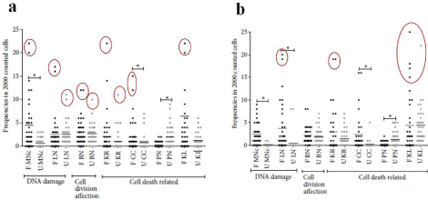
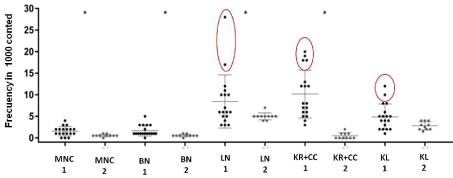
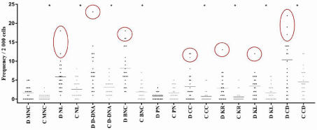
In the case of genomic instability, as it is unknown precisely the effects that may trigger in the future, although in any way they will be negative effects on health, identifying individuals whose biomarkers exceed the average as seen in Figures 1a, 1b, 2 and 3, in which they draw attention that the highest values at MNC or NA (inside red circle) are located in the study or exposed groups, but it is clear that each point represents a highly vulnerable person at genotoxic risk (Figure 4) and it is at this point that the heath provider should offer preventive measures such as antioxidant consumption, identify risk factors and implement protection measures or public policies aiming a environmental sanitation, all in order to avoid rapid deterioration of health..

    Figure 1: Frequencies of micronucleated cells and nuclear abnormalities in farmers and unexposed women (a) and children (b). F = farmers; U =Unexposed; MNc
= Micronucleated cells; LN = Cells with lobulated nucleus; BN = Binucleated cells; KR = Karyorrhexis; CC = Cells with condensed chromatin; PN = Pyknotic cells;
and KL = Karyolysis. *p ‹ 0.05. [21].

    Figure 2: Frequencies of micronucleated cells and nuclear abnormalities in Mexican with risk for cervicouterin cancer. MNc = Micronucleated cells; LN = Cells
with lobulated nucleus; BN = Binucleated cells; KR = Karyorrhexis; CC = Cells with condensed chromatin; PN = Pyknotic cells; and KL = Karyolysis. *p ‹ 0.05 . [6].

    Figure 3: Dispersion of micronucleated cells and nuclear abnormalities in dental surgeons and controls (D: Dental surgeon; C: Control; MNC: Micronucleated cells;
NL: Lobulated nucleus; D-DNA: DNA damage (MNC +LN), BNC: Binucleated cells (damage to cytokinesis); PN: Pyknotic nucleus; CC: Condensed chromatin; KR:
Karyorrhexis; KL: Karyolysisand; CD: Cell death (CC+KR+KL); *p‹0.05) [20].

    Figure 4: Microphotographs of MNi and NA, identified according to the HUMNxl scoring criteria. The figure shows a microphotographs of oral mucosa cells stained
with acridine orange at 100x optic amplification with a Carl Zeiss IVFL Axiostar Plus microscope, 450–490 nm fluorescence filters. a) Normal buccal cell without any
MNi or NA, b) A buccal cell with the presence of micronucleus (CMN) (white arrow) and c) a lobed-nuclei cell (LN); MN and LN both are DNA damage.
Acknowledgments
We thank the support to Lic. Ricardo Del Castillo Ruano and International Program of Medicine, of the Universidad Autónoma de Guadalajara.
References
- Hovhannisyan, G., Harutyunyan, T. Aroutiounian, R. Curr Genet Med Rep. 2018; 6: 1-11.
- Gratia M, Rodero MP, Conrad C, BouSamra E, Maurin M, Rice GI, etc. Bloom syndrome protein restrains innate immune sensing of micronuclei by cGAS. Exp Med. 2019; 5: 1199-1213.
- Cuello-Almarales DA, Almaguer-Mederos LE, Vázquez-Mojena Y, Almaguer- Gotay D, Zayas-Feria P, Laffita-Mesa JM, etc. Buccal Cell Micronucleus Frequency Is Significantly Elevated in Patients with Spinocerebellar Ataxia Type 2. Arch Med Res. 2017; 3: 297-302.
- Wu XY, Lu L. Vitamin B6 deficiency, genome instability and cancer. Asian Pac J Cancer Prev. 2012; 11: 5333-5338.
- Flores-García A, Torres-Bugarin O, Velarde Félix JS, Rangel-Villalobos H, Zepeda-Carrillo EA, Rodríguez-Trejo A, etc. Micronuclei and other nuclear anomalies in exfoliated buccal mucosa cells of Mexican women with breast cancer.Journal of Balkan Union of Oncology (JBUON). 2014: 4; 328-332.
- Flores-García A, Ruiz-Bernés S, Aguiar-García P, Benítez-Guerrero V, Valle- Solís MO, Molina-Noyola LD, etc. Micronúcleos y anormalidades nucleares en células de mucosa bucal de mujeres Mexicanas con factores de riesgo para cáncer cérvicouterino: Estudio piloto. Revista El Residente. 2018; 2: 56-61.
- Mihaljevic O, Zivancevic-Simonovic S, Milosevic-Djordjevic O, Djurdjevic P, Jovanovic D, Todorovic Z, etc. Apoptosis and genome instability in children with autoimmune diseases. Mutagenesis. 2018; 6: 351-357.
- Torres-Bugarin O, De Anda-Casillas A, Ramirez-Munoz MP, Sanchez-Corona J, Cantu JM, Zuniga G. Determination of diesel genotoxicity in firebreathers by micronuclei and nuclear abnormalities in buccal mucosa. Mutat Res. 1998; 3: 277-281.
- Petrozzi L, Lucetti C, Scarpato R, Gambaccini G, Trippi F, Bernardini S, etc. Cytogenetic alterations in lymphocytes of Alzheimer’s disease and Parkinson’s diseasepatients. Neurol Sci. 2002; 23: 97-98.
- Andreassi MG, Barale R, Iozzo P, Picano E. The association of micronucleus frequency with obesity, diabetes and cardiovascular disease. Mutagenesis. 2011; 1: 77-83.
- Thomas P, Fenech M. Buccal Cytome Biomarkers and Their Association with Plasma Folate, Vitamin B12 and Homocysteine in Alzheimer’s Disease. J Nutrigenet Nutrigenomics. 2015; 2: 57-69.
- Naga MB, Gour S, Nallagutta N, Ealla KK, Velidandla S, Manikya S. Buccal Micronucleus Cytome Assay in Sickle Cell Disease. J Clin Diagn Res. 2016; 6: 62-64.
- Borba TT, Molz P, Schlickmann DS, Santos C, Oliveira CF, Prá D, etc. Periodontitis: Genomic instability implications and associated risk factors. Mutat Res Genet Toxicol Environ Mutagen. 2019; 840: 20-23.
- Tadin A, Gavic L, Roguljic M, Jerkovic D, Zeljezic D. Nuclear morphological changes in gingival epithelial cells of patients with periodontitis. Clin Oral Investig. 2019; 26.
- Pastor S, Rodríguez-Ribera L, Corredor Z, da Silva Filho MI, Hemminki K, Coll E, etc. Levels of DNA damage (Micronuclei) in patients suffering from chronic kidney disease. Role of GST polymorphisms. Mutat Res Genet Toxicol Environ Mutagen. 2018; 836: 41-46.
- Diaz Jacques CE, de Souza HM, Sperotto NDM, Veríssimo RM, da Rosa HT, Moura DJ, etc. Hunter syndrome: Long-term idursulfase treatment does not protect patients against DNA oxidation and cytogenetic damage. Mutat Res Genet Toxicol Environ Mutagen. 2018; 835: 21-24.
- Karatayli R, GülZamani A, Gezginç K, Tuncez E, Soysal S, Karanfil F, etc. Micronuclei frequencies in lymphocytes and cervical cells of women with polycystic ovarian syndrome. Turk J Obstet Gynecol. 2017; 3: 151-155.
- Torres-Bugarín O, Covarrubias-Bugarín R, Zamora-Perez AL, Torres- Mendoza BMG, García-Ulloa M, Martínez-Sandoval FG- Anabolic androgenic steroids induce micronuclei in buccal mucosa cells of body builders.Br J Sports Med. 2007; 9: 592-596.
- Torres-Bugarín O, Macriz N, Zavala-Cerna MG, Flores-García A, Ramos- Ibarra ML. Procedimientos Básicos de la Prueba de Micronúcleos y Anormalidades Nucleares en Células Exfoliadas de Mucosa Oral. El Residente. 2013; 1: 4-11.
- Molina Noyola LD, Coronado Romo ME, Vázquez Alcaraz SJ, Izaguirre Pérez ME, Arellano-García E, Flores-García A, etc. Evaluation of genotoxicity and cytotoxicity amongst in dental surgeons and technicians by micronucleus assay. Dental, Oral and Craniofacial Res. 2019. 5: 1-5.
- Castañeda-Yslas IY, Arellano-García ME, García-Zarate MA, Ruiz-Ruiz B, Zavala-Cerna MG, Torres-Bugarín O. Biomonitoring with micronuclei test in buccal cells of female farmers and children exposed to pesticides of Maneadero agricultural valley, Baja California, Mexico. J Toxcol. 2016.
- Torres-Bugarín O, De Anda-Casillas A, Ramírez-Múñoz MP, Sánchez- Corona J, Cantú JM, Zúñiga G. Determination of diesel genotoxicity in firebreathers by micronuclei and nuclear abnormalities in buccal mucosa. Mutat Res. 1998; 3: 277-281.
- Jara-Ettinger AC, López-Tavera JC, Zavala-Cerna G, Torres-Bugarín O. Genotoxic Evaluation of Mexican Welders Occupationally Exposed to Welding-Fumes Using the Micronucleus Test on Exfoliated Oral Mucosa Cells: A Cross-Sectional, Case-Control Study. PLOS ONE. 2015.
- Torres Bugarín O, Ramos Ibarra ML, Carrillo Gómez CS, Zavala-Aguirre JL. Micronúcleos y. otras anormalidades nucleares en células de mucosa bucal como biomarcadores de genotoxicidad y citotoxicidad en personal expuesto a gases anestésicos. Revista Colombiana de Salud Ocupacional. 2016; 1. 4-8.
- Torres-Bugarín O, Ramos Ibarra ML. Micronúcleos y anormalidades nucleares en mucosa bucal para evaluar población en riesgo laboral por mutágenos. Rev Costarr Salud Pública. Ene-Jun. 2013; 1: 1-3.
- Torres-Bugarín O, Ramos Ibarra ML. Utilidad de la prueba de micronúcleos y anormalidades nucleares en células exfoliadas de mucosa oral en la evaluación de daño genotóxico y citotóxico. Inter J Morph. 2013; 2: 650-657.
- Torres-Bugarín O, Pacheco-Gutiérrez AG, Vázquez-Valls E, Ramos-Ibarra ML, Torres-Mendoza BM. Micronuclei and nuclear abnormalities in buccal mucosa cells in patients with anorexia and bulimia nervosa. Mutagenesis. 2014; 6: 427-431.
- Bonassi S, Coskun E, Ceppi M, Lando C, Bolognesi C, Burgaz S, etc. The HUman Micro Nucleus project on eXfoLiated buccal cells (HUMN(XL)): the role of life-style, host factors, occupational exposures, health status, and assay protocol. Mutat Res. 2011; 3: 88-97.
- Schmid W. Themicronucleus test. Mutat Res. 1975; 1: 9-15.
- Migliore L, Cocchi L, Scarpato R. Detection of the centromere in micronuclei by fluorescence in situ hybridization: its application to the human lymphocyte micronucleus assay after treatment with four suspected aneugens. Mutagenesis. 1996; 3: 285-290.
- Afshari AJ, McGregor PW, Allen JW, Fuscoe JC. Centromere analysis of micronuclei induced by 2-aminoanthraquinone in cultured mouse splenocytes using both a gamma-satellite DNA probe and anti-kinetochore antibody. Environ mol mutagen. 1994; 2: 96-102.
- Migliore L, Bevilacqua C, Scarpato R. Cytogenetic study and FISH analysis in lymphocytes of Systemic Lupus Erythematosus (SLE) and Systemic Sclerosis (SS) patients. Mutagenesis. 1999; 2: 227-231.
- Ramos-Remus C, Dorazco-Barragan G, Aceves-Avila FJ, Alcaraz-Lopez F, Fuentes-Ramirez F, Michel-Diaz J, etc. Genotoxicity assessment using micronuclei assay in rheumatoid arthritis patients. Clin Exp Rheu. 2002; 2: 208-212 .
- Rodríguez-Vázquez MS-OA, Ramos-Remus C, Zúñiga G, Torres-Bugarín O. Evaluación de la genotoxicidad de ciclofosfamida mediante prueba de micronucleos en pacientes con lupus eritematoso sistemico. Rev Mex Reumat. 2000; 2: 41-45.
- Bonassi S, Coskun E, Ceppi M, Lando C, Bolognesi C. The Human MicroNucleus project on exfoliated buccal cells (HUMNXL): The role of lifestyle, host factors, occupational exposures, health status, and assay protocol. Mut Res Rev. 2011; 3: 88-97.
- Heddle JA, Cimino MC, Hayashi M, Romagna F, Shelby MD, Tucker JD, etc. Micronuclei as an index of cytogenetic damage: past, present, and future. Environ Mol Muta. 1991; 4: 277-291.
- Migliore L, Coppedè F, Fenech M, Thomas P. Association of micronucleus frequency with neurodegenerative diseases. Mutagenesis. 2011; 1: 85-92.
- Torres-Bugarín O, Covarrubias-Bugarín R, Zamora-Perez AL, Torres- Mendoza BMG, García-Ulloa M, Martínez-Sandoval FG. Anabolic androgenic steroids induce micronuclei in buccal mucosa cells of bodybuilders. Br J Sports Med. 2007; 9: 592-596.
- Torres-Bugarín O, Fernández-García, Torres-Mendoza BM, Zavala-Aguirre JL, Nava-Zavala A, Zamora-Perez AL. Genetic profile of overweight and obese school-age children. Toxicol Environ Chem. 2009; 4: 789–795.
- Torres-Bugarín O, Macriz Romero N, Ramos Ibarra ML, Flores-García A, Valdez Aburto P, Zavala-Cerna MG. Genotoxic Effect in Autoimmune Diseases Evaluated by the Micronucleus Test Assay: Our Experience and Literature Review. Bio Med Research International. 2015.
- Torres-Bugarín O, Zamora-Pérez A, Esparza-Flores A, López-Guido B, Feria-Velasco A, Cantú JM, etc. Eritrocitos micronucleados en niños esplenectomizados con y sin quimioterapia. Bol Méd Hos Infan Méx. 1999; 4: 212-217.
- Torres-Bugarín O, Zavala-Cerna MG, Nava A, Flores-García A, Ramos- Ibarra ML. Potential uses, limitations and basic procedures of Micronuclei and nuclear abnormalities in buccal cells. Disease Markers. 2014.