
Mini Review
Austin J Pathol Lab Med. 2020; 7(1): 1027.
Uterine Adenosarcoma: Histological Aspects and Literature Review
Chadi F1,2*, Rouas L1,2 and Lamalmi N1,2
1Department of Gynecological and Pediatric Pathology, IbnSina University Hospital of Rabat, Morocco
2Faculty of Medicine and Pharmacy, Mohammed V University of Rabat, Morocco
*Corresponding author: Chadi F, Department of Gynecological and Pediatric Pathology, IbnSina University Hospital, Mohammed V University of Rabat; Villa 42 Al-Mountalak II Rue Al-Bahr Al-Aswad CYM Rabat, Morocco
Received: June 17, 2020; Accepted: July 15, 2020; Published: July 22, 2020
Abstract
Uterine adenosarcoma is an uncommon tumor that represents 8% of uterine sarcomas. It consists of a double component; a benign component made of a proliferative epithelium and a malignant component represented by a sarcomatous stroma. Its prognosis is relatively favorable after a total hysterectomy with bilateral annexectomy. The objective of this work is to report the histological aspects of uterine adenosarcoma discovered on anatomopathological examination of a polyploid formation delivered by the cervix.
Keywords: Adenosarcoma; Histology; Sarcoma; Uterus
Introduction
Uterine sarcomas represent less than 3% of malignant tumors of the female genital tract, including adenosarcoma, which represents 8%. It is a rare subtype of mixed epithelial and mesenchymal malignant tumor. Clinically, the symptomatology is polymorphic dominated by metrorrhagia. Surgery is the basis for the treatment of localized forms. Only the anatomopathological examination allows the positive diagnosis.
Observation
We report the observation of a 41-year-old patient, smoking and chronic alcoholic, appendectomy, having two vaginal deliveries, who presents for metrorrhagia. The hysteroscopy shows a polyploid formation delivered by the cervix measuring 5x6 cm.
Macroscopic examination showed several fragments weighing 76g and measuring between 1.5 and 4x3 cm in non-friable soft consistency.
Microscopic examination shows a mixed mesenchymal tumor made of a malignant stroma showing a papillary architecture made up of papillae composed of a stroma of variable cell density more pronounced in the subepithelial, giving a leafy appearance reminiscent of a phyllode tumor (Figure 1,2). This stroma contains mesenchymal cells showing moderate cytonuclear atypia with some rare images of mitosis and includes some rare regular endometrial glands and thick-walled vessels (Figure 3,4). The fragments are most often ulcerated on the surface or lined with an endometrial epithelium showing focal immature tubal, mucinous or squamous metaplasia (Figure 5, 6). On a fragment, there is a broad base for implantation of the polyp and a superficial infiltration of the myometrial tissue (Figure 7).
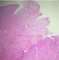
Figure 1: Leafy appearance reminiscent of a phyllode tumor HEx10.
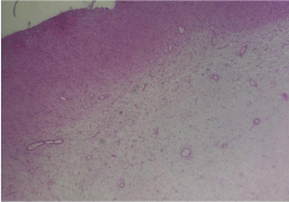
Figure 2: Cell density more pronounced in the subepithelial HEx10.
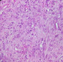
Figure 3: Cells showing moderate cytonuclear atypia with some rare images of mitosis HEx40.
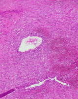
Figure 4: Regular endometrial glands HEx20.
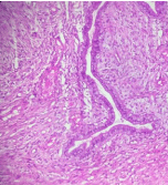
Figure 5: Mucinous metaplasia HEx20.
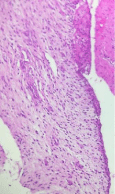
Figure 6: Squamous metaplasia HEx20.
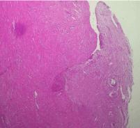
Figure 7: Superficial infiltration of the myometrial tissue HEx4.
Discussion
Uterine sarcomas are rare tumors, which represent 3 to 5% of malignant tumors of the uterus, characterized by significant histopathological and clinical diversity [1]. Uterine adenosarcoma is composed of a benign glandular component intimately associated with a sarcomatous stroma. The sarcomatous component is typically homologous and low-grade [2]. This neoplasm was first recognized as a distinct histologic variant in 1974 [3]. Uterine adenosarcoma understood a group of mixed mesenchymal tumors most commonly arising from the endometrium. However, adenosarcoma can arise from other gynecologic tissue. The second most common site for adenosarcoma is the ovaries [4]. It is a cancer of women in the post-menopausal period, exceptional cases have been found in young women under 40 [5]. The pathogenesis is not clearly explained [1]. Two etiological factors have been reported in the literature: pelvic irradiation and hormone therapy. The clinical symptomatology is very polymorphic, dominated by metrorrhagia (76%), it can be pelvic pain, vaginal discomfort, prolapse of recent appearance or sometimes the appearance of a mass pelvic [5]. Imaging findings are generally not specific to establish an adenosarcoma diagnosis. Features suggestive of uterine adenosarcoma are a regular well-demarcated mass that is hypointense and heterogeneous on T1, multiseptated cystic appearance on T2 [4]. Only the anatomopathological examination allows the positive diagnosis which highlights the two contingents malignant mesenchymal and benign epithelial. Macroscopically, uterine adenosarcoma usually presents as a huge polypous, grayish or yellowish formation showing when cut necrotic areas and many cystic formations with mucoid content [5]. In the largest series, the size of these tumors ranged from 1 to 17 cm in maximum dimension, with a mean size of 5cm [4]. The tumor can have an intra-cavitary, intra- luminal or cervical delivery [5]. Cases of multiple polyps have been reported [4].
Histologically, the epithelial glands are rounded or more commonly slit-like. The epithelial morphology is usually endometrioid, though mucinous, serous, and squamous epithelium have been noted as well. Occasionally, the epithelial component can have cellular atypia. The stromal or mesenchymal component is characterized by spindled and / or round cells that concentrate around the glandular elements forming peri -glandular cuffs. The stromal cells in these periglandular cuffs exhibit varying degrees of cellular atypia and increased mitotic activity. Up to 10 to 25% of adenosarcomas have heterologous elements including rhabdomyoblasts, sex cord-like stromal elements, chondrosarcoma elements, liposarcoma elements, or smooth muscle-derived elements. The most common immunohistochemical markers for adenosarcoma are CD10 and WT1, but these are not specific [4]. The differential diagnosis is made on the one hand with mixed malignant müllériennes tumors where the two components are malignant (carcinosarcomas and mülléroblastomas) and on the other hand with papillary adenofibroma characterized by a benign proliferation as well of the epithelial component as conjunctive [5]. Standard of care treatment is total hysterectomy with bilateralsalpingo -oophorectomy without lymphadenectomy [4]. Adjuvant treatments (chemotherapy and / or radiotherapy) have been instituted in advanced forms [5]. Limited evidence exists for the role of hormonal therapy in uterine adenosarcomas [4].
The majority of patients present with stage I disease, with a 5-year overall survival of 60 to 80%. Survival is influenced by the presence of myometrial invasion, sarcomatous overgrowth, lymphovascular invasion, necrosis, and the presence of heterologous elements including rhabdomyoblastic differentiation [4]. It is not very metastatic, often peritoneal and pulmonary; on the other hand, local recurrences are frequent and can reach 50% of cases [5].
Conclusion
Uterine adenosarcoma is a cancer characterized by its rarity. Its pathogenesis is still unknown. Histological examination alone allows the diagnosis. The prognosis remains relatively favorable after radical surgery.
References
- Bouzid N, KanounBelajouza S, Boussen H, Bouaouina N. Uterine sarcomas in Tunisia: retrospective study of 14 cases. Obstetrics & Fertility Gynecology. 2014; 42: 838-843.
- Carroll A, Ramirez PT, Westin SN, Soliman PT, Munsell MF, Nick AM, et al. Uterine adenosarcoma: An analysis on management, outcomes, and risk factors for recurrence. Gynecologic Oncology. 2014; 135: 455-461.
- Arend R, Bagaria M, Lewin SN, Sun X, Deutsch I, Burke WM, et al. Long-term outcome and natural history of uterine adenosarcomas. Gynecologic Oncology. 2010; 119: 305-308.
- NathensonMJ,Ravi V, Fleming N, Wang WL, Conley A. Uterine Adenosarcoma: A Review. CurrOncol Rep. 2016; 18: 68.
- Fatnassi R, Amri F. Adenosarcoma of the uterus: a case report. J GynecolObstetBiolReprod 2005; 34: 270-272.