Case Report
Austin J Pathol Lab Med. 2022; 9(1): 1034.
Spindle Cell Hemangioma of the Elbow: Case Report of an Underdiagnosed Entity
Essaoudi MA1,2*, Allaoui M1,2, EL Ochi MR1,2, Chahdi H1,2 and Oukabli M1,2
1Department of Pathology, Mohamed V Military Hospital, Hay Riad, Rabat, Morocco
2Faculty of Medicine and Pharmacy, Mohammed V University, Hay Riad, Rabat, Morocco
*Corresponding author: Mohamed Amine Essaoudi, Department of Pathology, Mohamed V Military Hospital, Hay Riad, Rabat, Morocco; Faculty of Medicine and Pharmacy, Mohammed V University, Hay Riad, Rabat, Morocco
Received: February 21, 2022; Accepted: March 12, 2022; Published: March 19, 2022
Abstract
Spindle cell hemangioma (SCH), formerly known as spindle cell hemangioendothelioma, was first described by Weiss and Enzinger in 1986. It is a unique vascular tumor, composed of cavernous spaces and kaposiform solid area. It almost affects the dermis and subcutis of distal extremities. Herein we describe a case of SCH in the right elbow of 9 years old boy. Because the reported cases in the literature are rare, it is suggested that SCH is an underdiagnosed tumor.
Keywords: Spindle cell hemangioma; Vascular tumor; Hemangioendothelioma
Introduction
Spindle cell hemangioma (SCH) is a rare tumor with unique histological features that affects the dermis and subcutis of the distal extremities; it affects rarely the proximal extremities, the trunk and head and neck region [1]. It presents as a solitary lesion or as multiple clustered nodules [2]. Its size ranges from a few millimeters to a few centimeters [3]. Herein we report a case of SCH occurred in the right elbow of 9 years old boy.
Case Presentation
A 9 years old boy presented with a 9 months history of a slow growing painful nodule in his right elbow. No local trauma or inflammation was reported. The patient visited a pediatric surgeon and the mass was diagnosed as hemangioma after an ultrasound’s examination.
Clinical examination revealed a 1.5 x 1 cm mass with intact overlying skin. The nodule was slightly painful and mobile. No palpable lymph node was found in the axillary region.
Surgical enucleation was performed under local anesthesia and the tumor was removed successfully.
On gross examination, there was a well-defined mass in the dermis with brow surface.
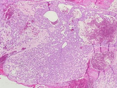
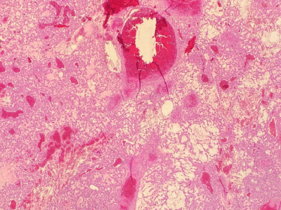
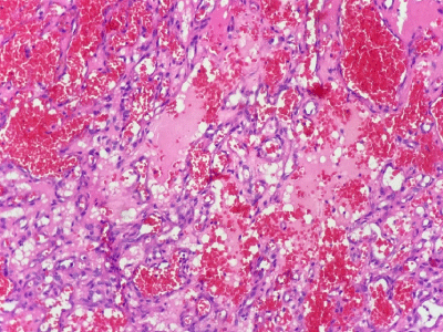
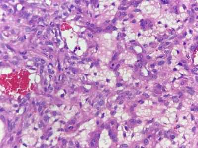
Histologically, the tumor combined two components (Figure 1), the first one was a cavernous irregular vascular space (Figure 2) and the second one was solid area with spindled zones and extravasated erythrocytes (Figure 3). The tumor showed also some vacuolated lipoblast-like cells (Figure 4), epithelioid endothelial cells and flattened vessels without significant nuclear atypia or mitotic activity.

Figure 1: Cavernous vascular spaces and solid areas (x50; H&E).

Figure 2: Cavernous irregular vessels (x100; H&E).

Figure 3: Spindle cells with extravasated erythrocytes (x250; H&E).

Figure 4: Vacuolated cells with lipoblast-like appearance (x400 H&E).
Immunohistochemistry results showed strong reactivity in cavernous area for CD34 antibody, whereas spindled cells were focally positive for smooth-muscle actin. Human-Herpes-Virus8 (HHV8) antibody was negative. From these findings, the diagnosis of SCH was made.
Discussion
SCH is a rare vascular tumor affecting almost exclusively the distal extremities, less often it can be seen in proximal extremities, trunk and head and neck [4,5].
Multifocal SCHs have been reported in association with Maffucci syndrome [1,2], Ollier disease [6] and Millory disease [3].
On microscopic examination, SCH has a characteristic biphasic pattern with many cavernous irregular vascular spaces lined by flattened endothelial cells; the tumor shows a solid proliferation of spindle cells without nuclear atypia or mitotic activity. The main helpful clue for the diagnosis is the vacuolated lipoblast-like cells.
SCH is considered as a neoplasm of intermediate malignancy [1]. Lack of nuclear atypia, mitotic activity and necrosis are not in favor of malignant tumor [7].
The treatment of choice consists on local excision with excellent prognosis. Addition of low-dose interferon alpha-2b and recombinant interleukin 2 with adjuvant radiotherapy has been successful in preventing and/or treating recurrences [8].
Conclusion
SCH is considered to be an uncommon and underrecognized vascular tumor that all general pathologist needs to be aware of it, because of high rate of recurrence and frequent association with other diseases. Surgical excision remains the treatment of choice.
References
- Weiss SW, Enzinger FM. Spindle cell hemangioendothelioma. A low-grade angiosarcoma resembling a cavernous hemangioma and Kaposi’s sarcoma. Am J Surg Pathol. 1986; 10: 521-530.
- Perkins P, Weiss SW. Spindle cell hemangioendothelioma: an analysis of 78 cases with reassessment of its pathogenesis and biologic behavior. Am J Surg Pathol. 1996: 1196-1204.
- Fletcher CDM, Beham A, Schmid C. Spindle cell haemangioendothelioma: a clinicopathological and immunohistochemical study indicative of a nonneoplastic lesion. Histopathology. 1991; 18: 291-301.
- Scott GA, Rosai J. Spindle cell hemangioendothelioma: report of seven additional cases of a recently described vascular neoplasm. Am J Dermatopathol. 1988; 10: 281-288.
- Baron JA, Raines J, Bangert J, Hansen RC. Persistent nodule on the nose. Arch Dermatol. 2002; 138: 259-264.
- Murakami I, Sarker AB, Teramoto N, Horie Y, Taguchi K, Akagi T. Spindle cell hemangioendothelioma: a report of two cases. Acta Pathol Jpn. 1993; 43: 529-534.
- Ding J, Hashimoto H, Imayama S, Tsuneyoshi M, Enjoji M. OOOOE 220 Tosios et al. Spindle cell hemangioendothelioma: probably a benign vascular lesion not a low-grade angiosarcoma. A clinicopathological, ultrastructural and immunohistochemical study. Virchows Arch A Pathol Anat. 1992; 420: 77-85.
- Setoyama M, Shimada H, Miyazono N, Baba Y, Kanzaki T. Spindle cell hemangioendothelioma: successful treatment with recombinant interleukin-2. Br J Dermatol. 2000; 142: 1234-1264.