
Case Report
Austin J Pathol Lab Med. 2022; 9(1): 1035.
Müllerian Cyst in the Posterior Mediastinum: A Case Report and Review of Literature
Rawan Alghamdi¹*, Rakan Mounla Ali² and Amna Almutrafi³
1Department of Anatomical Pathology, Prince Sultan Military Medical City, Riyadh, Saudi Arabia
2Department of Thoracic Surgery, National Guard Hospital, Riyadh, Saudi Arabia
3Department of Anatomical Pathology, National Guard Hospital, Riyadh, Saudi Arabia
*Corresponding author: Rawan Alghamdi, ¹Department of Anatomical Pathology, Prince Sultan Military Medical City, Riyadh, Saudi Arabia
Received: June 01, 2022; Accepted: June 30, 2022; Published: July 07, 2022
Abstract
A Müllerian cyst is a rare newly identified cyst of the posterior mediastinum. It is an important differential diagnosis to other more commonly known mediastinal neurogenic tumor or bronchogenic cyst. This current report represents a case of 51-year-old woman with an incidentally identified mediastinal Müllerian cyst with review of literature.
Keywords: Müllerian Cyst; Mediastinalcyst; Hattori’s Cyst
Introduction
A Müllerian cyst is a rare and benign tumor which is recently defined of the posterior mediastinum. It is crucial that it is differentiated from a neurogenic tumor, mesothelial cyst, or bronchogenic cyst arising in the posterior mediastinum. Hattori [1] was the first who assumed that cystic tumors may be caused by Müllerian ducts occurring in the posterior mediastinum, in 2005. Since then, several cases have been reported as mediastinal Müllerian cyst. This is a case report of a Müllerian-type cyst and review of the literature.
Case Presentation
A 51-year-oldwoman presented with mainly Cushing-like symptoms of 10 months duration. She underwent excision of a carcinoid tumor from the lingula of the left lung and a left paraspinal cyst. Incidental findings on the cervical spine magnetic resonance imaging (MRI) study revealed a left paraspinal cyst along the T7 and T8 vertebrae, and the suspicion was of a neurogenic cyst. Gross pathological examination of the cyst showed a translucent cyst (2.4 x 1.5 x 0.9 cm) filled with watery fluid (Figure 1).
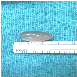
    Figure 1: Gross pathological examination of the cyst showed a translucent

cyst.
Microscopic examination revealed a cyst lined by cuboidal to ciliated columnar epithelium on an underlying fibrous stroma (Figures 2, 3, and 4). In addition, immunohistochemical staining demonstrated nuclear positivity for paired box protein 8 (PAX8), estrogen (ER), and progesterone (PR) (Figure 5). The cyst was diagnosed as a benign cyst lined by Müllerian epithelium consistent with Mülleriancyst, and the patient was seen at follow-up and was doing well with no further complications.
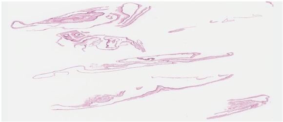
Figure 2: Low power examination: multiple fragments of the cyst wall.
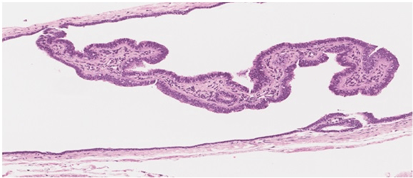
    Figure 3: Medium power examination: the cyst lining of cuboidal to ciliated

columnar epithelium on an underlying fibrous stroma.
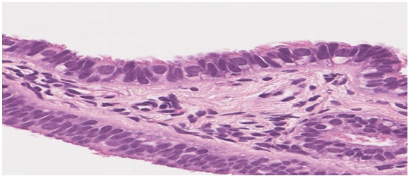
Figure 4: High power examination of the lining epithelium.
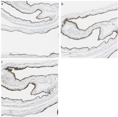
Figure 5: Nuclear positivity for PAX8 (a), estrogen (b), and progesterone (c).
Discussion
Müllerian cysts commonly develop in the male pelvis where the Müllerian duct is left behind, although female Müllerian cysts in the retroperitoneum and mediastinum have been discussed [2]. The first case of Müllerian cyst was published by Hattori [1] in 2005, and up tothe date of these report 26 cases of Müllerian cysts have been reported, including our case. Left thoracic cavity occurrence was noted in 15 cases (60%), and the majority were located around T4 (Table 1). The median size of the Müllerian cysts was about 30mm, the cyst was asymptomatic in 12 cases (48%), and associated with coughing in 6 cases (24%). On immunohistochemical staining, ER showed nuclear positivity in 23 cases (92%) and PR in 21 cases (84%). No recurrence after removal of the cysts was reported.
![]()
    Patient characteristics


    N =25


    Age

    46.8 (44–52)


    Gender

    Female

    26


    Male

    0


    Size (mm)

    30 (13–80)


    Laterality

    Left

    15


    Right

    9


    Bilateral

    1


    NA

    1


    Neighboring vertebrae

    T2

    2


    T3

    5


    T4

    10


    T5

    8


    T6

    4


    T7

    1


    T8

    2


    Multiple

    1


    NA

    2


    Symptoms

    Asymptomatic

    12


    Coughing

    6


    Chest pain

    5


    Genital bleeding

    2


    Dyspnea

    1


    Immunohistochemical study

    ER

    23


    PR

    21


    Abbreviations: ER, estrogen; PR, progesterone.
Table 1: Patient Characteristics.
The etiology of Müllerian cysts is unclear. Batt et al. suggested that the cyst is the choristoma from the primary Müllerian apparatus, similar to the one in the postulated pathogenesis for the Mayer- Rokitansky-Kuster-Hauser syndrome reported by Ludwig [3]. In stage 17 of the embryonic phase, the anlage of the Müllerian tube proliferates parallel to the Wolff tube on the caudal side. The lesion with the Müllerian duct epithelium left behind T3–T5 was found to be the source of the Müllerian duct and is believed to have proliferated due to abnormal hormonal stimulation, resulting in the formation of a cystic lesion. However, the etiology of Müllerian cysts located in the mediastinum should ideally be different from those in the retroperitoneum, considering that the latter often have endocervical differentiation, which is typically not seen in mediastinal cysts [4].
Hattori (1) suggested that the Müllerian cyst in the mediastinum is a new disease entity,which was misdiagnosed initially as a bronchogenic cyst due to its ciliated epithelium. Thomas-de- Montepreville reported that in 3 of the 9 cases of Müllerian cysts occurring in the mediastinum, benign serous cysts were initially misdiagnosed as pleuropericardial cysts [5]. In differentiating Between Müllerian-type cysts and bronchogenic cysts, the lack of cartilaginous structures and the positivity for ER and PR staining suggest the former.
Conclusion
The prognosis for Müllerian cyst tumors are presumed to be good, as there have been no reports of recurrence after surgical removal. One case of respiratory distress was noted due to multiple lesions and progression [6]. A small cystic tumor with a radiological diagnosis of bronchogenic cyst or neurogenic cysts in the posterior mediastinum can be correctly diagnosed based on morphology and immunohistochemical staining.
References
- Hattori H. High prevalence of estrogen and progesterone receptor expression in mediastinal cysts situated in the posterior mediastinum. Chest. 2005; 128(5): 3388-90.
- Schuhrke TD, Kaplan GW. Prostatic utricle cysts (müllerian duct cysts). The Journal of urology. 1978; 119(6): 765-7.
- Batt RE, Mhawech-Fauceglia P, Odunsi K, Yeh J. Pathogenesis of Mediastinal Paravertebral Müllerian Cysts of Hattori: Developmental Endosalpingiosis— Müllerianosis. International Journal of Gynecological Pathology. 2010; 29(6): 546-51.
- Miyawaki M, Takumi Y, Hashimoto T, Suehiro S, Osoegawa A, Sugio K. Mullerian cyst in the posterior mediastinum. J Jpn Assoc Chest Surg. 2013; 64: 614-9.
- Thomas-de-Montpréville V, Dulmet E. Cysts of the posterior mediastinum showing müllerian differentiation (Hattori’s cysts). Annals of diagnostic pathology. 2007; 11(6): 417-20.
- Skancke MD, Auzenne TD, Tabbara SO, Mortman KD. Thoracoscopic resection of multiple Müllerian cysts. The Annals of thoracic surgery. 2015; 100(5): 1898-900.