Case Report
Austin J Pathol Lab Med. 2022; 9(1): 1036.
Cutaneous Horn of Knee: A Rare Case Report
Bhutani N¹, Kamlesh² and Nadesan A¹*
1Department of Pathology North DMC & Hindu Rao Hospital, Delhi, India
2GB pant institute of Post Graduate Medical Education and Research (GIPMER), Delhi, India
*Corresponding author: Akhil Nadesan, Department of Pathology, North DMC & Hindu Rao Hospital, Delhi, India
Received: October 22, 2022; Accepted: November 15, 2022; Published: November 22, 2022
Abstract
Cutaneous horns are uncommon lesions consist of cornified elements, occurs predominantly in sun-exposed areas mainly in old patients. A six year old girl presented with painless non-tender horn-like protrusion over right knee. The lesion was removed via surgical excision and sent for histopathological examination which confirmed the diagnosis of cutaneous horn. Accurate diagnosis and early management are mandatory steps for the prevention of the risk of transformation to malignancy and psycho-social stress owing to the bizarre appearance.
Keywords: Cutaneous horn; Cornified; Histopathological; Sun-exposed; Malignancy
Introduction
In humans, animal horn-like protrusions arise from the skin and are known as cutaneous horns or cornu cutaneum. They are rare entities consisting of cornified elements that resemble the shape of horns [1]. The exact incidence and prevalence are not known yet, but these lesions frequently occur in the elderly age group, mainly over the sun-exposed areas, e.g., face, forearms. The incidence of premalignant or malignant horn is high among males [2].
Before 1670, the detailed work of Thomas Bartholin’s on cutaneous horn, people who were suffering from it had been considered to be associated with the supernatural, superstitious, magic, and the devil. These horned people were exhibited in the show. Among these most popular names is Mrs. Margaret Gryffith, who suffered from a forehead cutaneous horn that was noticed in her youth. Another wellknown name is Francois Trouvillou, who has had horny growth on his forehead since the age of seven [1].
However, to our best knowledge, in the Indian population, the first documented cutaneous corn by civil surgeon JM Richardson published in The Indian Medical Gazette in 1935. He removed a large horn from the head of a young male, which were about 5 inches in length and 3.5 inches in circumference at its middle [3].
Herein, we report a case of cutaneous horn in a 6-year-old girl over the right knee. This case report is very rare due to age, sex, and the presence of a lesion at an uncommon site.
Case Report
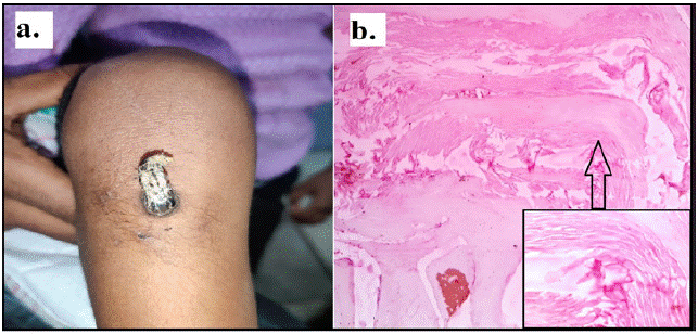
A 6-year-old girl presented to the dermatology outpatient clinic with complaints of a horn-like lesion that had been growing in size over her right knee for three years. On physical examination, a firm grayish-white cutaneous projection, measuring 2x0.5x0.2cm with a bluish red non-ulcerated base over the right knee, was present (Figure 1a). A history of discharge or pain was absent. On palpation, the lesion was non-tender and firm to hard in consistency. A history of medical illness, trauma, or surgery was absent. The family history of cutaneous horn was non-contributory. The systemic examination was within the normal limit. The patient underwent complete excision of the cutaneous horn under local anesthesia. The excised lesion was sent for histopathological examination. The postoperative period was uneventful. Multiple Hematoxylin and Eosin (H&E) stained microsections from the tissue biopsy show a marked degree of hyperkeratosis in the form of horn (a column of keratin) arising from the acanthotic epidermis (Figure 1b). There was no evidence of verruca, seborrhic keratosis, actinic keratosis, keratoacanthoma, or squamous cell carcinoma. Pathognomic features were suggestive of cutaneous horn.

    Figure 1a: A bluish red non-ulcerated, non-tender and firm to hard in consistency lesion over the right knee. b. H&E stained micro sections from the tissue biopsy

show a marked degree of hyperkeratosis in the form of horn (a column of keratin) arising from the acanthotic epidermis.
Discussion
Cutaneous horns are less frequently encountered entities that resemble animal horns. A cutaneous horn consists of cornified proliferative keratinocytes without a bony component, whereas an animal horn contains an osseous cast, which is a major difference between both entities [4]. Hence, histopathological examination of the tissue is the only tool to confirm the clinical diagnosis of cutaneous horn and to rule out other entities, including benign, premalignant, or malignant lesions, which are associated with cutaneous horn [5]. This signifies the importance of interdepartmental communication for better evaluation and management of the patient [4].
A study conducted by Mantese et al showed a total of 92 cutaneous horns among which benign lesions were most commonly encountered, including viral warts, keratoacanthoma, seborrhoeic keratosis, benign epithelial hyperplasia, and trichilemmoma. Most common premalignant primary cause of cutaneous horn is actinic keratosis, while squamous cell carcinoma is the most common malignant cause [6].
Malignant transformation is mostly seen in male patients at the base of the cutaneous horn. Other risk factors are old age, sunexposed areas, and giant horns. A study by Yu et al concluded that 61% of cutaneous horns originated from benign lesions and 39% from premalignant lesions or malignant epidermal lesions. This study also highlights that cutaneous horns were frequently noticed at the sun-exposed areas of the patient [7]. Our patient was a 6-year-old girl who presented with a right knee lesion, which is a non-sun-exposure area. These features make it a rare case of cutaneous horn. A few cases of cutaneous horn on the knee are reported in the literature [8].
The exact pathogenesis of cutaneous horn is still not known, but risk factors such as sun-exposure, cellular aging, and epithelial dysfunction play a significant role [2]. Cutaneous horns are more common in Caucasian patients and relatively rare among Asian and African. It is believed that pigmented skin provides protection from ultra-violet light. Hence, Asian and African races have a lower risk of cutaneous horn [9].
The earliest documented case of cutaneous horn in India was in 1935 by JM Richardson. The patient was a young, 18-yr-old male who had a soft tumour ten years ago which, in the next six years increased in size and ulcerated. The horny growth protruded from the aponeurosis of the occipito-frontalis and was easily removed. The horn was hard in consistency, measuring four and a half inches in circumference at the base and five inches in length from base to tip [3].
Accurate diagnosis and early management are mandatory steps for the prevention of the risk of transformation to malignancy and psycho-social stress owing to the bizarre appearance. Histopathological examination has a major role in confirmation of the clinical diagnosis as well as to rule out malignancy, metastasis, and other similar entities. A constellation of clinical presentation and histopathological analysis determines the final diagnosis [10].
Cutaneous horns can be treated surgically, medically, or via laser ablation, e.g. carbon dioxide and neodymium-doped yttrium aluminium garnet. However, in most cases, surgical excision remains the treatment of choice [11]. The current standard of treatment for cutaneous horns is complete excisional biopsy. In cases of premalignant or malignant tumors, wide local excision is preferred [12]. Prognosis of cutaneous horn depends on the underlying disease. In benign cases, surgical removal is indicated for cosmetic purposes [6].
Conclusion
Cutaneous horns are uncommon lesions that arise primarily from sun-exposed areas of the body in the old age group. But it can occur in young patient at non-sun-exposed areas as well. The exact pathogenesis is unknown, but ultraviolet radiation is thought to play a significant role. They are usually hard, non-tender cutaneous protrusions that resemble animal horns. The base of the cutaneous horn may show benign, premalignant, or malignant transformation. Cutaneous horns are managed with an interprofessional team approach. Surgical excision followed by histopathological analysis is mandatory for early accurate diagnosis and to prevent malignancy.
References
- Bondeson J. Cutaneous horns: A historical review. Am J Dermatopathol. 2001; 23: 362-69.
- Kneitz H, Motschenbacher S, Wobser M, Goebeler M. Photoletter to the editor: Giant cutaneous horn associated with squamous cell carcinoma. J Dermatol Case Rep. 2015; 9: 27-8.
- Richardson JM. An unusually large horny growth. Indian Medical Gazette. 1935; 70: 150.
- Thiers BH, Strat N, Snyder AN, Zito PM. Cutaneous Horn. StatPearls Publishing. 2021.
- Cristobal MC, Urbina F, Espinoza A. Cutaneous horn malignant melanoma. Dermatol Surg. 2007; 33: 997-9.
- Mantese SA, Diogo PM, Rocha A, Berbert AL, Ferreira AK, et al. Cutaneous horn: a retrospective histopathological study of 222 cases. An Bras Dermatol. 2010; 85: 157-63.
- RCH Yu, et al. A histopathological study of 643 cutaneous horns. British Journal of Dermatology. 1990; 123: 46-47.
- Al-Zacko SM, Mohammad A. Cutaneous Horn Arising From a Burn Scar: A Case Report and Review of Literature. J Burn Care Res. 2018; 39: 168-70.
- Garg G, Goyal S. Cutaneous horn mimicking animal horn: case report & brief review Innovative. J Med Health Sci. 2013; 3: 258–9.
- W Leppard, Loungani R, Saylors B, Delaney K. Mythology to reality: case report on a giant cutaneous horn of the scalp in an African American female. Journal of Plastic, Reconstructive & Aesthetic Surgery. 2014; 67: 22–24.
- Narang T, Kanwar AJ. Ectopic nail with polydactyly. J Am Acad Dermatol. 2005; 53: 1092-3.
- Nahhas AF, Scarbrough CA, Trotter S. A Review of the Global Guidelines on Surgical Margins for Nonmelanoma Skin Cancers. J Clin Aesthet Dermatol. 2017; 10: 37-46.