Research Article
J Pediatri Endocrinol.2016; 1(2): 1007.
Ethnicity and Excess Body Weight Impact on Pubertal Onset in Girls: A Longitudinal Study of Hormonal and Bone Maturation Changes
Chen K, Corpus D, Zhong C, Rabii K and Klein KO*
Department of Pediatric Endocrinology, Rady Children’s Hospital and University of California, San Diego, USA
*Corresponding author: Klein KO, Department of Pediatric Endocrinology, Rady Children’s Hospital and University of California, San Diego, USA
Received: August 28, 2016; Accepted: October 03, 2016; Published: October 06, 2016
Abstract
Objective: Define the relative effects of ethnic background and excess body weight on peri-pubescent girls’ hormonal profiles, skeletal maturation and age of pubertal onset.
Methods: 28 girls, comprising 4 groups (Hispanic and Caucasian with and without BMI>85%) were studied every 6 months approaching pubertal onset.
Results: Age of onset of breast development varied by ethnicity and by BMI. Girls with greater BMI reached breast stage 2 at younger ages. Ethnicity and excess body weight appear to independently affect pubertal onset as BMI alone did not explain the differences in breast onset. Ethnicity may play more of a role than BMI. Hispanic girls reached breast onset earlier than Caucasian girls. Bone maturation was advanced prior to puberty in girls with BMI>85th%, but did not accelerate as puberty approached. No hormonal measures were robust enough to predict pubertal onset.
Conclusion: Our small subset confirms previous work showing that Hispanic girls on average enter puberty earlier that Caucasian girls and that ethnicity is a stronger influence on pubertal onset than weight status. The results also portray the subtle changes in hormonal variables prior to breast development, as well as the pattern of advanced bone maturation in girls with higher BMI in the peripubertal years.
What is already known about this topic?
- Ethnicity influences pubertal onset.
- BMI influences pubertal onset.
- Longitudinal study of hormonal changes approaching puberty.
- Shorter interval of study of hormonal changes and detection of breast onset.
- Single observer in longitudinal study.
- Biro FM, Galvez MP, Greenspan LC, Succop PA, Vangeepuram N, Pinney SM, et al. Pubertal assessment method and baseline characteristics in a mixed longitudinal study of girls. Pediatrics. 2010; 126: 583-590.
- Herman-Giddens ME, Slora EJ, Wasserman RC, et al. Pediatrics. 1997; 99: 505-512.
- Crocker MK, Stern EA, Sedaka NM, Shomaker LB, Brady SM, Ali AH, et al. Sexual dimorphisms in the associations of BMI and body fat with indices of pubertal development in girls and boys. J ClinEndocrinol Metab. 2014; 99: 1519-1529.
- Kaplowitz PB, Slora EJ, Wasserman RC, Pedlow SE, Herman-Giddens ME. Earlier onset of puberty in girls: relation to increased body mass index and race. Pediatrics. 2001; 108: 347-353.
- Marshall WA, Tanner JM. Variations in pattern of pubertal changes in girls. Arch Dis Child. 1969; 44: 291-303.
- Greulich WW, Pyle I. Radiographic atlas of skeletal development of the hand and wrist. Stanford, CA: Standford University Press. 1959.
- Klein KO, Baron J, Colli MJ, McDonnell DP, Cutler GB Jr. Estrogen levels in childhood determined by an ultra-sensitive recombinant cell bioassay. J Clin Invest. 1994; 94: 2475-2480.
- Anderson DC, Hopper BR, Lasley BL, Yen SSC. A simple method for assay of eight steroids in small volumes of plasma. Steroids. 1976; 28: 179-196.
- Yen SSC, Llerena 0, Little B, Pearson OH. Disappearance rates of endogenous luteinizing hormone and chorionic gonadotropin in man. J ClinEndocrinolMetab. 1968; 28: 1763-1767.
- Biro FM, Greenspan LC, Galvez MP, Pinney SM, Teitelbaum S, Gayle C, et al. Onset of breast development in a longitudinal cohort. Pediatrics. 2013; 132: 1019-1027.
- Neely EK, Wilson DM, Lee PA, Stene M, Hintz RL. Spontaneous serum gonadotropin concentrations in the evaluation of precocious puberty. J Pediatr. 1995. 127: 47-52.
- Kimm SY, Barton BA, Obarzanek E, McMahon RP, Sabry ZI, Waclawiw MA, et al. Racial Divergence in adiposity during adolescence: the NHLBI growth and healthy study. Pediatrics. 2001; 107.
- Rosenfield RL, Lipton RB, Drum ML. Thelarche, Pubarche and Menarche Attainment in Children with Normal and Elevated Body Mass Index. Pediatrics. 2009; 123: 84-88.
- Klein KO, Larmore KA, Lancey DE, Brown JM, Considine RV, Hassink SG. Effect of Obesity on Estradiol Level and its Relationship to Leptin, Bone Maturation and Bone Mineral Density in Children. J ClinEndocrinolMetab. 1998; 83: 3469-3475.
- Klein KO, Newfield RS, Hassink SG. Bone maturation along the spectrum from normal weight to obesity: a complex interplay of sex, growth factors and weight gain. JPediatrEndocrinolMetab. 2016; 29: 311-318.
- Sopher AB, Jean AM, Zwany SK, Winston DM, Pomeranz CB, Bell JJ, et al. Bone age advancement in prepubertal children with obesity and premature adrenarche: possible potentiating factors. Obesity (Silver Spring). 2011; 19: 1259-1264.
- Polito C, Toro DA, Collini R, Cimmaruta E, D’Alfonso C, Giudice DG. Advanced RUS and normal carpel bone age in childhood obesity. Int J Obesity. 1995; 19: 506-507.
- Sims EK, Addo OY, Gollenberg AL, Himes JH, Hediger ML, Lee PA. Inhibin B and luteinizing hormone levels in girls aged 6-11 years from NHANES III, 1988-1994. Clin Endocrinol (Oxf). 2012; 77: 555-563.
- McCartney CR, Prendergast KA, Blank SK, Helm KD, Chhabra S, Marshall C. Maturation of Luteinizing Hormone (Gonadotropin–Releasing Hormone) Secretion across Puberty: Evidence for Altered Regulation in Obese Peripubertal Girls. J ClinEndocrinolMetab. 2009; 94: 56-66.
What does this study add?
Keywords: Ethnicity; Obesity; Bone age; Pubertal onset
Introduction
Across ethnicities, several large studies now report that puberty is starting earlier than it used to for girls [1,2]. With childhood obesity being an increasing problem in the United States, obesity has been discussed as one of the causes of a possible trend toward earlier puberty in children [3,4]. The age of normal pubertal onset in African American, Hispanic and Caucasian girls has been controversial. The traditional view of puberty is that it usually begins between the ages of 8-13 years in girls and 9-14 years in boys. The prevailing view over the last 9 years has been that puberty begins earlier in African American and Hispanic girls as compared to Caucasian girls [1,2].
The onset of puberty has physical and hormonal definitions. Gonadotropin-Releasing Hormone (GnRH) pulses are activated at puberty. This regulates production and release of Luteinizing Hormone (LH) and Follicle-Stimulating Hormone (FSH) causing gonadal maturation and release of sex steroids. Estradiol, the predominant estrogen during a girl’s reproductive years, promotes the development of secondary sex characteristics at puberty. Estradiol also drives the pubertal growth spurt and bone maturation. Estradiol is predominantly produced by aromatization of testosterone in the ovaries, but lesser amounts are derived from adrenal testosterone. Moreover, fat cells are known to convert testosterone and precursors into estradiol. Sex Hormone-Binding Globulin (SHBG) is a glycoprotein that binds and inhibits testosterone and estradiol. The level of SHBG in the bloodstream is thus correlated to the bioavailability of these sex hormones. Dehydroepiandrosterone Sulfate (DHEAS) is used as an indicator for adrenarche. We studied these markers of puberty to identify the immediate prepubertal period.
This study was designed to further delineate ethnic differences and the impact of excess body weight on the timing of pubertal onset, as well as to determine whether the hormonal profile is reflective of the onset of puberty or degree of bone maturation. Girls were followed longitudinally as they approached pubertal onset.
Methods
Subjects
Volunteers were recruited through advertisement and from the Endocrinology clinic at Rady Children’s Hospital San Diego. 28 prepubertal Caucasian and Hispanic girls were recruited and followed every 6 months until they reached onset of puberty. Eight were Caucasian with BMI<85%, 6 were Caucasian with BMI>85th%, 7 were Hispanic with BMI<85% and 7 were Hispanic with BMI>85th%. Ethnicity was determined by history of 4 grandparents, with all 4 grandparents being the same ethnicity. The girls were ages 8-10 at the first visit and 9-12 at the last visit. Girls were classified by BMI greater than or equal to, or less than the 85th percentile. All had normal physical examinations and no significant past medical illness other than excess body weight. No girl was taking any medications at the time of the study. Girls with any endocrine cause of excess body weight, such as hypothyroidism or Cushing’s syndrome were not included. The study was approved by the joint Institutional Review Board at Rady Children’s Hospital and University of California, San Diego, where the girls were studied. Consent was given by one parent and assent by each child.
Study design
Girls were evaluated every 6 months. Evaluations included measurement of height by a Harpender Stadiometer, weight and pubertal staging by a single observer (KOK). Waist and hip circumference were measured to the nearest 0.1 cm. Pubertal staging was assessed based on a modification of Tanner’s classification [5] evaluated by palpation. Breast stage 2 or more was considered to be the marker of pubertal onset. If there was any question whether the breast tissue was true breast tissue as opposed to adipose tissue, the girls was seen again in follow up 6 months later to confirm. This was only needed in 2 of the girls and no outliers were observed in those girl’s data. Most girls were followed for 2 years, but a few were in the study for 3-4 years prior to breast onset.
Body Mass Index (BMI) was calculated based on height and weight measurements and growth velocity was calculated based on changes in height between two consecutive visits.
Bone age was assessed at each visit and read blinded by a single observer (KOK) according to the method of Greulich and Pyle [6]. Laboratory studies were performed at each visit including, estradiol by recombinant cell bioassay, estradiol by GCMSMS, LH, FSH, SHBG, DHEAS and testosterone.
Hormonal measurements
Estrogen assay: The bioassay for estradiol has been previously described in detail [7]. Briefly, it uses recombinant yeast cells containing plasmids coding for the estrogen receptor and coding fora β-galactosidase reporter. The sensitivity of the bioassay was 0.07-0.7 pmol/L (0.02-0.2 pg/mL) during the time frame of the study. The intraassay and interassay Coefficients of Variation (CV) ranged from 10% at 7 pmol/L (2 pg/mL) to 10-50% at 0.7 pmol/L (0.2 pg/mL) during the time frame of the study.
Other assays: LH, FSH, SHBG and ultrasensitive estradiol were measured at Endocrine Sciences (Calabasas, CA). LH was measured by chemiluminescent assay. Intra and inter assay CV were 5.35% and 7.07% maximum, respectively. The sensitivity was 0.3 ml U/ ml. FSH was measured by chemiluminescent assay. Intra and inter assay CV were 6.8% and 6.9% maximum, respectively. The sensitivity was 0.5 m U/ml. ultrasensitive estradiol was measured by GCMSMS. All CV’s were less than 17.8% and assay sensitivity was 0.63 pg/ ml.SHBG was measured by Electrochemiluminescence Immunoassay (ECLIA). Intra and inter assay CV were 2.9% and 3.3% maximum, respectively. The functional sensitivity was at least 0.74 nmol/L. DHEAS was measured by RIA after enzymolysis of the DHEAS. Intra and inter assay CV were 6.9% and 8.4% maximum, respectively. The sensitivity was 10 ug/dL. Serum concentrations of testosterone and DHEAS were measured by well-established RIA [8,9] using column chromatographic separation in assays as previously published with intra-assay CV less than 7%. (Diagnostic Systems Laboratories, Inc). Any value less than the detection limit were set equal to the detection limit for purposes of analysis. This occurred for E2 in 2 girls at time- 18 months and 1 girl at 12 months and for LH in 3 girls at 18 months and 2 girls at 12 months.
Statistical methods and data analysis: Data was analyzed by setting the onset of breast development to time 0 in all girls so that comparisons were made relative to pubertal onset. Unadjusted analyses used paired t tests to compare differences between Caucasian and Hispanic girls, as well as between girls of different BMI groups. ANOVA was used to compare differences across all four groups. Correlations were done using simple linear regression analysis. Differences were considered significant at P<0.05.
Results
Indices of pubertal onset
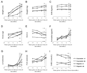
Age, height, weight and bone age all increase significantly across time in the 18 months leading up to breast onset, as expected in all girls (p<0.05) (Table 1 & Figure 1). Weight, BMI, waist and hip circumference are all greater in the groups with BMI>85% than the groups with BMI<85%, as expected and confirming the weight categories (Table 1). BMI was relatively stable in all girls during the 18 months of participation in this study, allowing easier comparisons between groups. Waist and hip circumference changes paralleled BMI and did not contribute any different information.
    Caucasian normal weight
    Caucasian BMI>85%
    Hispanic normal weight
    Hispanic


    BMI>85%


    Age
    time-18 mo
    9.65±1.13d
    9.43±1.18d
    8.87±0.77d
    8.65±0.28


    -12 mo
    10.09±0.93d
    9.96±1.00
    9.52±0.69
    9.09±0.90d


    -6 mo
    10.64±0.87
    10.45±1.14
    9.96±0.77
    10.05±1.02


    breast onset
    11.25±0.96
    11.01±0.91
    10.45±0.77
    10.50±1.05


    Height
    time-18 mo
    135.32±7.63
    137.42±6.67d
    134.8±2.6
    138.9±5.09d


    -12 mo
    139.54±6.60
    142.70±8.14
    135.32±4.73
    139.76±3.91d


    -6 mo
    143.28±6.66
    146.16±9.63
    136.93±4.06
    143.56±3.09**


    breast onset
    146.91±7.78
    148.87±8.40
    140.51±4.13
    146.50±3.02**


    Weight
    time-18 mo
    29.88±3.89d
    43.66±8.08**
    31.07±3.35
    49.6±2.69**


    -12 mo
    31.34±3.92
    45.38±7.79**
    31.6±3.70
    54.22±6.40**


    -6 mo
    34.22±4.80
    51.08±8.11**
    31.77±3.27
    55.94±9.66**


    breast onset
    35.55±4.43
    53.18±7.14**
    33.29±4.56
    59.71±10.13**


    BMI
    time-18 mo
    16.27±0.89
    22.95±2.54**
    17.07±1.35
    25.71±0.49**


    -12 mo
    16.06±1.32
    22.30±3.45**
    17.21±0.99
    27.75±3.00**#


    -6 mo
    16.63±1.72
    23.85±2.62**
    16.92±1.21
    27.10±4.34**


    breast onset
    16.43±1.14
    24.00±2.66**
    16.83±1.93
    27.78±4.38**


    Growth Velocity
    time-18 mo
    5.74±1.32
    4.83±0.33
    5.74±1.72
    6


    -12 mo
    6.48±0.36
    6.22±2.31
    6.27±0.63
    5.30±1.27


    -6 mo
    4.62±1.14d
    6.24±2.66
    3.68±2.32d
    6.37±1.16*


    breast onset
    6.85±0.70
    5.40±1.45
    7.41±0.45
    6.89±0.89


    Rate of weight gain
    time-18 mo
    1.15±1.85
    8.37±4.29
    4.76±5.06
    9.9


    -12 mo
    2.65±2.51
    4.39±4.95
    4±2.85
    2.5±2.97


    -6 mo
    3.20±2.34
    9.6±10.5
    3.24±2.89
    6.40±4.29


    breast onset
    4±2.56
    6.7±3.84
    3.54±4.87
    8.63±2.71*


    Bone Age
    time-18 mo
    9.00±1.39
    10.27±0.90
    9.22±0.67d
    9.92±0.83


    -12 mo
    9.26±1.30
    10.39±0.86
    10.17±0.83
    10.27±0.83


    -6 mo
    9.78±1.17
    10.8±0.45
    9.78±1.11
    10.62±0.81


    breast onset
    10.35±1.00
    10.83±0.41
    10.5±0.58
    11.12±1.16


    BA/CA
    time-18 mo
    0.93±0.09
    1.10±0.12*
    1.04±0.05
    1.15±0.13


    -12 mo
    0.92±0.13
    1.05±0.10
    1.07±0.13
    1.13±0.04


    -6 mo
    0.92±0.10
    1.04±0.09*
    0.99±0.15
    1.06±0.07


    breast onset
    0.92±0.07
    0.99±0.07
    1.01±0.09
    1.06±0.09


    Waist circumference
    time-18 mo
    59.90±2.10
    76.08±6.35**
    60.8±3.30
    87.5**


    -12 mo
    60.65±4.33
    77±10.31**
    61.42±3.63
    87.83±9.01**


    -6 mo
    60.83±6.31
    79.88±5.30**
    61.60±3.44
    85.87±8.84**


    breast onset
    60.27±4.32
    78.33±9.22**
    63.60±8.24
    87.24±10.03**


    Hip circumference
    time-18 mo
    69.98±3.55d
    83.12±9.02*
    73.9±5.18
    90*


    -12 mo
    71.90±4.43
    84.58±5.41**
    72.4±5.59
    91.38±2.93**


    -6 mo
    73.79±5.43
    87.3±11.42*
    73.07±3.54
    94.27±8.84**


    breast onset
    75.77±3.31
    90.52±7.01**
    76.39±8.24
    96.39±10.03**


    Data are represented as the mean±standard deviation.
      *p<0.05 vs. normal weight, same ethnicity; **p<0.01 vs. normal weight, same ethnicity; #p<0.05 vs. Caucasian; ##p<0.01 vs. Caucasian; dp<0.05 vs. time of breast onset; n as in    methods, except at time-18 m; n is missing 3 in Caucasian<85%, missing 1    in Caucasian>85%, missing 2 in Hispanic<85% and missing 4 in Hispanic>85%.
Table 1: Clinical characteristics and bone age by group.

    Figure 1:  Prevalence of breast maturation in each cohort at each age.
Hispanic participants reached breast maturation earlier than Caucasian
participants. Caucasian BMI<85% (◊); Caucasian BMI>85% (□); Hispanic
BMI<85% (Δ); Hispanic BMI>85% (○).
When all girls are taken together, hormonal indices of puberty also increased across time leading up to breast onset including: LH, FSH, E2 and T (Table 2 & Figure 1). There was also a significant increase in these same variables between 6 months prior to breast onset and breast onset (p<0.05). When analyzed by groups, the same trends are seen, but not all reached significance (Table 3 & Figure 1). Considering each ethnic group or each weight category, the same hormonal changes were also seen, but not all reached significance (Table 4).
    Estradiol (GCMS) (pg/ml)
    time-18 mo
    2.58±1.72*


    -12 mo
    4.47±3.18*


    -6 mo
    4.88±4.76*


    breast onset
    8.68±7.62


    FSH (mlU/ml)
    time-18 mo
    1.92±0.99*


    -12 mo
    2.15±0.88*


    -6 mo
    2.46±1.11*


    breast onset
    3.26±1.67


    DHEAS (ug/dl)
    time-18 mo
    592.79±665.77


    -12 mo
    556.48±654.93


    -6 mo
    634.88±636.95


    breast onset
    391.19±571.25


    Testosterone (ng/dl)
    time-18 mo
    5.44±2.12*


    -12 mo
    6.96±3.26*


    -6 mo
    6.56±2.73**


    breast onset
    10.65±6.61


    E2 Bioassay
    time-18 mo
    19.26±8.79*


    - 12 mo
    21.50±16.74*


    - 6 mo
    20.59±20.26*


    breast onset
    37.12±17.83


    LH (mlU/ml)
    time-18 mo
    0.06±0.10*


    -12 mo
    0.04±0.04*#


    -6 mo
    0.13±0.19*


    breast onset
    0.42±0.67


    SHBG (nmol/L)
    time-18 mo
    76.79±43.11


    -12 mo
    70.52±43.45


    -6 mo
    67.24±46.43


    breast onset
    57.40±34.51


    Data are represented as the mean ± standard deviation.
      *p<0.05 vs. time of breast onset; **p<0.01 vs. time of breast onset; #p<0.05 vs. time-6 mo n as in methods, except at time-18 m; n is missing 3 in    Caucasian<85%, missing 1 in Caucasian>85%, missing 2 in Hispanic<85%    and missing 4 in Hispanic>85%.
Table 2: Hormonal parameters of all participants.
    Caucasian normal weight
    Caucasian with BMI>85%
    Hispanic normal weight
    Hispanic with BMI>85%


    E2 Bioassay
    time-18 mo
    19.53±7.97d
    16.01±11.35d
    22.28±6.26
    20.57±14.53


    -12 mo
    28.77±20.19
    19.23±13.79d
    17.64±17.13
    18.81±18.18


    -6 mo
    21.97±10.75d
    31.71±3.10
    24.03±34.89
    10.82±7.58d


    breast onset
    38.43±14.15
    49.01±17.89†
    15.05±9.50
    34.62±8.08


    Estradiol (GCMS)
    time-18 mo
    2.12±0.73d
    3.97±2.14
    2.73±2.66
    1.40±0.57


    -12 mo
    4.94±3.37
    3.75±0.76
    3±1.61
    5.8±5.71


    -6 mo
    4.41±2.91
    3.62±2.02
    6.30±8.75
    5.16±4.07


    breast onset
    6.79±4.57
    11.9±9.50†
    7.48±9.27
    9.29±8.08


    LH
    time-18 mo
    0.03±0.02
    0.08±0.12
    0.13±0.15
    0.03±0.01


    -12 mo
    0.05±0.03
    0.04±0.06
    0.05±0.03
    0.03±0.01


    -6 mo
    0.05±0.04
    0.18±0.32
    0.12±0.14
    0.21±0.24


    breast onset
    0.47±0.74
    0.31±0.45
    0.12±0.12
    0.85±1.00


    FSH
    time-18 mo
    1.70±0.63
    1.90±1.01
    2.73±1.50
    1.33±0.67


    -12 mo
    2.63±0.75
    1.57±0.55*
    2.46±1.11
    1.86±0.85


    -6 mo
    2.06±0.67
    1.87±0.87
    3.47±1.37#
    2.4±0.94


    breast onset
    2.84±1.62
    3.08±1.95
    3.09±0.81
    4.03±2.17


    SHBG
    time-18 mo
    101.89±38.72
    41.15±9.27**
    112.34±28.45
    22.9


    -12 mo
    106.45±47.30
    38.56±11.73**
    96.21±22.40
    33.87±10.86**


    -6 mo
    104.69±53.16
    33.98±12.09*
    79.94±18.92
    25.40±8.36**


    breast onset
    84.53±33.52
    43.85±28.33*
    78.66±17.13
    23.65±10.14**


    DHEAS
    time-18 mo
    1011.99±912.74
    404.86±466.81
    391.59±194.97
    40


    - 2 mo
    1036.53±886.02
    467.17±447.62
    372.15±459.40
    175.92±285.21


    -6 mo
    966.24±803.72
    502.38±521.15
    358.09±412.64
    561.34±564.43


    breast onset
    577.29±817.48
    105.17±41.34
    352.19±453.44
    483.60±620.42


    Testosterone
    time-18 mo
    5.56±1.43d
    7.43±2.14d
    3.67±1.10
    3.85±1.91


    -12 mo
    9.1±4.94
    6.68±1.61d
    5.38±1.38
    5.87±2.72


    -6 mo
    7.22±3.45
    6.88±2.12d
    5.99±1.82
    6.04±3.15


    breast onset
    11.63±5.52
    15.6±6.11
    6.01±2.10#
    10.77±8.71


    Data are represented as the mean ± standard deviation.
      *p<0.05 vs. normal weight, same ethnicity; **p<0.01 vs. normal weight, same ethnicity; #p<0.05 vs. Caucasian; ##p<0.01 vs. Caucasian; dp<0.05 vs. time of breast onset n as in    methods, except at time-8 m; n is missing 3 in Caucasian<85%, missing 1 in    Caucasian>85%, missing 2 in Hispanic<85%, and missing 4 in    Hispanic>85%.
Table 3: Hormonal parameters by group.
DHEAS did not increase over the time period of this study (Table 2), which was consistent with not all girls having pubic hair development prior to breast onset. SHBG decreased across time in all girls together (Table 2). SHBG was lower in girls with BMI<85% than in girls with BMI>85th%, as expected and this was independent of ethnicity. Estradiol was also assessed by an ultrasensitive recombinant cell bioassay for further comparison with the GCMSMS assay. While the 2 assays showed strong correlation (R=0.66, p<0.01), the GCMSMS assay proved more consistent and was able to better detect increasing trends of estradiol levels while approaching puberty.
The intervals for measuring growth velocity and variation between girls did not allow for accurate assessment over the short time in the individual groups (Table 1).
Timing of pubertal onset
Our small cohort had onset of breast development slightly laterthan reported in the previous large NHANES III study groups [10], but with a similar difference between Caucasian and Hispanic groups of about 1 year (average age 10.45 y for Hispanic girls with BMI>85th%, 10.5 y for Hispanic girls with BMI<85% vs. 11 y and 11.25 y for Caucasian girls with BMI>85% and BMI<85%, respectively).
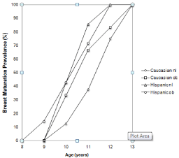
Similar to previous studies [10], girls with BMI>85th% reached breast onset earlier than those with BMI<85th% (Figure 2). Ethnicity played a greater role on determining breast onset than did weight status. Hispanic girls reached breast maturation earlier than Caucasian participants, with even Hispanic girls with BMI<85% reaching puberty earlier than Caucasian girls with BMI>85th%. 40% of Hispanic girls regardless of BMI group had breasts by age 10 years, compared to 30% of Caucasian girls with BMI>85% and only 10% of Caucasian girls with BMI<85%. A greater percentage of Hispanic girls had breast development at each age, with 100% by age 12 years. In Caucasian girls, a greater percentage of girls with BMI>85% had breast development at each age compared girls of normal weight, with 100% of both groups having breast development by age 13 years.

    Figure 2: Prevalence of breast maturation in each cohort at each age.
Hispanic participants reached breast maturation earlier than Caucasian
participants. Caucasian BMI<85% (◊); Caucasian BMI>85% (□); Hispanic
BMI<85% (Δ); Hispanic BMI>85% (○).
Predictors of pubertal onset
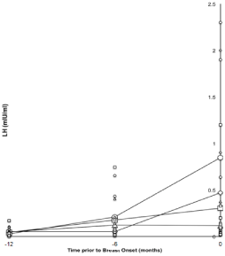
There was an increasing trend in LH values as participants approached puberty. LH levels in all girls showed a gradual increase from 18 months before puberty to 6 months before puberty and then a more dramatic increase from 6 months before puberty to the onset of puberty (Figure 3). LH ranged from 0.02-2.3 IU/L at the time of breast onset. Half of the girls had LH levels greater than 0.1 IU/L and 40% had levels greater than 0.3 IU/L, a cutoff proposed by Neely [11]. 12 months prior to the onset of breast development, only 3/28 (10%) had an LH level greater than 0.1 IU/L. Six months prior to detection of breast development, 10/28 (35%) girls had LH levels greater than 0.1 IU/L, with 4 of those greater than 0.3 IU/L (14%) and none had LH levels greater than 1 IU/L.

    Figure 3:  LH pediatric laboratory measurements for all participants and
averages for the 4 cohorts. There is an increasing trend in LH values as
participants approach puberty. Caucasian BMI<85% (◊); Caucasian
BMI>85% (□); Hispanic BMI<85% (Δ); Hispanic BMI>85% (○).
Age of onset of breasts was not directly correlated with BMI by regression analysis across all girls. On average estradiol levels were greater than 6 pg/mL at observation of breast development and less than 6 pg/mL prior to breast development (Figure 1).
Bone maturation
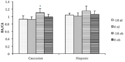
Girls with BMI>85th% have higher BA/CA at all time points compared to girls with BMI<85%, although this only reached statistical significance (p<0.05) at 6 and 18 months prior to breast onset, probably due to sample size. BA/CA was also higher in Hispanic girls compared to Caucasian girls at all time points, but only reached statistical significance at 12 months prior to breast onset and at the time of breast onset (p<0.05) (Table 4). BA/CA was highest in all groups 18 months prior to breast onset and decreased closer to onset (Tables 1 & 4 and Figure 4). Higher BMI correlated with higher BA/CA in all girls together (R=0.4, p<0.1). Caucasian girls with BMI<85% have a consistent ratio of BA to CA just under one, but the other 3 groups have higher ratios that trend downward as puberty approaches, such that bone age at the time of breast onset, is similar between all groups (Table 1 & Figure 4).
    Caucasian participants
    Hispanic participants


    Age (years)
    time-18 mo
    9.54±1.10**
    8.79±0.58**


    -12 mo
    10.03±0.93**
    9.31±0.79**


    -6 mo
    10.58±0.93
    10.00±0.87


    breast onset
    11.15±0.91
    10.47±0.88


    Height (cm)
    time-18 mo
    136.37±6.85**
    136.44±3.86**


    -12 mo
    141±7.21*
    137.54±4.71**


    -6 mo
    144.31±7.61
    140.24±4.88


    breast onset
    147.75±7.79
    143.51±4.66


    Weight (kg)
    time-18 mo
    36.77±9.41
    38.48±10.51


    -12 mo
    37.82±9.27
    42.91±12.90


    -6 mo
    40.24±10.23
    43.86±2.24


    breast onset
    43.11±10.59
    46.5±15.65


    BMI (kg/m2)
    time-18 mo
    19.61±3.95
    20.52±4.83


    -12 mo
    18.94±4.04
    22.48±5.94


    -6 mo
    19.21±4.10
    22.01±6.11


    breast onset
    19.6±4.30
    22.31±6.55


    DHEAS (ug/dl)
    time-18 mo
    708.43±754.66
    303.69±237.17


    -12 mo
    773.75±750.51
    274.04±375.03


    -6 mo
    823.52±740.41
    459.71±486.57


    breast onset
    359.44±628.40
    422.95±531.77


    Testosterone (ng/dl)
    time-18 mo
    6.39±1.93
    3.74±1.23#


    -12 mo
    7.89±3.72*
    5.56±1.81


    -6 mo
    7.1±2.96**
    6.01±2.47


    breast onset
    13.28±5.86
    8.39±6.57


    Bone Age
    time-18 mo
    9.63±1.29*
    9.50±0.74*


    -12 mo
    9.78±1.22
    10.22±0.78


    -6 mo
    10.14±1.08
    10.20±1.03


    breast onset
    10.56±0.82
    10.81±0.94


    Waist circumference (cm)
    time-18 mo
    67.99±9.62
    71.48±14.81


    -12 mo
    68.20±11.21
    73.16±15.19


    -6 mo
    67.64±11.08
    74.66±14.23


    breast onset
    68.01±11.35
    75.42±15.11


    E2 Bioassay
    time-18 mo
    17.96±9.14**
    21.60±8.56


    -12 mo
    24.00±17.22*
    18.16±16.47


    -6 mo
    23.74±10.44**
    17.93±26.08


    breast onset
    42.27±15.65
    29.03±19.18


    Estradiol (GCMS) (pg/ml)
    time-18 mo
    2.81±1.59*
    2.2±2.04


    -12 mo
    4.39±2.51*
    4.6±4.40


    -6 mo
    4.13±2.57*
    5.68±6.37


    breast onset
    8.92±7.15
    8.45±8.32


    LH (mlU/ml)
    time-18 mo
    0.05±0.09
    0.09±0.12


    -12 mo
    0.05±0.05
    0.04±0.03


    -6 mo
    0.10±0.19
    0.17±0.20


    breast onset
    0.39±0.61
    0.45±0.75


    FSH (mlU/ml)
    time-18 mo
    1.80±0.80
    2.17±1.35


    -12 mo
    2.14±0.84
    2.16±0.98*


    -6 mo
    1.99±0.72
    2.94±1.26#


    breast onset
    2.95±1.71
    3.56±1.65


    SHBG (nmol/L)
    time-18 mo
    71.52±41.59
    89.98±50.39


    -12 mo
    75.11±49.17
    64.54±36.34


    -6 mo
    82.93±55.45
    52.67±31.60


    breast onset
    65.76±36.63
    49.04±31.44


    BA/CA
    time-18 mo
    1.02±0.13
    1.08±0.10


    -12 mo
    0.98±0.13
    1.10±0.10#


    -6 mo
    0.96±0.11
    1.03±0.12


    breast onset
    0.95±0.08
    1.04±0.09#


    Hip circumference (cm)
    time-18 mo
    76.55±9.47**
    80.34±9.55


    -12 mo
    77.75±8.08
    80.83±10.90


    -6 mo
    78.61±10.17
    84.48±13.49


    breast onset
    68.01±11.35
    85.89±12.81


    Data are represented as the mean±standard deviation.
      *p<0.05 vs. time of breast onset, same ethnicity; **p<0.01 vs. time of breast onset, same ethnicity; #p<0.05 vs. Caucasian n as in methods, except    at time-18 m; n is missing 3 in Caucasian<85%, missing 1 in Caucasian>85%,    missing 2 in Hispanic<85% and missing 4 in Hispanic>85%.
Table 4: Characteristics by ethnic groups.

    Figure 4:  Bone age to chronological age ratios for Caucasian and Hispanic
participants at 18 months prior to puberty and at the visit with breast onset.
Caucasian girls with BMI>85% had a significantly higher BA/CA at 18 months
before puberty compared to the time of onset of puberty (p<0.05). BMI< 85%
at 18 months prior to puberty (speckled bars); BMI<85% at puberty (gray
bars); BMI>85% at 18 months prior to puberty (gray striped bars); BMI>85%
at puberty (black striped bars).
Discussion
We confirm the finding that pubertal onset is earlier in Hispanic girls than in Caucasian girls and earlier in girls with BMI>85% compared to those with BMI<85% [12]. Ethnicity was a larger contributor to pubertal onset than BMI with Hispanic girls with BMI<85% entering puberty prior to Caucasian girls with BMI>85%. Rosenfield et al. [13] likewise concluded from the NHANES III survey that adiposity and ethnicity were independently associated with earlier pubertal development in girls.
Bone maturation was accelerated in girls with BMI>85% 18 months prior to pubertal onset but was closer to chronological age by onset of puberty, suggesting that the weight gain accelerated bone maturation and may have started the process toward pubertal onset, but did not continue to accelerate. In fact, BA/CA decreases as puberty approaches.
We and others have previously described increased bone maturation in children with excess body weight compared to those without excess body weight [14-17].
LH has been the most sensitive marker of pubertal onset so far in the study of puberty. In the present study, the onset of breasts was detected within 6 months of their appearance. Of note, most of the LH levels were still less than 1 IU/L at first detection of breast development, a cutoff for puberty suggested by Biro and others [10], indicating that LH is not a sensitive enough marker to define pubertal onset.
Obviously biochemical changes happen prior to physical changes, but are not able to be measured accurately enough, or variability among girls sensitivity is too great to generalize to rigorous cutoff values. However, the trend of increasing LH levels is clearly seen and most dramatic the 6 months prior to pubertal onset, supporting both a slowly increasing hormonal milieu and an “on switch” for puberty. Not only do Hispanic girls and girls with BMI>85% enter puberty earlier than Caucasian girls or girls with BMI<85%, but Hispanic girls and girls with BMI>85% have higher levels of LH at onset of puberty compared to Caucasian girls and girls with BMI<85%. This is interesting since the same LH levels could be responsible for pubertal onset regardless of timing, but that does not seem to be the case. This was similarly suggested by Sims, et al. [18] suggesting the value of this finding.
While this is a small cohort, it has the advantage of a single observer for assessment of breast onset and bone age. Confirmation of previous findings in this limited number of participants selected at random supports the robustness of ethnic and weight status variation impact on pubertal onset.
Another strength of this study, is assessment every 6 months (rather than annually as in some previous reports) to detect the earliest sign of breast development. A weakness is that the subjects were not followed past onset of breast development to see if any had regression, as is sometimes seen and reported. However, history was carefully taken for exogenous hormone or phytoestrogen substances that could cause temporary breast development. If there was any question whether the breast tissue was true breast tissue as opposed to adipose tissue, the girl was seen again in follow up to confirm.
Crocker, et al. [3] reported earlier breast onset with increased testosterone levels and increased bone age in girls with excess body weight compared to girls without excess body weight, without activation of the hypothalamic-pituitary-gonadal pulse generator. They suggest that advanced breast development observed in early pubertal girls with excess body weight may be primarily due to peripheral conversion of inactive androgens to more bioactive estrogens by aromatase in adipose tissues. The present study did not look at gonadotropin pulsatility, but did see increased morning LH levels and increased testosterone levels. McCartney [19] also reported decreased LH pulse frequency, amplitude and day-night changes in girls with excess body weight compared to those without excess body weight.
The results of this small subset confirm the role of excess body weight and ethnicity in the timing of the onset of puberty. Hispanic girls enter puberty on average earlier that Caucasian girls and ethnicity is a stronger influence on pubertal onset than weight status. These results also portray the slowly increasing changes in hormonal variables during the months preceding detection of breast development, with a greater rate of hormonal change in the 6 months prior to onset of breast development. The pattern of advancement in bone maturation in girls with higher BMI appears to be greatest in the immediate peripubertal period, with BA/CA decreasing as puberty begins.
Acknowledgement
We are grateful to Jeff Wong for his expert technical assistance. Testosterone, SHBG and DHEAS assays were performed by him at the University of California, San Diego.
References