Case Report
J Pediatr & Child Health Care. 2016; 1(1): 1009.
Sudden Systemic Embolism in an Infant with Supraventricular Tachycardia
Matteo Castagno¹, Saracco Paola²*, Sara Zanetta¹, Gabriella Agnoletti3 and Davide Marini3,4
¹Division of Pediatrics, Department of Health Sciences, Università del Piemonte Orientale, Italy
²Pediatric Hematology, Department of Pediatrics, Citta’ della Salute e della Scienza, Italy
³Division of Pediatric Cardiology, Citta’ della Salute e della Scienza, Italy
4Department of Public Health and Pediatrics, University of Turin, Italy
*Corresponding author: Saracco Paola, Pediatric Hematology, Department of Pediatrics, Citta’ della Salute e della Scienza, Italy
Received: July 14, 2016; Accepted: August 18, 2016; Published: August 19, 2016
Abstract
We report the case of a 5-month-old male who arrived at the emergency room with vomit, crying spells, difficulty in breastfeeding and deterioration of general condition because of Supraventricular Tachycardia (SVT). At the Emergency Department (ED) tachycardia was easily stopped with adenosine. Surprisingly, echocardiogram revealed a large and a floating mass of unclear etiology in the left atrial appendage. After one hour, the infant presented sudden and progressive lower limb pallor with the disappearance of femoral pulses, and echocardiography showed that the thrombus had vanished from the left atrial appendage. Contrast-enhanced Computed Tomography (CT) showed a complete occlusion of the superior mesenteric artery and the abdominal aorta below the renal arteries. The patient was rapidly addressed to catheterization theatre and the thrombus was then successfully removed, restoring patency of arteries involved.
Keywords: Infant; Supraventricular Tachycardia; Systemic Embolism; Thrombus; Left Atrial Appendage
Case Presentation
A 5-month-old male was referred to the Emergency Department (ED) of our tertiary care hospital because of vomit, crying spells, difficulty in breastfeeding and deterioration of general condition, transferred from another hospital, where he had been admitted for acute infectious gastroenteritis.
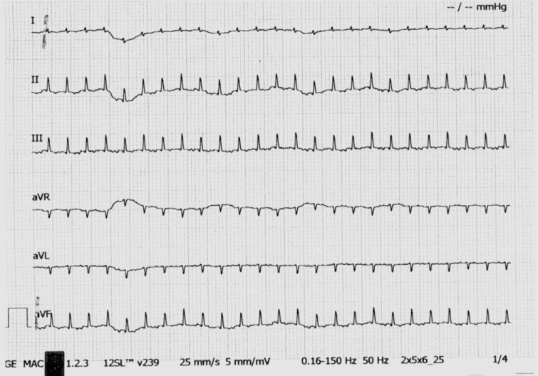
Physical examination noticed poor general condition, dyspnea, tachycardia, and hepatomegaly. It was performed an Electrocardiogram (EKG) that indicated a narrow QRS tachycardia at 303/min (Supraventricular Tachycardia (SVT), unresponsive at diving reflex (Figure 1). The tachycardia was definitively stopped with adenosine 1mg bolus.

Figure 1: Supraventricular Tachycardia (SVT).
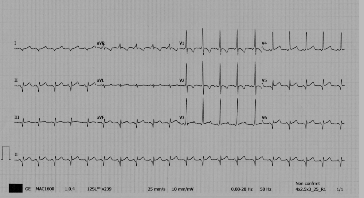
The electrocardiogram in sinus rhythm showed ventricular preexcitation by left accessory pathway of Kent (Wolff-Parkinson-White syndrome (WPW syndrome)) (Figure 2).

Figure 2: Ventricular pre-excitation by left accessory pathway of Kent.
In the child’s medical history, there was scarce information of a birth completed by abdominal caesarean delivery for fetal tachycardia (in another hospital) and maternal hyperthyroidism during pregnancy.
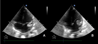
In sinus rhythm, the infant was pale and with signs of low cardiac output. Echocardiography was performed by the on call cardiologist, which revealed anatomically normal heart with reduced ejection fraction (EF 40-45% M-mode), and the presence of a large and floating mass (13mm x 7mm) in the left atrial appendage, protruding in the mitral valve, of unclear etiology (Figure 3).

Figure 3: The mass in the left appendage during the tele-systolic phase and protruding in the mitral annulus during the early protodiastolic filling.
In suspicion of thrombosis, intravenous heparin (20 U/Kg/h) was started.
After one hour, the infant presented lower limb pallor with the disappearance of femoral pulses, and echocardiography showed that the thrombus had vanished from the left atrial appendage.
Contrast-enhanced Computed Tomography (CT) showed the abdominal aorta below the renal arteries occluded by the embolized thrombus (Figure 4, Panel A and B). Additionally, a minor thrombus was embolized in the middle tract of the superior mesenteric artery (Figure 4, Panel C). The patient was rapidly addressed to the catheterization theatre and the thrombus was percutaneously removed, restoring patency of involved arteries. Histological analysis confirmed that the material removed was of thrombotic origin.

    Figure 4:  The thrombus embolized into the abdominal aorta provoking a long aortic occlusion beginning immediately below the renal arteries origin (red arrow,
Panel A). Please note the ischemia of the inferior pole of the right kidney (red arrow): The thrombus extended until the iliac bifurcation, involving the origin of a
lower polar accessory arterial artery (Panel B). The thrombus embolized in the superior mesenteric artery (Panel C).
Routine blood testing and thrombophilia screening resulted normal by age. Protein C and protein S were normal in both parents. The research of mutations, as factor V Leiden (R506A), Prothrombin (G20210A) and MTHFR (C667T) was negative. Screening for metabolic diseases resulted negative (urine organic acids, acylcarnitine and homocysteine).
The patient was discharged in perfect clinical conditions two days after the procedure. After 6 months, the infant is still in prophylaxis with propranolol with no recurrence of arrhythmia or cardiac thrombosis; Low Molecular Weight Heparin (LMWH) was stopped after 12 weeks.
Discussion
We report a case of sudden systemic embolization of a left atrial thrombus occurring after the resumption of sinus rhythm in an infant with SVT.
In adult, management of atrial fibrillation and atrial flutter in adults requires transesophageal echocardiography to screen the left atrial appendage for thrombi before to recur to pharmacological or electrical conversion to sinus rhythm [1]. This because it is well known that the conversion in sinus rhythm may provoke systemic embolism of pre-existent left atrial thrombi, a fearsome complication that may be prevented by initiating anticoagulation therapy before resumption of sinus rhythm.
Nowadays, Pediatric Advanced Life Support (PALS) courses train physicians in ED to recognize and treat SVT without a formal indication to provide an echocardiography evaluation before [2] and this normally occurs in clinical practice in many centers.
Indeed, cardiac thrombosis is very uncommon in infants: more frequently it occurs as a complication of central venous catheters [3]. This condition mostly affects the right atrium, but occasionally, when the catheter crosses the patent foramen ovale, it may occur into the left atrium.
Even if left appendage thrombosis has been already reported in infants after a paroxysmal SVT associated with intermittent Wolff-Parkinson-White syndrome [4,5], when an isolated mass is found in the heart at echocardiographic evaluation, the diagnosis is challenging and uncertain. Potentially, also every cardiac tumor may be responsible of SVT.
The association between cardiac rhabdomyomas, WPW syndrome and sustained supraventricular tachycardia is well documented [6]. The rhabdomyomas constitute up to 80% of all primary cardiac tumors in <1 year children and it is most frequently diagnosed in newborn and infants [7]. In general, they are multiple, well circumscribed, non-capsulated, intramural and they occur anywhere within the heart, but most commonly involving the ventricle. In the present case the echographic aspect of floating mass in the left appendage made this diagnosis very unlikely.
Myxomas constitute about 50% of primary cardiac tumors in patient of all ages [8], most often they are diagnosed in the second to sixth decades of life, but rarely may be found in newborns and infants [9,10]. Cardiac myxomas are single left atrial tumors in about 75% of cases. Typically myxomas have pedicle morphology, protruding from the fossa ovalis; only occasionally, this tumor takes its origin from other segments of the atrial septum or atrial free wall. Myxoma in the left appendage is very rare, although it has been described [11].
To the best of our knowledge, there are no predictive factors to assess the risk of systemic embolization, in case of unidentified isolated cardiac mass in children. Systemic thrombolysis may be indicated in the presence of intracardiac large floating mass, but the risk of paradoxical embolization and of bleeding must be considered. In our case, embolization occurred under heparin infusion one hour after the resumption of sinus rhythm. Aortic occlusion provoked progressive ischemia of the limbs, of the abdomen, and of the inferior pole of the right kidney. The possibility to recur rapidly to on-call physicians with specific expertise in Interventional catheterization allowed to restore aortic patency and to discharge the infant very quickly, without the need of invasive and challenging abdominal surgery.
Although still controversial, a full thrombophilia screening may be recommended in any infant with thrombosis, to determine future risk of recurrence and timing of prolonging anticoagulation, as in case of major thrombophilia (inherited PC or PS deficiency, homozygous or double heterozigous mutations).
Recurrence risk of SVT in WPW syndrome is higher during the first year of life [12], therefore propranolol therapy has been undertaken until this age.
Conclusion
In conclusion, our case raises the question if all children presenting SVT should undergo early echocardiography evaluation before reduction to sinus rhythm. Moreover, this dramatic experience convinced us that all patients with undefined left heart mass should be addressed and managed in centers capable to prompt interventional catheterization in case of sudden systemic embolization.
References
- Anderson JL, Halperin JL, Albert NM, Bozkurt B, Brindis RG, Creager MA, et al. AHA/ACC/HRS Guideline for the Management of Patients With Atrial Fibrillation: Executive Summary A Report of the American College of Cardiology/American Heart Association Task Force on Practice Guidelines and the Heart Rhythm Society. J Am Coll Cardiol. 2014; 64: 2246-2280.
- Kleinman ME, Chameides L, Schexnayder SM, Samson RA, Hazinski MF, Atkins DL, et al. American Heart Association Guidelines for Cardiopulmonary Resuscitation and Emergency Cardiovascular Care: Pediatric Advanced Life Support. Circulation. 2010; 122: 876-908.
- Ferrari F, Vagnarelli F, Gargano G, Roversi MF, Biagioni O, Ranzi A, et al. Early intracardiac thrombosis in preterm infants and thrombolysis with recombinant tissue type plasminogen activator. Arch Dis Child Fetal Neonatal Ed. 2001; 85: 66-72.
- Russo G, Trappan A, Benettoni A. Unusual left atrial appendage thrombus in a neonate with normal heart after sustained supraventricular tachycardia. Int J Cardiol. 2008; 131: 17-19.
- Amirtha Ganesh B, Ravi Cherian M. Left Atrial Thrombus in a Neonate with Normal Heart after Sustained Supraventricular Tachycardia. J Clin Diagn Res. 2014; 8: PD01-PD02.
- O’Callaghan FJ, Clarke AC, Joffe H, B Keeton, R Martin, A Salmon, et al. Tuberousis complex and Wolff–Parkinson-White syndrome. Arch Dis Child. 1998; 78: 159-162.
- Isaacs H. Fetal and neonatal cardiac tumors. Pediatr Cardiol. 2004; 25: 252- 273.
- Talley JD, Wenger NK. Atrial myxoma: Overview, recognition and management. Compr Ther. 1987; 13: 12-18.
- Dianzumba SS, Char G. Large calcifed right atrial myxoma in a newborn: rare cause of neonatal death. Br Heart J. 1982; 48: 177-179.
- Jals J, Ek J, Sandnes J. Cardiac myxoma as the cause of death in an infant. Acta Paediatr Scand. 1990; 79: 999-1000.
- Malik L, Borgohain S, Gupta A, Grover V, Gupta VK. Left atrial appendage myxoma masquerading as left atrial appendage thrombus. Asian Cardiovasc Thorac Ann. 2013; 21: 205-207.
- Riggs TW, Byrd JA, Weinhouse E. Recurrence risk of supraventricular tachycardia in pediatric patients. Cardiology. 1999; 91: 25-30.