Case Report
J Pediatr & Child Health Care. 2021; 6(1): 1040.
Ultrasound-Guided Caudal Epidural Block for Bilateral Testicular Ectopy and Circumcision in Child above 6 Years with Joubert Syndrome
Fekih Hassen Amjed*, Ben Fraj Asma, Blaiti Hajer, Ben Slimen Ahmed, Kaddour Refka, Trifa Mehdi
Department of Anaesthesia and Intensive Care, Children Hospital Bechir Hamza, Faculty of Medicine of Tunis, University of Tunis El Manar, Tunis, Tunisia
*Corresponding author: Fekih Hassen Amjed, Department of Anaesthesia and Intensive Care, Children Hospital Bechir Hamza, Bab Saadoun 1007 Tunis, Tunisia
Received: April 14, 2021; Accepted: April 30, 2021; Published: May 07, 2021
Abstract
Background: Abnormal respiratory control has been clearly documented in infants and children with Joubert Syndrome (JS) by polygraphic recordings, characterized by episodes of apnea, tachypnea, and/or hyperpnea and the risk of recurrence of these episodes increase probably in perioperative period. In those cases, the choice of anesthesia technique and postoperative pain protocol, constitute a challenge for anesthesiologists.
Case: We describe a case of successful ultrasound-guided Caudal Epidural Block (CEB) for children above 6 years with Joubert Syndrome undergoing bilateral testicular ectopy and circumcision avoiding opioid-use.
Conclusions: Despite the difficulty to achieve CEB in child above 6 years, the ultrasound-guided can increase the success providing effective analgesia in-patient with a high-risk of respiratory failure as child with JS.
Keywords: Analgesia; Caudal epidural block; Joubert syndrome; Paediatric anaesthesia; Regional anaesthesia.
Categories: Anaesthesiology, Paediatric, Paediatric Surgery.
Introduction
Joubert Syndrome (JS) is a recessive neurodevelopmental disorder with structural abnormalities of the brainstem, central respiratory control problems, airway hypotonia and swallow dysfunction, so individuals with JS should be considered high risk for anaesthesia [1]. Caudal Epidural Block (CEB) as epidural anaesthesia of the cauda equine roots, is a common paediatric regional technique and provides a high quality of intraoperative and early postoperative analgesia. Avoiding opioid-use in-patient with a high-risk of respiratory failure as child with JS, CEB constitute an interesting alternative whenever the site of surgery allows it. However, superficial anatomic landmarks traditionally used for caudal placement are less predictable and unreliable [2]. The use of real-time ultrasound for direct visualization of injection will confirm CEB and potentially essentially eliminate block failure [3,4].
Herein, we would like to report a case of ultrasound-guided CEB, used for perioperative analgesia in six years and half-old child with JS undergoing cure of bilateral testicular ectopy and circumcision. Written informed consent was obtained from the child’s legal guardian for all procedures and for use of case data in this report.
Case Presentation
We report a case of a six years and half-old child with JS, scheduled for cure of bilateral testicular ectopy and circumcision. The bodyweight was 16 kg. The patient had hypotonia, and mental retardation. The anaesthesia assessment did not found any other abnormality. The chest X-ray showed normal lung parenchyma appearance with a normal cardiothoracic ratio and the laboratory analyses were within normal limits. The choice of the type of anaesthesia and analgesia was explained to the parent in detail and written informed consent for use of case data was signed.
Upon arrival in the operating room, basic monitoring was applied. Baseline parameters were documented: HR-110 bpm, SpO2- 98% and BP-90/60 mmHg. Intravenous access was established under Sevoflurane anaesthesia induction. After 60 mg intravenous bolus of propofol, a laryngeal mask was inserted and the child was putted on lateral position with flexed hips to performing ultrasound-guided CEB.
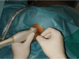
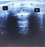
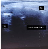
The ultrasound transducer was first placed transversely at the midline to obtain the transverse view of Sacral Hiatus (SH) (Figure 1). Under this view, the Block Needle (BN) was inserted using the “out of plane” technique. The BN can be visualized in real time, piercing the SCL, entering the SH (Figure 2). At this level, the ultrasound transducer was rotated 90 degrees to obtain the longitudinal view of SH. After verifying absence of spontaneous reflux of blood or cerebro-spinal-fluid, 12 ml (0.75 ml/kg) of 0.25% Levo-Bupivacaine was injected slowly (over about one minute), without resistance (Figure 3).

Figure 1: Transverse placement of the ultrasound.

    Figure 2: Transverse ultrasound view of the sacral hiatus. BN: block needle;
BS: base of sacrum; SC: sacral cornua; SCL: sacro-coccygeal ligament; SH:
sacral hiatus.

    Figure 3: Longitudinal ultrasound view of sacral hiatus.
BN: Block Needle; BS: Base of Sacrum; SCL: Sacro-Coccygeal Ligament;
SH: Sacral Hiatus.
General anaesthesia was maintained using Sevoflurane. The procedure lasted a total of 45 minutes and there were no complications. The emergence from anaesthesia was uneventful.
In recovery room, no desaturation occurred after oxygen weaning and the child did not need rescue analgesics. The “Objective Pain Score” was marked hourly during the first 24 hours, and it was less than three. The child never showed sign of respiratory distress and his respiratory rate ranged between 15 and 20 per minute with blood saturation stayed above 95% without oxygen. Standing and walking were possible at day one and the child left the hospital at this moment with prescription of oral Paracetamol.
Discussion
Individuals with JS often have complex medical histories [1]. The abnormalities of respiratory control are likely to be due to hypoplasia of the cerebellar vermis and brainstem [5]. The pre-anaesthetic consultation allows time for appropriate clinical evaluation and any required additional testing, ensuring that the anaesthesiology professional and facility are as prepared as possible [1,6]. For our case, the anaesthesia assessment shown hypotonia and mental retardation without any signs of respiratory irregularity. The chest X-ray and the laboratory analyses were within normal limits.
Additionally, patients with JS are extremely sensitive to the respiratory depressant effects of anaesthetic agents [5,7]. Consequently, avoidance of opioids and neuromuscular-blocking drugs using regional anaesthesia would seem reasonable [7-9]. Regional anaesthesia is strongly recommended, preferably a neuraxial block in high-risk patients [7,10]. The application of combined general and CEB should be successful and safe for child scheduled for cure of bilateral testicular ectopy and circumcision.
The fact of the difficulty to access to caudal epidural space in child above six years, we chose to use ultrasound to identify the site of needle entry, guide insertion of the needle into this space and potentially essentially eliminate block failure [4].
Our report demonstrate the potential efficiency of using regional anaesthesia in this challenging patient avoiding opioid-use. Despite the difficulty to achieve CEB in child above 6 years old, the ultrasoundguided can increase the success providing effective analgesia inpatient with a high-risk of respiratory failure as a child with JS.
References
- Bachmann-Gagescu R, Dempsey JC, Bulgheroni S, Chen ML, D’Arrigo S, Glass IA, et al. Healthcare recommendations for Joubert syndrome. Am J Med Genet A. 2020; 182: 229-249.
- Sheng-Chin Kao, Chia-Shiang Lin. Caudal Epidural Block: An Updated Review of Anatomy and Techniques. Biomed Res Int. 2017; 2017: 9217145.
- Shafy SZ, Hakim M, Villalobos MA, Pearson GD, Veneziano G, Tobias JD. Caudal epidural block instead of general anesthesia in an adult with Duchenne muscular dystrophy. Local Reg Anesth. 2018; 11: 75-80.
- Adler AC, Belon CA, Guffey DM, Minard CG, Patel NV, Chandrakantan A. Real-Time Ultrasound Improves Accuracy of Caudal Block in Children. Anesth Analg. 2020; 130: 1002-1007.
- Habre W, Sims C, D’Souza M. Anaesthetic management of children with Joubert syndrome. Paediatr Anaesth. 1997; 7: 251-253.
- Platis CM, Kachko L, Trabikin E, Simhi E, Bahar M, Katz J. Postoperative respiratory complications in Joubert Syndrome. Paediatr Anaesth. 2006; 16: 799-800.
- Galante D, Meola S, Cinnella G, Dambrosio M. Regional Caudal blockade in a pediatric patient affected by the Joubert syndrome. Acta Anaesthesiol Scand. 2009; 53: 693-694.
- Vodopich DJ, Gordon GJ. Anesthetic management in Joubert syndrome. Paediatr Anaesth. 2004; 14: 871-873.
- Laitselart P, Serey K, Ponsin P, Daban J-L. Ultrasound guided supra-inguinal fascia iliaca block for total hip arthroplasty in a patient with Joubert Syndrome: An efficient block for a patient with a high risk of post-operative respiratory failure. J Clin Anesth. 2019; 57: 3-4.
- Walther-Larsen S, Rasmussen LS. The former preterm infant and risk of postoperative apnoea: recommendations for management. Acta Anaesthesiol Scand. 2006; 50: 888-893.