Case Series
Austin J Pediatr. 2015; 2(1): 1020.
Hand, Foot and Mouth Disease in Children of Niš City: A Report of Six Cases
Djordjevic G¹ and Vlajkovic S²*
¹Department of Pediatrics, Health Center Niš, Serbia
²Department of Anatomy, Faculty of Medicine, University of Niš, Serbia
*Corresponding author: Vlajkovic S, Department of Anatomy, Faculty of Medicine, University of Nis, 18000 Nis, Serbia
Received: August 23, 2015; Accepted: September 18, 2015; Published: September 29, 2015
Abstract
Hand, foot and mouth disease (HFMD) is an acute viral disease that occurs mostly in young children. It is characterized by fever, oral ulcerations and papulovesicular rash on the palms and soles. The disease usually has a mild clinical course, but complications can occur, even resulting in death.
This work presented cases of hand, foot and mouth disease which the author met in the pediatric outpatient practice.
Cases of HFMD occured from mid-September to mid-December. In two families, the disease first occurred in one child, and two days later in another child of the same family. All patients had mild illness, with rapid recovery.
In our country HFMD occurs sporadically and we use this opportunity to present our experiences and it is necessary to monitor the prevalence of the disease in order to timely respond and prevent epidemics, which is the experience of many countries in the world.
Keywords: Hand foot and mouth disease; Papulovesicular lesions; Children
Introduction
Hand, foot and mouth disease (HFMD) usually affects children [1-8] mostly aged up to 5 years [1,2,5,6,8,9]. The disease has a seasonal character; typically occurs in the summer and early fall [5,7,9-12]. Human enteroviruses are proven causes of the disease, especially enterovirus 71 (EV71) and Coxsackievirus A16 (CVA16), as well as some others human enteroviruses – CVA4-CVA6, and CVA10 [1,3–14]. Clinical manifestations of HFMD are: fever; sore throat with oral ulcerations (enanthema); papulovesicular rash (exanthema) on the palms and soles [1-15]. These clinical manifestations are mainly used for early diagnosis of disease [4]. Painful ulcerations in the mouth present a major problem in children, especially younger ones, because they make difficulties to input adequate amounts of food and liquids, which can lead to dehydration of the child [4,8,12,15]. HFMD is usually self-limiting, mild disease, but there may be serious complications such as encephalitis, aseptic meningitis, acute flaccid paralysis, myocarditis, pneumonia, and even death. These complications are more frequently associated with infection of EV71 [1,3-7,9,10,12,15,16]. In some cases, HFMD may be associated with the loss (two or more) of nails – onychomadesis, which may be related to infection with the virus CVA6 [10,11,13]. The disease may be transmitted by direct contact with an infected person or contaminated objects through the air (coughing, sneezing) and the fecal-oral route [2,5,8,12]. Period of incubation lasts for 3–6 days [12], while the contagious period lasts even several weeks from the beginning of the infection [8,12]. A person with HFMD is most infectious during the first week of the disease [8]. Diagnosis is based on: (1) the clinical presentation [4,14]; (2) the isolation and detection of the virus from throat swabs, nasal swabs, rectal swabs, feces, vesicular fluid, serum or cerebrospinal fluid [3,4,9,10]; (3) the increase (fourfold) of specific antibodies [5,7]. Currently, there is no specific treatment for this disease. Patients receive supportive therapy [8,15]. There are frequent outbreaks of HFMD in many countries in the world today: Thailand, Taiwan, Australia, China, USA, Finland, France and Spain [1,7,9-11,13].
The aim of this study was to present cases of HFMD in children observed during outpatient pediatric practice of Nišava District in Niš, Serbia.
Methods
Presented data are based on anamnesis, physical examination and medical record data. Skin and mouth lesions are photographed with written consent of the parents and permission of children. Clinical presentation of presented patients was compared with available literature data.
Cases Presentation
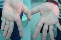
Case 1: (five-year-old boy). In mid-September 2014, the parents have brought the boy to the examination due to sore throat and skin lesions on the palms and feet. The boy didn’t have a fever. Lesions were not found in the oral cavity or throat, but were present as macular rush on the palms and soles (Figure 1), the boy was in a good general condition. Supportive therapy was advised (analgesic and skin care). As the skin lesions withdrew in a week, additional therapy was not indicated on the control examination.

Figure 1: Macular rush on the palms and soles in a 5-year-old boy.
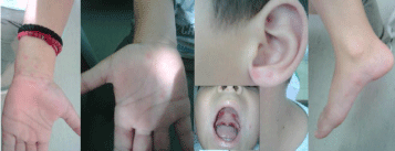
Case 2: (seven-year-old boy, the older brother of the previously described boy). Two days after the disease appeared in younger brother, a sore throat and skin lesions on the palms, soles of the feet and ear lobe appeared in his older brother. The boy was in a good general condition, without fever, but with ulcerations on the hard and soft palates, and perioral papular rash; papulovesicular rash was present on the palms, earlobes and feet (Figure 2). Laboratory analyzes showed leukocytosis (Le 15,800). Despite the leukocytosis, antibiotics were not given; for two days later, repeated laboratory findings were within normal limits. He was advised supportive therapy, and the boy completely recovered.

    Figure 2:  Ulcerations on the hard and soft palates, perioral papular rash and
papulovesicular rash on the palms, earlobes and feet in a 7-year-old boy.
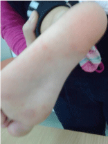
Case 3: (two year-old-girl). In mid-September 2014, the parents have brought the girl to the pediatrician due to fever of 39.5°C lasting four days, and lesions on the skin of soles (Figure 3), palms, around the lips and buttocks. The girl allowed only a photo of one sole. Skin lesions occurred when the fever stopped. The girl was exhausted and tearful; she refused food and fluid intake. Six months before that, the girl had febrile seizures, but they were not repeated now, although the girl was febrile. On examination, the girl was in good general condition, although with mild dehydration; on the soft palate were several vesicles, and on the mentioned parts of the body was present papulovesicular rash. With supportive care, the girl recovered without complications.

Figure 3: Macular rush on the sole in a 2-year-old girl.
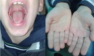
Case 4: (nine year-old-boy). In early October 2014, in a boy’s palms appeared a rash and he had a sore throat. Although he was in a good general condition, on the soft palate were present vesicular lesions (vesicular lesions were presented on the soft palate), and papular rash was on the palms (Figure 4). The disease was fully withdrawn, without any specific therapy.

    Figure 4:  Vesicular lesions of the soft palate and papular rash on the palms
in a 9-year-old boy.
Case 5: (three and a half year-old-girl; the younger sister of the previously described boy). Two days after the disease has occurred at his brother, sore throat, cough and skin lesions on hands occurred in a girl. The girl consumed less food and fluid because of sore throat. The inspection showed that the girl was in a good general condition, with signs of mild dehydration; several vesicular lesions were on the soft palate, and a papulovesicular rash was on the palms and soles. These lesions were not photo documented, because the girl did not permit. An analgesic was advised, as well as oral electrolyte fluid for rehydration. The parents are advised to nourish the girl’s skin. In a few days the girl felt better and fully recovered.
Case 6: (two year-old-girl). The parents brought the child in early December 2014 to a pediatrician, because of skin lesions on the fingers. Clinical examination found an intensive hyperemia of the throat, perioral papules and vesicular lesions on the lateral side of the fingers and toes (Figure 5). The girl was in good general condition. At follow-up, nine days after the examination, there were no scars or other signs of previous illnesses.

    Figure 5:  Perioral papules and vesicular lesions of the fingers of hands in
a 2-year-old girl.
Discussion
It is considered that HFMD is an acute viral disease that affects mainly young children [1–8], which was our experience, too. Age of our patients was among 2–9 year: two children aged 2 years; by one patient aged 3.5, 5, 7 and 9 years. The first patients came in mid-September, and the last at the beginning of December 2014. In the literature, there are also data showing that disease occurred in the summer and autumn [2,3,9,12]. According to international experience, the disease is characterized by fever [3–9,12,14,15], and its occurrence in the prodromal period [8,11]. In contrast to previous experience, our patients were without fever, except 2 year-old girl who had fever (39.5°C) for 4 days. In two families, the disease first occurred in one child, and two days later in another child of the same family. Sore throat and skin lesions on the palms and soles were typical for our patients. In the practice of other authors, localization of lesions in the mouth in patients with HFMD was variously defined and described. Some authors have described the lesions localization on the tongue and buccal mucosa [7,13,15]; or on the palate [8,9], as well as in our four cases; or in any of previously mentioned localization [1].
Leukocytosis (Le > 15,000) was also described in a certain percentage of patients [15]. Our patients had mild clinical picture, were treated at home with supportive therapy and the disease went without complications. Due to the fact that the disease was mild, there was no need for additional tests and that is the reason why the cause of HFMD in our patients was not determined.
In our country HFMD occurs sporadically and there have been no epidemics such as those described in the literature [1,3, 6,7,9,11,13,15], and therefore HFMD is not subject to the obligation for report of infectious diseases.
Conclusion
As the disease occurred in two children in two families, it can be confirmed that the disease is transmitted through contact and saliva, after a short incubation period up to 48 hours, in the late summer and during the autumn.
Taught by the international experiences, the authors consider that HFMD cases should be registered not only in the medical records, but also in this way.
Case reports underline the importance of prevention, the importance of preventing the spread of disease and the occurrence of complications, but also the exchange of experiences in relation to the subjective and objective status of patients with HFMD and treatment of patients in these cases.
References
- McMinn PC. An overview of the evolution of enterovirus 71 and its clinical and public health significance. FEMS Microbiol Rev. 2002; 26: 91–107.
- Kaye D. Severe hand, foot, and mouth disease associated with Coxsackievirus A6. MMWR 2012; 61: 213.
- Osterback R, Vuorinen T, Linna M, Susi P, Hyypiä T, Waris M. Coxsackievirus A6 and hand, foot, and mouth disease, Finland. Emerg Infect Dis. 2009; 15: 1485–1488.
- Huang WC, Huang LM, Lu CY, Cheng AL, Chang LY. Atypical hand-foot-mouth disease in children: a hospital based prospective cohort study. Virol J. 2013; 10: 209.
- He SJ, Han JF, Ding XX, Wang YD, Qin CF. Characterization of enterovirus 71 and coxsackievirus A16 isolated in hand, foot, and mouth disease patients in Guangdong, 2010. Int J Infect Dis. 2013; 17: 1025–1030.
- Wu Y, Yeo A, Phoon MC, Tan EL, Poh CL, Quak SH, et al. The largest outbreak of hand; foot and mouth disease in Singapore 2008: The role of enterovirus 71 and coxsackievirus A strains. Int J Infect Dis. 2010; 14: 1076–1081.
- Mirand A, Henquell C, Archimbaud C, Ughetto S, Antona D, Bailly JL, et al. Outbreak of hand, foot and mouth disease/herpangina associated with coxsackievirus A6 and A10 infections in 2010, France: a large citywide, prospective observational study. Clin Microbiol Infect. 2012; 18: 110–118.
- Centers for disease control and prevention. Hand, foot and mouth disease (HFMD).
- Puenpa J, Chieochansin T, Linsuwanon P, Korkong S, Thongkomplew S, Vichaiwattana P, et al., Hand, foot and mouth disease caused by Coxsackievirus A6, Thailand, 2012. Emerg Infect Dis. 2013; 19: 641–643.
- Bracho MA, González-Candelas F, Valero A, Córdoba J, Salazar A. Enterovirus co-infections and onychomadesis after hand, foot and mouth disease, Spain, 2008. Emerg Infect Dis. 2011; 17: 2223-2231.
- Flett K, Youngster I, Huang J, McAdam A, Sandora TJ, Rennick M, et al. Hand, foot and mouth disease caused by Coxsackie virus A6. Emerg Infect Dis. 2012; 18: 1702–1704.
- Aronson SS, Shope TR. Managing infectious diseases in child care and schools: A quick reference guide, 2nd edn. American academy of pediatrics 2009.
- Chen YJ, Chang SC, Tsao KC, Shih SR, Yang SL, Lin TY, et al. Comparative genomic analysis of Coxsackievirus A6 strains of different clinical disease entities. PLoS ONE. 2012; 7: 52432.
- Goksugur N, Goksugur S. Hand, foot and mouth disease. N Engl J Med. 2010; 362: e49.
- Lo SH, Huang YC, Huang CG, Tsao KC, Li WC, Hsieh YC, et al. Clinical and epidemiologic features of Coxsackievirus A6 infection in children in northern Taiwan between 2004 and 2009. J Microbiol Immunol Infect. 2011; 44: 252–257.
- Huang W-C, Huang L-M, Kao C-L, Lu C-Y, Shao P-L, Cheng A-L, et al. Seroprevalence of enterovirus 71 and no evidence of crossprotection of enterovirus 71 antibody against the other enteroviruses in kindergarten children in Taipei city. J Microbiol Immunol Infect. 2012; 45: 96–101.