
Case Report
Austin Pediatr. 2016; 3(4): 1042.
Retrieval of Broken Instrument from Primary Molar and Preservation using Lesion Sterilization and Tissue Repair: A Case Report
Mujawar S and Tiwari G*
Department of Pedodontics & Preventive Dentistry, ACPM Dental College, Dhule, India
*Corresponding author: Tiwari G, Department of Pedodontics & Preventive Dentistry, ACPM Dental College, Opposite Jawahar Sutgirni, Morane, Dhule-424001, India
Received: September 27, 2016; Accepted: October 17, 2016; Published: October 19, 2016
Abstract
Background: Breakage of endodontic instrument within root canal of a primary tooth is a mishap that needs to be rectified, either by retrieval or creating a circumvent around it, failing which it can become a nidus of infection causing dentoalveolar abscess and pathologic mobility. Primary tooth loss at an early age compromises function and stability of dental arch, hence, the tooth should be preserved. LSTR sterilizes the canal space, aids in rapid repair of damaged periradicular tissues and restores healthy state of tooth.
Case Report: A 6-year-old girl reported with dentoalveolar abscess of mandibular deciduous left second molar with history of attempted pulpectomy aborted halfway. Radiographs revealed breakage of S1 Protaper rotary file in distobuccal canal, pathologic root resorption and furcal radiolucency. This compromised tooth was saved by manual retrieval of broken instrument and promoting healing using L.S.T.R. Purchase point adjacent to broken file was created in the canal and instrument was retrieved using H files. Triple Antibiotic Paste was used as intracanal medicament. In subsequent visits tooth was obturated with Metapex and restored with preformed stainless steel crown. Follow-up after a month, at 3, 6, 14, 18 and 24 months reported patient to be asymptomatic.
Conclusion: Manual instrument retrieval with Triple Antibiotic Paste proves to be a savior of teeth with severe infections to deliver smiles in pediatric patients.
Keywords: L.S.T.R; Endodontics; Instrument retrieval; Triple Antibiotic Paste
Abbreviations
LSTR: Lesion Sterilization and Tissue Repair; TAP: Triple Antibiotic Paste
Introduction
Breakage of instrument in deciduous tooth canal is a serious complication as inability to retrieve it hinders optimal preparation and obturation leading to failure [1,2]. It may lead to abscess formation, root resorption and pathologic premature mobility, which may alter the eruption of succedaneous tooth. Most clinicians go ahead with extraction of such primary teeth 2 followed by a space maintainer. However, loss of a primary tooth leads to compromised masticatory function in addition to trauma of extraction to the child and carers1. This report demonstrates retrieval of a broken instrument from primary molar followed by restoring its health using LSTR (Lesion Sterilization and Tissue Repair).
Case Presentation
A 6 year old female reported with pain in her lower left back tooth since a month. She had developed a round to oval, fluctuant swelling, 1cm x 1cm on the buccal aspect of deciduous mandibular left second molar (Figure 1) since four days. Parents gave history of attempted pulp therapy two months back that was aborted midway due to breakage of instrument.
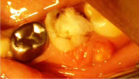
    Figure 1:  Deciduous mandibular left second molar showing swelling on
buccal aspect.
The tooth was mobile. Intraoral periapical radiographs at different angulations were taken to determine exact location of broken instrument and type of instrument separated. The radiographs revealed fracture of Protaper S1 file in the middle third of the distobuccal canal (Figure 2). Pathologic root resorption and irregular radiolucency was noted in the inter-radicular furcation area suggestive of dentoalveolar abscess with the same.
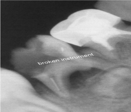
    Figure 2:  Instrument breakage in the disto-buccal canal with pathologic
resorption and furcal radiolucency.
Inspite of poor prognosis of the tooth, its preservation was carried out. Access was gained into the root canal system and the abscess was drained. Next, coronal flaring of the distobuccal canal was done to enhance access to the broken file using Protaper SX rotary file (Dentsply). The other canal orifices were packed with cotton pellets to prevent the broken file from jumping into another canal accidentally. Pathfinder file was used to obtain a purchase point in between the distobuccal canal wall and the broken file. At the purchase point, with hand 8-K file circumferential filing was carried out. The space was further enlarged with 10-K, 15-K, 20-K hand files. Simultaneous copious irrigation with normal saline was done. This procedure was repeated till the densely packed file became loose in the canal. By circumferential filing and continuous forceful irrigation, the broken file came to lie freely within the irrigant in the canal and was then retrieved (Figure 3) by engaging with H file. After file retrieval, the canals were irrigated thoroughly with 0.12% chlorhexidine solution.
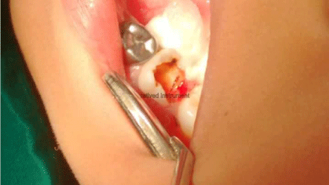
Figure 3: Retrieved instrument.
Working lengths were determined using electronic apex locator and biomechanical preparation was performed with rotary 4 % Hero shaper file of Micromegga along with EDTA. Copious irrigation with chlorhexidine followed by normal saline was done. Root canals were dried with absorbent points and Triple Antibiotic Paste (TAP) – a combination of Metronidazole, Ciprofloxacin and Minocycline mixed in 1:1:1 ratio using glycerine as a vehicle, was placed as an intracanal medicament using lentulo spirals for 21 days. Temporary restoration was given with zinc oxide eugenol cement.
At one week, patient was relieved with resolution of abscess and reduction in mobility of the tooth. After three weeks, the tooth was asymptomatic. The canals were irrigated, thoroughly dried and obturation was done with Metapex; a calcium hydroxide and iodoform based obturating material. At four weeks, tooth was absolutely asymptomatic with no sign of mobility. Permanent restoration was done with composite light cure resin (3M) and preformed metal crown was placed (Kids Crown) (Figure 4).
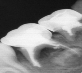
Figure 4: Post-obturation radiograph with Stainless Steel crown in place.
All procedures undertaken were in accordance with guidelines of the institutional ethical committee. Parents were informed about the procedure to be undertaken and written informed consent was obtained prior to the procedure.
Follow up was done after a month, at 3, 6, 14, 18 and 24 months with patient being asymptomatic with no discomfort (Figure 5).
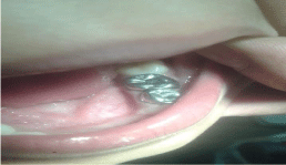
Figure 5: 18 months Follow-up.
Discussion
Instrument retrieval from root canal depends on the experience and skill of the operator and the anatomical factors of the root canals, success rate reported being 55–79% [3]. Manual retrieval of broken instrument, though time consuming, is convenient in deciduous teeth because of inherently wider and straighter root canals [4]. It also prevents further damage to the tooth and adjacent tissues [3]. Simultaneous use of irrigants warrants canal disinfection and provides medium for easy movement of files within the blocked canal [5].
LSTR is based on the concept that if dentinal, pulpal or periradicular lesions are disinfected repair of damaged tissues can be enhanced6. TAP utilizes the synergistic action of three antibiotics with wide antibacterial spectrum against endodontic bacteria to eliminate infection at the immediate vicinity [4,6]. The combination has been shown to penetrate efficiently through dentine and disinfect the lesion [7]. Therefore, TAP was utilized in this case.
By retrieval of the instrument, the nidus of infection was removed and TAP eliminated the rest of pathogens. With no pathogens to disturb the process of healing, repair of periradicular tissues was accelerated and in three weeks time the tooth was sound enough to bear the load of occlusion.
Combination of early intervention in retrieving the broken instrument and disinfection of the root canal and periradicular tissues with antimicrobials brought favourable outcomes.
Conclusion
Extraction should always be sort as the last remedy to instrument breakage in deciduous teeth and measures should be taken to preserve the deciduous tooth until its normal exfoliation to guide permanent dentition into proper function. LSTR proves to be a boon in restoring health of compromised teeth.
References
- Patel JJ, Morawala A, Talathi R, Shirol D. Retrieval of a broken instrument from root canal in primary anterior teeth. Univ Res J Dent. 2015; 5: 203-206.
- Spili P, Parashos P, Messer HH. The impact of instrument fracture on outcome of endodontic treatment. J Endo. 2005; 31: 845–850.
- Kunhappan S, Kunhappan N, Patil S, Agarwal P. Retrieval of separated instrument with instrument removal system. J Int Clin Dent Res Organ. 2012; 4: 21-24.
- Taneja S, Kumari M, Parkash H. Nonsurgical healing of large periradicular lesions using a atriple antibiotic paste: A case series. J Cont Cli Dent. 2010; 1: 31-35.
- Torabinejad M, Lemon RR. Procedural accidents. Walton RE, Torabinejad M, editors. In: Principles and practice of endodontics. 3rd ed. Philadelphia: Saunders. 2002; 310–330.
- Hoshino E, Takushige T. LSTR 3Mix-MP method-better and efficient clinical procedures of lesion sterilization and tissue repair (LSTR) therapy. Dent Rev. 1998; 666: 57–106.
- Fernandes M, Ida de Ataide. Nonsurgical management of periapical lesions. J Conserv Dent. 2010; 13: 240–245.