
Case Report
Austin Pediatr. 2016; 3(5): 1047.
Pseudopapilar Solid Tumor of Pancreas, as a Finding in Pediatric Patient
Delgado YS*, Azuela OC, Quinto AG, Zepeda NC and Viana EH
General Mexican Hospital, ¨Dr. Eduardo Liceaga, Mexico
*Corresponding author: Sánchez Delgado Y, General Mexican Hospital ¨Dr. Eduardo Liceaga, Mexico
Received: November 08, 2016; Accepted: December 19, 2016; Published: December 21, 2016
Introduction
The pseudopapillary solid tumor, predominantly in young women in the third decade of life. It represents less than 1% of all pancreatic neoplasms [1] is also called Frantz-Gruber tumor was first described in 1959. The anatomy and histological elements include solid, cystic and pseudopapillary components; as well as being producers of mucin, present nonspecific clinical symptoms so the diagnosis is a finding against the persistent vague abdominal symptoms; however, described capsule ruptured acute peritonitis [2]. Hemoperitoneum and malignant potential of 14%. Preoperative diagnosis may be referred to as pancreatic pseudo cysts in patients with no history or risk factors for pancreatitis with normal serum amylase and a solid component of a cystic cavity without communication between the cyst and pancreatic ducts [3]. La surgical resection is performed pancreatoduodenectomy if the lesion is at the level of head-neck or distal pancreatectomy with splenectomy should be in the queue. Greater than 90% at 5 years, survival about 15% of patients develop metastatic disease to the liver, peritoneum and lymph nodes [4,5].
Purpose
Describe the incidental finding of a solid tumor pseudopapillary as abdominal tumor.
Methods
Was reported a case of abdominal tumor in the Pediatric General Surgery General Hospital of Mexico.
Clinical Presentation
Female patient 17 years of age having increased volume in the right upper quadrant of 6 months of evolution, which is progressive up to 10x10cm, after normal pregnancy. Referred early satiety, postprandial nausea and occasional vomiting, denies and weight loss. No chronic degenerative history, exposure to organic dusts or previous pictures of pancreatitis. Physical examination: Abdomen with tumor located in hypochondrium and right flank of 15x15x15 cm, indurated, defined, painless, motionless, well-defined edges, without vascular compromise, no evidence of peritoneal irritation. Laboratory and images reported (Figure 1 and Figure 2): 1.99 alpha-fetoprotein, carcinoembryonic antigen 1.29, beta fraction of chorionic gonadotrophin 0.23. Pancreatoduodenectomy was performed classically finding as tumor pancreatic head solid aspect of 20x20 cm surgical finding no evidence of metastasis, firm and loose adhesions, through unidentifiable Wirsung, cystic artery 10x1 mm, cistico of 10x3 mm, coledoco 6mm, root mesentery with no evidence of tumor. Figure 3 pathology results: pseudopapillary solid tumor in the pancreatic head (Figure 4). In the postoperative period, the patient has adequate clinical evolution, without evidence of bleeding, controlled pancreatic fistula presented with obstructive jaundice pattern, which was resolved with percutaneous catheter placement, he was discharged without complications.
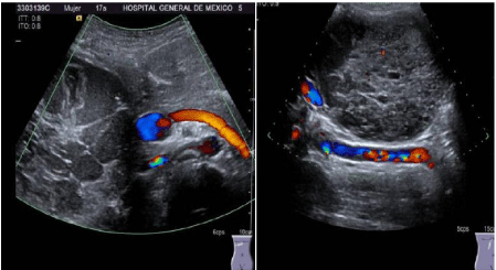
    Figure 1:  HEAD OF PANCREA Sis identified heterogenous lesion, with
rounded morphology, encapsulated with thickened wall of 3.4mm. Showing
regular and well defined edges, with approximated imensions of 0.6x9.3x12.0
cm. With volume of 629.6c.
CABEZADEPáNCREAS se identificales ión heterogénea, demorfología
redondeada, encapsulada con pared engrosadade 3.4mm. Most
randobordes regular es bien definidos, con dimensiones aproximadas de
10.6x9.3x12.0cm. convolumend e 629.6cc.
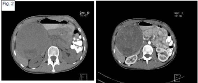
    Figure 2:  Round encapsulated thin image, with regular and well defined
edges, wall with thickness of 3.4mm. Its interior presents a heterogeneous
attenuation pattern, lesions hows relations by its superior surface with the
visceral face of the liver and biliary vesicle to which it molds, compresses the
portion pyloric and duo denumantrumin its first, second and third portion to
which it collapses And demonstrating filiform passage of the oral medium to
the rest of the small intestine.
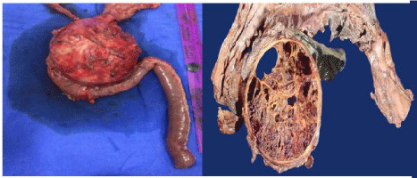
    Figure 3:  Piece 71cm and length, the gastric antrum portion is 8.0cm, the
duodenum measures 20cm, the proximal portion of the jejunum measures
23cm, the gall bladder measures 6.9x4.5x3.8cm, apportion of the head of
the Pancreas measuring 4.7x3.0x3.0cm and adjacent to the pancreas and
duodenum there is a nodular lesion measuring 13.0x10.0x8.0cm, the outer
surface is smooth and bright brown.
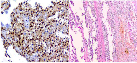
    Figure 4:  To the left the histological aspect or responds to a neoplasm
characterized by fibrous septa bordered by several layers of cells with little
cohesiveness, eosinophilic cytoplasm to clear, round nuclei without atypia
with fine chromatin (HyE staining). On the right, nuclear immune expression
for B-catenin is observed in neo plastic cells.
Conclusion
The total resection of the pseudo papillary pancreatic tumor with pancreatoduodenectomy is the surgical procedure of choice for the low malignant potential and the high survival rate after surgery. In pediatric patients following the finding of an abdominal tumor, clinical and imaging data are important in order to plan a timely surgical intervention that affects their prognosi.
References
- Fritz S, Warshaw AL, Thayer SP. Management of Mucin-Producing Cystic Neoplasms of the Pancreasv. The oncologist. 2009; 14: 125-136.
- Chakrabarti S, Ghosh S, Sarkar R. Solid Pseudopapillary Tumour of Extrapancreatic Origin Presenting as Mesenteric Cystic Mass: A Diagnostic Dilemma. J Clin Diagn Res. 2016; 10: 01-02.
- Castellano-Megias VM, Andres CI, Lopez-Alonso G, Colina-Ruizdelgado F. Pathological features and diagnosis of intraductal papillary mucinous neoplasm of the pancreas. World J Gastrointest Oncol. 2014; 6: 311-324.
- Papavramidis T, Papavramidis S. Solid pseudopapillary tumors of the pancreas: review of 718 patients reported in English literature. J Am Coll Surg. 2005; 200: 965-972.
- Cao Z, Zhang TP, Zhao YP. Diagnosis and treatment of solid pseudo papillary neoplasm’s of the pancreas. Zhonghua Wai Ke Za Zhi. 2016; 54: 550-552.