Case Report
Austin Pediatr. 2020; 7(1): 1073.
Neonatal Cerebral Sinovenous Thrombosis: A Rare Case Report and Literature Review
Wu YJ1, Chen PJ1, Chen YL2,4, Chen SJ4 and Wang AC1-4*
1Department of Pediatrics, Taoyuan Armed Forces General Hospital, Taiwan
2Department of Radiology, Taoyuan Armed Forces General Hospital, Taiwan
3Department of Life Sciences, National Central University, Taiwan
4National Defense Medical Center, Neihu , Taiwan
*Corresponding author: Wang AC, Department of Pediatrics, Taoyuan Armed Forces General Hospital, Taiwan
Received: February 17, 2020; Accepted: March 14, 2020; Published: March 21, 2020
Abstract
Neonatal cerebral sinovenous thrombosis is a rare and potentially lifethreatening disorder associated with various long-term neurological deficits. The pathogenesis of cerebral sinovenous thrombosis in neonates is still unclear. Many potential risk factors have been identified, such as gestational or delivery complications or neonatal comorbid conditions including dehydration, sepsis, or cardiac defects. A correct diagnosis is often delayed due to the subtle presentation of the disorder, leading to delayed treatment with poor outcomes. Herein, we report a preterm female neonate who was born only with the presentation of intrapartum maternal fever. Routine brain sonography showed intraventricular hemorrhage. In a further study, brain magnetic resonance imaging revealed neonatal multiple sinovenous thrombosis. To prevent potential thrombosis development and ameliorate possible thrombosis-related problems, the infant immediately received anticoagulation therapy. At the 3-month followup, developmental milestones were within the normal range, and the follow-up brain MRI scans also showed normal results. In conclusion, early recognition and proper treatment may yield a better prognosis for neonatal cerebral sinovenous thrombosis, especially when patients exhibit any possible risk factors, which should alert healthcare professionals.
Keywords: Perinatal stroke; Maternal fever; Perinatal infection; Cerebral sinovenous thrombosis; Coagulopathy
Introduction
Cerebral sinovenous thrombosis during the neonatal period is a relatively rare disorder, and its clinic presentations are often obscure, making early and correct diagnosis difficult. The estimated incidence of the disorder is 0.67 per 100,000 children, with approximately half of the cases occurring in the neonatal period [1]. Etiologies of cerebral sinovenous thrombosis are diverse. Some risk factors include dehydration, sepsis, or cardiac disorders [2,3]. Some authors have also identified hypercoagulation risk factors such as polycythemia, protein C deficiency, protein S deficiency, antithrombin III deficiency, factor V Leiden, G20210A prothrombin gene mutation, and antiphospholipid antibody [4-7]. Moreover, obstetric risk factors including chorioamnionitis, preeclampsia, and gestational diabetes mellitus all may link to neonatal cerebral sinovenous thrombosis [8].
Herein, we report a case of a preterm neonate with a history of intrapartum maternal fever and who presented neonatal fever and lethargy after birth and who was diagnosed with multiple neonatal cerebral sinovenous thrombosis and intraparenchymal hemorrhage. This case might be due to intrapartum fever, which causes an unstable neonatal hemodyanamic status, leading to sinovenous thrombosis formation. Written informed consent was obtained from the parents to publish the case report, including the use of images and other relevant information.
Case Presentation
A preterm female neonate was delivered by normal spontaneous vaginal delivery at the gestational age of 34 and 5/7 weeks to a 28-year- old gravida 1, para 0, mother, who had an uncomplicated pregnancy. At birth, the neonate’s weight was 2870g (90th percentile), length was 47.5 cm (70%), and head circumference was 34.5 cm (75%). No family history of coagulopathy was detected.
Throughout the labor course, no fetal distress was recorded, rupture of the membranes occurred within 18 h before her birth, and her doctor prescribed ampicillin (150mg/kg/day) and gentamicin (5mg/kg/day) due to intrapartum fever.
The amniotic fluid was clear, and no meconium or microorganisms (Gram staining) were detected after her delivery. The neonate’s initial Apgar score was 8 at 1 min and 9 at 5 min. Subsequently, the infant showed lethargy, decreased activity, and fever. A full sepsis work-up, including blood and urine, were within the normal range. However, the Cerebrospinal Fluid (CSF) was bloody and was xanthochromic after centrifuging. The differential cell count of CSF showed a red blood cell count of 2.1 × 10⁵ cells/μL and a white blood cell count of 9.0 × 10³ cells/μL (30% segmented neutrophils and 70% monocytes). CSF total protein and glucose were 1358mg/dL and 36mg/dL, respectively, but the CSF culture grew no bacteria. Serum biochemistry data were within the normal range, including liver function, blood urea nitrogen, creatinine, glucose, free calcium, and electrolytes. Hematological tests revealed a hemoglobin level of 14.7g/ dL and a platelet count of 22,900/μL.
Her brain sonography showed grade II, bilateral intraventricular hemorrhages (Figure 1). Other imaging study results such as those of echocardiography, abdominal ultrasound, Doppler ultrasound of the renal vessels, and chest radiography were normal.

    Figure 1: Brain sonography showed grade II, bilateral intraventricular
hemorrhages.
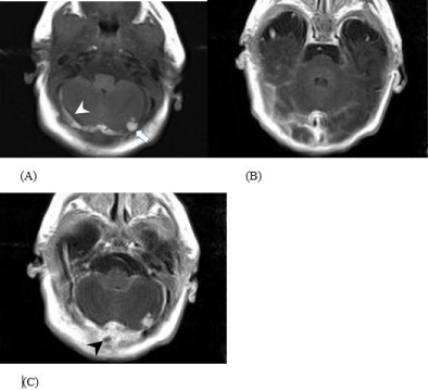
Magnetic Resonance Imaging (MRI) of the brain revealed multiple cerebral sinovenous thrombosis in the bilateral transverse sinuses and the straight sinus, with confluence of the sinuses. Additionally, intraparenchymal hemorrhages in the right posterior periventricular white matter and the left cerebellum were identified (Figure 2). Disseminated intravascular coagulation panel showed a D-dimer level of 955 ng/mL (normal range <500 ng/mL), fibrin degradation products of 10–40 μg/dL (normal range < 10 μg/dL), and fibrinogen of 555 mg/dL (normal range = 125-300 mg/dL). The coagulation testing results, including activated partial thromboplastin time, prothrombin time, levels of coagulation inhibitors (antithrombin III, protein S, and protein C), and antiphospholipid antibodies, were within normal limits. Screening for hyperhomocysteinemia was negative. Anticoagulation therapy (low molecular weight heparin) was prescribed immediately.

    Figure 2: Intraparenchymal hemorrhages in the right posterior periventricular
white matter and the left cerebellum.
Five months later, a follow-up brain MRI scan displayed a freely flowing cerebral sinovenous system without any sequalae of sinovenous thrombosis. Clinically, the infant appeared well and seizure-free after anticonvulsant and anticoagulation therapy. No other neurological problems were observed. Moreover, on assessment, her development was normal.
Discussion
Sinovenous thrombosis of the brain is a serious disease, and correct diagnosis is often delayed due to its subtle presentations. Most reported neonatal cases are in term neonates1 and rarely in preterm neonates. Cases of severe, multiple neonatal cerebral sinovenous thrombosis with intraparenchymal hemorrhage are few [11-14].
Immature physiological hemostatic systems are profoundly influenced by age. In this study, intrapartum maternal fever was associated with a number of apparently transient adverse effects in the newborn [9,10]. Despite the lack of a definite infectious source from her mother and the patient, intrapartum maternal fever and neonatal fever were recorded, which feasibly contributed to an unstable hemodynamic status and unbalanced hemostasis, leading to the deep sinovenous system to be prone to bleeding and clotting and intraparenchymal hemorrhage within the left cerebellum [15].
Moreover, intraventricular hemorrhage in sinovenous thrombosis has rarely been addressed. The possible explanation is that the deep venous system drains the choroidal, atrial, and thalamostriate veins; once deep venous blood clot formation is observed in the sinovneous system, this can lead to increased venous pressure and disturb the venous return and therefore predispose the patient to major bleeding along the caudothalamic groove, eventually resulting in intravenous hemorrhage, as observed in our case.
The role of thrombophilia in sinovenous thrombosis in the brain in the neonatal period and childhood has attracted huge attention. Prothrombotic disorders are found in 20% of cases of neonatal cerebral sinovenous thrombosis [1]. An analysis of the multifactorial origin of cerebral sinovenous thrombosis in children indicates that most thrombosis resulted from underlying prothrombotic risk factors, such as factor V Leiden deficiency and deficiency of proteins C and S. Genetic mutations such as those of factor V Leiden and prothrombin G20210A are common in whites but still undocumented among Taiwanese or Chinese populations [17,18].
Cranial Doppler ultrasound might provide an initial assessment of infants suspected of having superior sagittal sinus thrombosis [19]. However, in this case, multiple deep sinovenous thrombosis and intraparenchymal hemorrhage were involved, and the locations of lesions let the cranial Doppler ultrasound having limited usefulness. Furthermore, the highly echogenic tentorium and cerebellar vermis make localization of intracerebellar hemorrhage difficult [20,21]. By contrast, a brain MRI scan can not only delineate the extent and location of neonatal brain parenchymal changes and sinovenous thrombosis but also analyze the stages of brain intraparenchymal hemorrhage and the phases of sinovenous thrombosis [22].
In conclusion, if the neonate has any accompanying risk factors, either with or without fetal distress or clinical symptoms, physicians should be cautious of the effects of sinovenous thrombosis. Early MRI is strongly recommended if sinovenous thrombosis is suspected because this approach can better identify the presence, size, and location of lesions and provide early intensive treatment with favorable outcomes.
Ethics Statement
This patient and her parents provided all of the clinical and laboratory information and samples and agreed to the case publication.
Consent to Publish
Written informed consent was obtained from the patient for publication of this case report and any accompanying images.
Conflict of Interest Statement
The authors declare that the research was conducted in the absence of any commercial or financial relationships that could be construed as a potential conflict of interest.
Acknowledgments
This work was supported by the Taoyuan Armed Forces General Hospital, Taiwan, Grant AFTGH 10732 and Grant AFTGH 10829 to AC Wang.
References
- DeVeber G, Andrew M, Adams C, Bjornson B, Booth F, Buckley DJ, et al. Cerebral Sinovenous Thrombosis In Children New England Journal Of Medicine. 2001; 345: 417-23.
- Karen SC, Bodensteiner JB, Connolly PJ, and Bhuwan P. Garg Cerebral Venous Thrombosis In Children Journal Of Child Neurology. 2001; 16: 574- 80.
- Hanigan WC, Rossi LJ, McLean JM, and Wright RM. MRI Of Cerebral Vein Thrombosis In Infancy: A Case Report Neurology. 1986; 36: 1354.
- Yukuo K, Kuriyama M, Sudo M, Konishi K, Hayakawa K, and Ishii Y. Superior Sagittal Sinus Thrombosis In Neonates Pediatric Neurology. 1987; 3: 222-25.
- Richard M, and Neumann A. Neonatal Purpura Fulminans Due To Homozygous Protein C or Protein S Deficiencies. Seminars In Thrombosis And Hemostasis. 1990; 16: 299-309.
- Seligsohn U, and Lubetsky A. Genetic Susceptibility To Venous Thrombosis New England Journal Of Medicine. 2001; 344: 1222-31.
- de Klerk OL, de Vries TW, and Sinnige LGF. An Unusual Cause Of Neonatal Seizures In A Newborn Infant. PEDIATRICS. 1997; 100: 8.
- Yvonne WW, Hamrick SEG, Miller SP, Haward MF, Lai MC, Callen PW, et al. Ferriero Intraventricular Hemorrhage In Term Neonates Caused By Sinovenous Thrombosis Annals Of Neurology. 2003; 54: 123-6.
- Ellice L, Lang J, Richardson DK, Frigoletto FD, Heffner LJ, and Cohen A. Intrapartum Maternal Fever And Neonatal Outcome. Pediatrics. 2000; 105:8- 13.
- Hisayo OM, Glaser B, Niemann WH, and James LS. Increased Uterine Activity And Fetal Deterioration During Maternal Hyperthermia. American Journal Of Obstetrics And Gynecology. 1975; 121: 531-38.
- Wu YW, Miller SP, K Chin, Collins AE, Lomeli SC, Chuang NA, et al. Multiple Risk Factors In Neonatal Sinovenous Thrombosis Neurology. 2002; 59: 438- 40.
- Orville TB, and George M Hass Dural Sinus Thrombosis In Early Life The Journal Of Pediatrics. 1937; 11: 755-71.
- Ramenghi LA, Gill BJ, Tanner SF, Martinez D, Arthur R, and Levene MI. Cerebral Venous Thrombosis, Intraventricular Haemorrhage And White Matter Lesions In A Preterm Newborn With Factor V (Leiden) Mutation Neuropediatrics. 2002; 33: 97-9.
- Clode N, Gouveia R, and Graça LM. Prenatal Diagnosis Of Thrombosis Of The Dural Sinuses International Journal Of Gynecology & Obstetrics. 2005; 91: 172-4.
- Michael DW, Chalmers EA, and Gibson BES. The Investigation And Management Of Neonatal Haemostasis And Thrombosis British Journal Of Haematology. 2002; 119: 295-309.
- Limperopoulos C. Cerebellar Hemorrhage In The Preterm Infant: Ultrasonographic Findings And Risk Factors PEDIATRICS. 2005; 116 : 717- 24.
- Sebire G. Cerebral Venous Sinus Thrombosis In Children: Risk Factors, Presentation, Diagnosis And Outcome Brain. 2005; 128: 477-89.
- Chen TY, Su WC, and Tsao CJ. Incidence Of Thrombophilia Detected In Southern Taiwanese Patients With Venous Thrombosis Annals Of Hematology. 2003; 82 : 114-17.
- Lam AH. Doppler Imaging Of Superior Sagittal Sinus Thrombosis Journal Of Ultrasound In Medicine. 1995; 14: 41-6.
- Serfontein CL, Rom S and Stein S. Posterior Fossa Subdural Hemorrhage In The Newborn Pediatrics. 1980; 65: 40-3.
- Reeder JD, Setzer ES and Kaude JV. Ultrasonographic detection of perinatal intracerebellar hemorrhage. Pediatrics. 1982; 70: 385-86.
- Connor SEJ, and Jarosz JM. Magnetic Resonance Imaging Of Cerebral Venous Sinus Thrombosis. Clinical Radiology. 2002; 57: 449-61.