Research Article
Phys Med Rehabil Int. 2024; 11(2): 1228.
Surgical Treatment of Chronic Boutonniere Deformity of Fingers
Rasha Yossery Saleh, MD*
Department of Orthopedic, Faculty of Medicine, Menoufia University, Egypt
*Corresponding author: Rasha Yossery Saleh Professor of orthopedics, Faculty of Medicine, Menoufia University, 32511, Egypt. Tel: + 201003961071 Email: drnagwan80@gmail.com
Received: March 05, 2024 Accepted: April 11, 2024 Published: April 18, 2024
Abstract
Background: Boutonniere deformity is a result of injury of the central slip of the extensor tendon associated with volar migration of the lateral bands on both sides. Patients had deformity and change of function of joint resulting from over- extension of distal interphalangeal joint. We aimed to assess the surgical release of chronic boutonniere deformity using open dorsal release.
Methods: Sixty patients with 60 trauma-flexed deformed fingers were prospectively evaluated and managed by releasing the extensor tendon up to the oblique retinacular ligament insertion and elevating the lateral bands dorsal to proximal interphalangeal joint and tightening the central slip of extensor tendon. All fingers had no open injury. All patients were followed up from 9 to 18 months.
Results: Preoperatively Proximal Interphalangeal joint (PIP) extension lag was 70 degree and postoperatively improved to 8-degree, preoperative Distal Interphalangeal Joint (DIP) motion was 15degree of hyperextension, post-operative, DIP active flexion was 70 degrees. At the last follow-up showing 55fingers (91.67%) had excellent hand grip and Total Active Motion score (TAM), 3 (5%) had good and 2 had fair result (3.33%).
Conclusion: Open dorsal release showed excellent results. The extensor tendon freely mobile and act very well and the DIP joint had good flexion motion and this technique was simple and had long-time of good results.
Keywords: Boutonniere Deformity; Extensor Tendon; Lateral Bands.
Introduction
The loss of normal flexion function mechanism of proximal interphalangeal joint, leading to flexion deformity of proximal interphalangeal joint also injury of central band associated with volar migration of lateral band on both sides [1,2]. So no balance between flexor and extensor function of joints [3-5]. The muscles of hand transfer their function to the lateral bands leading to hyperextension of the distal interphalangeal joint [6-8].
Volar migration of the lateral bands and shorting of oblique retinacular ligament makes the proximal interphalangeal joint in flexed position and the distal interphalangeal joint in extended position [9,10]. Acute deformity and early injury were improved by splinting and physiotherapy, if failed need surgical repair of central band and immobilization for three weeks with physiotherapy holding PIP joint in full extension against resistance with flexion of DIP joint [11,12].
The best choice of treatment is according to state of all joints structures as bone integrity and tendon function [13,14]. The stiff flexed joint had another treatment as release soft tissue’s structure and release of capsule and joint [13,14]. Several methods to improve chronic flexion deformity as substitution of the central slip by the lateral band’s tendon graft, tenotomy of the central slip by smith and one lateral slip is mobilized and detached distally and repair of the central slip [14,15]. This study demonstrated the outcome of surgical release of the extensor expansion performed proximal to insertion of the oblique retinacular ligaments with dorsal lifting of the lateral bands and tightening with central band.
Patients and Methods
Patients
This was a prospective study; sixty patients with 60 fingers had boutonniere deformity and thirty males and thirty females. The affected deformed fingers were thirty middle, twenty indexes and ten rings. All fingers had intact skin with chronic in jury of extensor expansion. The mean of age was 30 (range: 15- 48) years. The mean time laps from trauma to treatment were four months. The average follow-up period was 12 (range: 9-18 months). All cases were subjected to clinical and radiological examination and preoperative data were obtained from hospital records. In this study affected fingers had mobile joint and no arthritic changes. The non-traumatic causes of boutonniere deformity were excluded. Results were estimated by evaluation of range of motion of all affected joint. Strength of grip were measured by Sphygmomanometer (for both grip and pinch) and also total active motion score (TAM score) [16] shown in Supplementary Table 1.
    Studied    variables

    Pre-operative N    (%)

    Post-operative N    (%)

    Test

    P-value


    TAM (N=60)


    Poor

    10 (10.67%)

    0 (0.0 %)

    Mc Nemar

    0.001*


    Fair

    50 (83.33%)

    2 (3.33 %)

    X2


    Good

    0 (0.0%)

    3 (5.00 %)

    15.4


    Excellent

    0 (0.0%)

    55 (91.67 %)


    Hand grip (N=60)


    Poor

    10 (10.67%)

    0 (0.0 %)

    Mc Nemar

    0.002*


    Fair

    50 (33.33%)

    2 (3.33 %)

    X2


    Good

    0 (0.0%)

    3 (5.00 %)

    17.2


    Excellent

    0 (0.0%)

    55 (91.67 %)


    *Statistically significant
Table 1: Preoperative and postoperative of TAM and handgrip of affected fingers.
Methods
All patients were subjected to two maneuvers of physiotherapy; the first maneuver was active extension of proximal interphalangeal joint to stretch contracted volar structures and help lateral band go dorsally and the second maneuver was flexion of distal interphalangeal joint to regain original length of contracted structure as lateral band and ligaments. This maneuver should be done 2 weeks before open release and postoperative to obtain excellent result.
Methods of Treatment
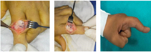
Surgical technique: By a posterior approach over the proximal interphalangeal joint and distal interphalangeal joints, the volar plate must expose to release the lateral bands, then the extensor expansion is divided to terminal part of the triangular ligament but before the insertion of the oblique retinacular ligament at the distal interphalangeal joint. The lateral bands were transferred dorsal to the axis of the proximal interphalangeal joint and tied to each other, tightening of central tendon with release of volar structure via the same dorsal incision showed in [Figure 1. 1(A),1(B),1(C) and 1(D)]. No K wire fixation is needed in this technique. Postoperatively, the (PIP) joint is splinted in extension for three weeks using a volar splint leaving the (DIP) joint free. Then, active range of motion exercises is begun for both joints showed in Figure 2 (A, B, C).

Figure 1: (A) Extension defect of PIP joint. (B) Showing division of the extensor expansion transversely proximal to the oblique retinacular ligament and division of the transverse retinacular ligament. (C): Showing division of the extensor expansion transversely proximal to the oblique retinacular ligament and division of the transverse retinacular ligament. (D): Intra-operative photography showing lateral bands are mobilized dorsally posterior to the axis of the (P.I.P) joint and sutured to each other, to the central tendon insertion, and the posterior capsule of the (P.I.P) joint.

Figure 2: (A): showing retinacular ligament. (B): Intra-operative photography shows a division of the extensor expansion transversely proximal to the oblique retinacular ligament. (C): Intra-operative photography shows that lateral bands are mobilized dorsally posterior to the axis of the (P.I.P) joint and sutured to each other, to the central tendon insertion, the posterior capsule of the (P.I.P) joint.
Methods of Evaluation
1-Total active motion score [16]
Total active motion = total active flexion (MCP joint +PIP +DIP) – total extension defect (MCP + PIP +DIP)
2-Hand grip: was evaluated as ratio (%) comparing with normal by using sphygmomanometer.
3- Range of motion: may be active or passive, it was assessed by the goniometer.
4- Total extension lag (deficit): it was assessed by the goniometer.
Statistical Analysis
Descriptive analyses as percentage (%), median, mean, range, t-test and Standard Deviation (SD) used to compare quantitative data, while qualitative data was asses by the Chi square test and also Mc Nemar X2 to compare between one group with multiple factors. A two-sided P value <0.05 was considered statistically significant.
Results
Sixty affected deformed fingers were treated by open posterior release. There were 30 males and 30 females. The deformity was posttraumatic. The mean age was 30 (15- 48 years), the mean duration of follow- up was 12 (range: 9-18 months) and the meantime interval between initial injury and surgery was four months. Two fingers had superficial infection were treated by strong antimicrobial agent. Two fingers had stiffness because patient was not obeyed order and was not wear splint. Grip was improved significantly (p 0:001) shown in (Table 1).
The mean of extension lag of PIP joint before surgery was 60 (range: 40 - 100) and improvement was 7 (range: 0-15) shown in (Table 2).
Affected finger (30)
Preoperative PIP joint extension lag
Postoperative joint extension lag PIP
Test
P-value
X+SD Range
8.34 + 13.6 (50-100)
4.1 + 8.1 (11-15)
# Paired test
1.890.01*
*Statistically significant # = Paired test
Table 2: Preoperative and Post-operative PIP joint extension lag affection of affected fingers.
Table 3 showed DIP range of motion before surgery was 10 (range: 7-15) of hyperextension. Post-surgery DIP active flexion was 75(range: 50-80).
The studied fingers
N = 30Test
P-value
DIP flexion
Preoperative N (%)
Postoperative N (%)
0-15 degree of flexion
(Poor grip)50 (83.33%)
0 (0.0 %)
Mc Nemar X2
19.30.003*
16-60 degree
(Good grip)10(16.67%)
5 (8.33%)
>60 degree (Excellent grip)
0 (0.0%)
55 (91.67%)
*Statistically significant
Table 3: Preoperative DIP flexion and postoperative improvement DIP joint flexion.
Patients were considered normal and had no any complain at 3 months after surgery. At the last visit at 12 months (range: 9-18 months), there were 55 (91.67%) excellent results, 3 (5.00%) good and 2 (3.33%) fairs because of the stiffness of both joints. There was statistically significant between hands grip improvement and gap between trauma and operation (Table 4).
Time-lapse
Pre-operative hand grip Degree (N)
Postoperative hand grip Degree (N)
Test
P-value
0-3 months
Fair
9
Poor
6
Excellent
40
Good
10
Mc
Nemar X2
18.30.002*
4-6 months
Fair
5
Poor
2
Excellent
55
Good
3
7- 12months
Fair
4
Poor
2
Excellent
55
Good
3
Fair
2
> 12months
Poor
2
Poor
2
*Statistically significant
Table 4: Handgrip according to the time lapse between original trauma and operation.
Discussion
The treatment of old neglected boutonniere deformity has poor result. The first line of treatment is non-operative treatment and physiotherapy for six weeks as by Souter [17] who showed that non-operative splint had 75% success result and 50% success in patients with operation.
In this study, at the final visit at 12 months (range: 9-18 months), there were 55 (91.67%) excellent results, 3 (5.00%) good and 2 (3.33%) fairs because of the stiffness of both PIP and DIP joints and postoperative mean of extension lag was 7 (range: 0-15) and post-surgery DIP active flexion was 75 (range: 50-80) and returned normal, had no any complain for 3 months post-surgery. Towfik et al 2005 showed 73% excellent results by central slip were reconstructed by palmaris tendon and physiotherapy was directed to extend PIP joint and flexion of DIP joint. Extensor tendon tenotomy was important to regain DIP joint flexion and cancelled action of tight lateral band [18].
Results of Le Bellec et al. 2001 reported that result of distal extensor tenotomy was reconstructed by resection of central band and relocation of lateral band posterior to joint in moderate flexion of PIP joint had 90% excellent and good results and poor results in 10% because in two patients not obeyed instruction as physiotherapy, in one patient because of septic osteoarthritis and in last finger due to suture was ruptured. Distal tenotomy was done to regain normal DIP joints motion was good in three affected finger (20-70) degrees [19].
Dubois, et al 2017 study had ten affected fingers with boutionniere deformity, postoperative result of release as following active range of DIP joint flexion was 80 degrees, and active extension lag was 8degrees (range 0-20) [20].
Lee JK, et al 2021study was reported that all patients presented with boutionniere deformity, preoperative parameter of joint, mean of extension deficit of PIP was (43.5°) was improved after surgery by 21.9° at the final visit (p < 0.001). The mean of swan neck deformity of the DIP joint before release was 19.2° and postoperatively improved by 0.8° flexion deformity (p < 0.001). The mean of total active motion was 220.4° (range, 160°-260°), only one finger had fair result and 23.1% had poor result. The average Strickland formula score was 70 (range, 28.6-97.1) [21].
In final follow-up, 10 fingers (77%) had excellent results, good in 2 fingers, fair result in 3 fingers and poor in one finger [21].Liu YJ, et al 2021 study had before surgery parameter of PIP joint extension lag was 48.0° ± 5.0° and after surgery parameter improved by 10.9° ± 9.3°. The DIP joint active flexion range before surgery was 34.4° ± 8.0°with postoperative result improvement was 71.4° ± 8.6° according to the Souter score were eleven excellent results, five good results and 2 poor s result [22].
Conclusion
In the current study, the result showed that this modified technique gave (83.33%) excellent, (10%) good, and 6.67% fair results. The extensor tendon acted mainly on the PIP joint and allowed the DIP joint to flex freely. The procedure is simple and provides long-term good results.
Author Statements
Institutional Review Board Statement
This research has been approved by the authors’ affiliated institutions.
Informed Consent Statement
Consent was obtained from all subjects were included in the research.
Data Availability Statement
The data presented in this research are available on request from the corresponding author.
Acknowledgments
The authors express their gratitude to all cases participated in the research.
Competing Interests
There is no conflict of the interests
Ethical Approval
This research was matched with the Menoufia University and Faculty of Medicine ethical standards.
References
- Elzinga K, Chung KC. Managing swan neck and Boutonniere def ormity. Clinic Plast Surg. 2019; 46: 329-37.
- Duzgun S, Duran A, Keskin E, Buyukogan H. Chronic Boutonniere deformity: cross-lateral band technique using Palmaris longus autograft. J Hand Surg Am. 2017; 42: e1-661.
- Khoo SL, Fernandes SV. Revisiting the Curtis procedure for boutonniere deformity correction. J Plast Surg. 2015; 4: 1800-4.
- Rogozinski J, Johnson RM. Vascularized tendon graft to the central slip using a finger fillet flap. Surg case Report. 2020: rjaa192.
- Haerle M, lotter O, Mertz I, Buschmeier N. The traumatic boutonniere deformity. Orthopade. 2008: 37: 1194-201.
- Frederick M, Azar S, Terry C, James H, Beat S. Operative technique of boutonniere deformity. Campbell’s operative Orthopedic –E- Book. 2016: 3671-6.
- Stauble DT, Kammer E, Wintsch K, Noever G, Operative treatment of the chronic post-traumatic boutonniere deformity by the technique of Pieper, Handchir Mikrochir Plast Chir. 2002; 34: 36-40.
- Burton RI. Extensor tendons-late reconstruction. In: Green DP, ed. Operative Hand Surgery. New York, NY: Churchill Livingstone. 1988: 2073-2116.
- Meadows SE, Schneider LH, Sherwyn JH. Treatment of the chronic Boutonniere deformity by extensor tenotomy. Hand Clin. 1995; 11: 441-7.
- Suzuki K. Reconstruction of post-traumatic boutonnière deformity. Hand. 1973; 5: 145-8.
- Urbaniak JR, Hayes MG. Chronic boutonnière deformity–an anatomic reconstruction. J Hand Surg. 1981; 6: 379-83.
- Caroli A, Zanasi S, Squarzina PB, Guerra M, Pancaldi G. Operative treatment of the post-traumatic boutonnière deformity: a modification of the direct anatomical technique repair J Hand Surg. 1990; 15: 410-5.
- Ohshio I, Ogino T, Minami A, Kato H. Reconstruction of the central slip by the transverse retinacular ligament for boutonnière deformity. J Hand Surg. 1990; 15: 407- 9.
- Massengill JB. The boutonniere deformity. Hand Clin. 1992; 8: 787-801.
- Curtis RM, Reid RL, Provost JM. A staged technique for the repair of the traumatic boutonniere deformiry. J Hand Surg Am. 1983; 8: 167-71.
- Hung LK, Wong JW, Yeung PC. Active mobilization after flexor tendon repair: comparison of results following injuries in zone 2 and other zones. J Orthop Surg. 2005; 13: 158-63.
- Souter WA. The problem of Boutonniere deformity. Clinc Orthop Relat Resch. 1974; 104: 116-33.
- Towfigh H, Gruber P. Surgical treatment of the Boutonniere deformity. Oper Orthop Traumatol. 2005; 17: 66-78.
- Le Bellec Y, Loy S, Touam C, Alnot JY, Masmejean E. Surgical treatment for boutonniere deformity of the fingers. Retrospective study of 47 patients. Chirurgie de la Main. 2001; 20: 362-7.
- Dubois E, Teboul F, Bihelt T, Gooubier JN. chronic boutonniere deformity, supple, or stiff: anew surgical technique with early mobilization in 11cases. Tech Hand Surg. 2017; 21: 37-40.
- Lee JK, Lee S, Kim M, Joscho JW, Hans H. Anatomic repair of central slip with anchor suture augmentation for treatment of established boutonniere deformity. Clin Ortho Surg. 2021; 13: 243-51.
- Liu YJ, Ding XH, Jiao HS, Ren SQ, Zhang HX. Y shaped tendon graft –A technique in the reconstruction of post-traumatic chronic Boutionniere. J Hand Surg Am. 2021; 46: 712.e1-712.e6.