
Research Article
Phys Med Rehabil Int. 2024; 11(4): 1237.
Effects of Radial Extracorporeal Shockwave Therapy on Knee Osteoarthritis Based on Soft Tissue Surgery Theory: A Multicenter, Randomized, Double-Blind, Placebo-Controlled Clinical Study
Wang YH1,2; Wang SH1; Liu RG1*
1Fuzhou University Affiliated Provincial Hospital, Department of Pain Management, Fujian Provincial Hospital, China
2Department of Anesthesiology, Gansu Provincial Hospital, PR China
*Corresponding author: Liu RG, Fuzhou University Affiliated Provincial Hospital, Department of Pain Management, Fujian Provincial Hospital, Fuzhou 350001, Fujian Province, China. Tel: 13706988238 Email: rgfw88@sina.com
Received: August 05, 2024 Accepted: September 03, 2024 Published: September 11, 2024
Abstract
Objectives: To investigate the effects of the radial extracorporeal shockwave therapy (rESWT) on the patients with Knee Osteoarthritis (KOA) and explore whether improvement of rESWT plus treatment sites based on the soft tissue surgery theory is superior to that of conventional rESWT for KOA.
Methods: 240 patients were randomly and equally divided into control group (C group), conventional treatment group (CT group), and modified treatment group (MT group). Patients in three groups were separately treated with sham rESWT, conventional rESWT, and rESWT plus treatment sites based on the soft tissue surgery theory once a week for 4 weeks. The outcomes were evaluated with the Numerical Rating Scale (NRS), Western Ontario and McMaster Universities Osteoarthritis index (WOMAC), and a 36-item short-form health survey (SF-36).
Results: Overall analysis, every outcome among the three groups showed statistical differences (P < 0.05). Compared with those of the MT group, the NRS of the other two groups were higher (P < 0.05). Compared with the C group, the NRS and WOMAC total scores in the other two groups were lower, while the scores of SF-36 were higher (P < 0.05). The WOMAC pain score in the MT group was lower than that in the CT group at the 3rd and 6th months after treatment (P < 0.05).
Conclusions: rESWT could effectively relieve pain and improve the knee function and living quality of KOA patients. According to the theory, soft tissue surgery can further enhance the therapeutic efficacy for KOA patients.
Keywords: Knee osteoarthritis; Extracorporeal shock wave therapy; Theory of soft tissue surgery
Introduction
Osteoarthritis (OA) is a kind of joint degenerative disease that can seriously affect patients' quality of life, and knee osteoarthritis (KOA) is the most common clinical type in middle-aged and elderly people. The prevalence of KOA is about 8.1%, and it is higher in women (10.3%) than in men (5.7%). The high incidence age of patients is 40-75 years old, and the incidence rate gradually increases with age [1-3]. KOA is a musculoskeletal disease that is often manifested as pain, deformity, and dysfunction of the knee joint. KOA can even be as disabling as diabetes, which significantly reduces the patient's quality of life [4]. With the combined effects of aging and an increasing obesity population, the prevalence of KOA is further increasing, placing a large and increasing burden on affected individuals, the healthcare system, and the socioeconomic costs of treatment [3]. Chronic pain and limited function in activities of daily living frequently plague patients with KOA, many patients are unable to perform their jobs due to pain. Therefore, the main goal of KOA management is to control pain, improve joint function, and ultimately improve the quality of life [5]. At present, non-invasive treatment methods are highly recommended and considered as a first-line treatment for KOA [6]. On the one hand, the organ function of the elderly is impaired, and their tolerance to surgical treatment is significantly decreased. For these patients, more conservative treatment methods are often used [7-9]. Physical therapy, on the other hand, is thought to have fewer side effects than non-steroidal anti-inflammatory drugs and intra-articular injections and to be more beneficial than exercise therapy in reducing joint pain and improving joint function [10].
Radial extracorporeal shock wave therapy (rESWT) is a non-invasive physical therapy technique emerging in recent years. rESWT has the advantages of being noninvasive, with few complications, and no need for hospitalization. rESWT is favored for the treatment of soft tissue pain due to its favorable anti-inflammatory effect [11]. Several studies [12,13] described the positive effects of rESWT in knee OA. Nevertheless, in one prior clinical trial [14], rESWT was not efficient for managing patients with disabling pain as a result of primary knee OA. How to improve the efficacy of rESWT in treating KOA and maintain long-term improvement has been a growing concern, with the focus mainly on energy, frequency, treatment cycle, and so on [15]. However, the study on adjusting the treatment site of rESWT to improve efficacy is rarely mentioned.
In the past few years, according to the new understanding proposed in the soft tissue surgery theory created by Dr. Xuan Zheren, we used rESWT by increasing the treatment sites such as the thigh root and the lateral hip region to seek an elevated efficacy. However, our preliminary study has some shortcomings, such as a lack of a blind method, one single center, and a too small sample size. Here, we conduct this multicenter, prospective, double-blind randomized controlled trial to further validate its efficacy.
Methods
Trail Design
This double-blind, randomized, controlled trial with a parallel group design was conducted domestically at 3 medical centers in China. Patient enrollment took place from April 2021 to July 2022.
Participants
The initial sample size calculation was based on the primary efficacy outcome, defined as the difference between the modified rESWT and the conventional rESWT measured by the change from baseline to 6 months in the Western Ontario and McMaster Universities osteoarthritis indexs (WOMAC) score. Assuming the significance level of 5%, the test power of 80%, and the loss of follow-up rate of 10%. Using these parameters, we calculated that we needed a minimum 79 participants in each group by PASS 11 [16,17]. A total of 240 patients with KOA were randomly assigned to receive each treatment group with the use of a computer-generated random list. Among them, there were 74 males and 166 females, aged 62.10 ± 10.56 years, course of disease 7.84 ± 6.00 months, unilateral lesions in 149 patients, and bilateral lesions in 91 patients. Prior to the start of the trial, all patients gave their written informed consent to participate in the study.
Inclusion Criteria
Eligible patients were men and women with over a 6-month history of symptoms of knee osteoarthritis. Participants with clinical knee osteoarthritis were diagnosed by rehabilitation physicians in accordance with the 2018 Chinese Guidelines for the Diagnosis and Treatment of Osteoarthritis [18]. In this research, presentable knee OA patients, defined as Kellgren-Lawrence classification grade I-III, were included [19]. Patients had knee pain on most days of Numerical Rating Scales (NRS) > 4 on their worst knee over the past month. The side with more severe symptoms was selected as the target knee in patients with bilateral knee osteoarthritis. When the symptoms of the 2 knees were similar, the right knee was selected as the target knee for evaluation. Moreover, there are highly sensitive tenderness points at the attachment of the adductor muscles and hip abductor in the patient.
Exclusion Criteria
Key exclusion criteria included the previous joint replacement, a history of joint injection, physical therapy in the last month, joint infection, tumor, or any major concomitant diseases that could interfere with participation in the trial. Participants with a history of diagnosis of significant neurologic or psychiatric impairments would be excluded in view of their difficulty in objectively answering the questionnaire.
Interventions
240 subjects were randomly divided into the control group (Group C), the convention treatment group (Group CT), and the modified treatment group (Group MT). 80 cases were engaged in each group. The following three treatment sites were selected, including the area around the knee joint (Site 1), the root of the thigh (Site 2), and the lateral hip area (Site 3). In Group C, the three sites were treated with placebo therapy. The D15 probe was used at site 1, and the D20 probe was used at sites 2 and 3. The parameters of therapy included a total of 4000 pulses of 10 Hz frequency at 5.0 bars of pneumatic pressure (prevent blind method failure, only increase the probe vibration sensation, and no impact energy into the treatment area). The first 2000 pulses were distributed to site 1. The remaining 2000 pulses are evenly divided into two parts, impacting site 2 and site 3, respectively. In Group CT, only Site 1 was treated with shockwave therapy, and Site 2 and Site 3 were treated with placebo therapy. For the treatment of Site 1, the D15 probe was selected, and the treatment handle mounted with the bullet was used to select the impact energy of 1.5 bar and the impact frequency of 10 Hz, totaling 2000 pulses. For treatment of Site 2 and Site 3, the D20 probe was used, and the bullet was taken out for placebo treatment; the treatment energy was set at 5.0 bar; the impact frequency was 10 Hz; 1000 pulses for each site, once a week, four times as a course of treatment. In Group MT, the D15 probe was used in part 1, and the D20 probe is used in parts 2 and 3. The impact probe installed with the bullet was used, and the impact frequency was selected as 10 Hz. Impact energy of 1.5 bar was set for Site 1 (2000 pulses) and Site 2 (1000 pulses), while 2.0 bar was set for Site 3 (1000 pulses). The treatment cycle was once a week for 4 weeks in all groups. No anesthetic drugs or sedatives were used, and the adverse reactions of patients were observed and recorded.
Blind Method
Each research unit consisted of an extracorporeal shock wave therapy device (STORZ MP-100, Switzerland), subjects, shock wave operators, device debuggers, and data managers. The subjects were randomly assigned to the corresponding group according to the established experimental protocol. During the treatment process, both the operator and the subject wore soundproof earplugs (Ohrfrienden, Germany) and noise-reducing earphones (Boss, Germany) to prevent different sounds from affecting the implementation of the blinding method. The device debuggers were responsible for replacing the shock wave probes, loading bullets, and setting parameters without the knowledge of the operator and subject. To prevent parameter leakage, it was necessary to cover the instrument screen with an opaque cloth after debugging. The data managers responsible for data collection and analysis were not involved in the treatment process and could not distinguish the groups of subjects when evaluating the results. After all results statistics were completed, the blind could be unblinded. After the unblinded process, the control group was given four times of supplementary therapy.
Outcome Measures
All patients were evaluated by an investigator who was blind to the group allocation and data acquisition at baseline, 1 week, 1 month, 3 months, and 6 months after intervention, respectively. The NRS and WOMAC were recorded for each study unit before treatment and at 1 week, 1 month, 3 months, and 6 months after treatment. 36-item Short-Form health survey (SF-36) scores were recorded before treatment and at 1, 3, and 6 months after treatment. Knee pain intensity was evaluated with NRS, which consists of 11 numbers with the same interval between 0 and 10, with marking endpoints as no pain and worst possible pain. WOMAC was a three-part questionnaire, which consisted of 24 questions and probed clinically important symptoms in the areas of pain, stiffness, and physical function for patients with OA of the knee. SF-36 was used for evaluating the quality of life, including the physical component and mental component.
For the subjects with NRS 4 in a resting state, they were encouraged to take celecoxib for pain relief (specification: 200 mg / tablet). For subjects taking painkillers, they need to stop the medication for one day before evaluation, and the amount of painkillers should be recorded. Adverse events such as dizziness, palpitations, chest tightness, local ecchymosis, and hematoma were recorded. The dosage of analgesics and adverse events were noticed closely throughout the entire trial process.
Statistical Analysis
IBM SPSS Statistics 23 statistical software was used for statistical analysis. The measurement data of continuous normal distribution were expressed as mean ± standard deviation (SDX±), while the measurement data of non-normal distribution were expressed as median (M) and interquartile distance (IQR). Analysis of variance (ANOVA) was used for comparison of continuous variables. Repeated measure analysis of variance was used to compare the results of NRS, WOMAC total score and SF-36 scale before and after treatment. Kruskal-Wallis rank sum test was used to compare WOMAC pain score, WOMAC stiffness score, and WOMAC activity function score among three groups. WOMAC pain score, WOMAC stiffness score, and WOMAC activity function score were compared within the group by Friedman test.
Results
Characteristics of the Patients
Figure 1. shows the flow of enrollment in the study. A total of 240 patients with knee osteoarthritis were included in this study, of which 29 dropped out during follow-up period. The general characteristics of the patients in each group are displayed in table 1. There are no statistical differences concerning age, gender, duration of complaint, lesion side, body mass index, NRS, WOMAC score and SF-36 score among the three groups.
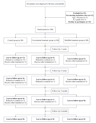
Figure 1: Recruitment and flow of participants through the trial.
![]()
    Characteristic
    MT group (n=76)
    CT group (n=75)
    C group (n=60)
    P Value


    Age, y
    62.75±10.65
    61.84±10.49
    60.80±10.14
    0.558


    Male gender, %
    18.42%
    30.67%
    25.00%
    0.217


    Body mass index, kg/m2
    23.34±2.54
    23.36±2.69
    23.48±2.08
    0.941


    Duration of complaints,    m
    7.79±6.26
    8.33±6.24
    8.03±6.57
    0.870


    Unilateral pain, %
    61.84%
    66.67%
    65.00%
    0.821


    NRS
    5.30±0.88
    5.13±0.91
    5.05±0.87
    0.234


    WOMAC


    Total points
    30.00±13.39
    28.61±13.06
    29.18±7.19
    0.770


    Pain score
    6.11±2.83
    5.67±3.28
    5.50±2.21
    0.428


    Stiffness score
    2.42±1.84
    2.39±1.79
    2.58±1.23
    0.755


    Activity function score
    21.47±10.82
    20.56±9.84
    21.10±5.70
    0.831


    SF-36


    PCS
    50.03±11.67
    52.17±14.46
    54.13±10.84
    0.166


    MCS
    60.36±15.86
    64.44±13.89
    65.81±11.91
    0.061


    Note: Data are given    as mean ± SD unless otherwise indicated.
Table 1: General data of the three groups of patients.
Outcomes
NRS
The results of the repeated measures ANOVA demonstrated significant differences in NRS between patients in the MT group, CT group and the C group (P < 0.001). Additionally, significant differences were observed at each time point (P < 0.001), and a cross-over effect was identified between the groups and time (P < 0.001). Three groups experienced a decrease in NRS, but the decrease was more pronounced in the MT group. The NRS in CT group showed an upward trend from the 1st month to the 6th month after the treatment, indicating that the pain may recur. The NRS in C group at all time points after treatment were worse than those in MT group and CT group (all P < 0.001). NRS scores in MT group were better than those in CT group at 3 and 6 months after treatment (all P < 0.001) (Figure 2).
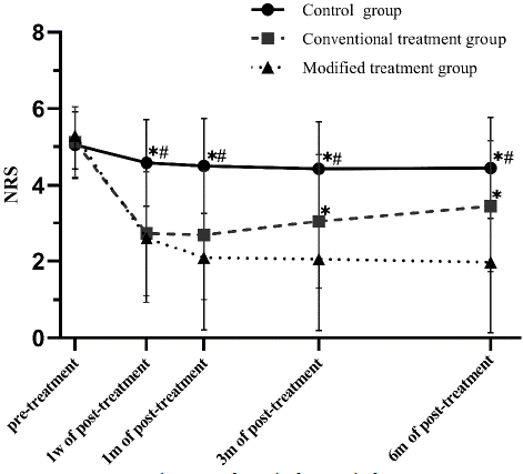
    Figure 2: Variation diagram of NRS before and after treatment (Score, SDX±).
*P<0.05, NRS compared with the modified treatment group; #P<0.05, NRS compared with the conventional treatment group.
WOMAC total Scores
The results of the repeated measures ANOVA demonstrated significant differences in WOMAC total scores between patients in the MT group, CT group and the C group (P < 0.001). Additionally, significant differences were observed at each time point (P < 0.001), and a cross-over effect was identified between the groups and time (P < 0.001). Three groups experienced a decrease in WOMAC total scores, but the decrease was more pronounced in the MT group. There were significant differences of the WOMAC total scores at 6th month after treatment among the three groups, however, those in MT group were superior to in CT group (P < 0.001) (Figure 3).
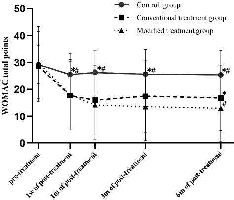
    Figure 3: Variation diagram of the total points of WOMAC before and after treatment (Score, SDX±).
*P<0.05, WOMAC scores compared with the modified treatment group; #P<0.05, WOMAC scores compared with conventional treatment group.
WOMAC Pain Scores
In CT group and MT group, there were significant differences in WOMAC pain scores at all time points (all P< 0.001), and the WOMAC pain scores after treatment were significantly lower than those before treatment. In MT group, the WOMAC pain score after treatment was significantly lower than that in the 1st week after treatment, and the difference was statistically significant (1st month of post-treatment P = 0.018, 3rd month of post-treatment P = 0.001, 6th month of post-treatment P < 0.001). WOMAC pain scores in the MT group were lower than those in the CT group at 3rd month and 6th month after the end of treatment, with statistical significance (3rd of post-treatment P = 0.027, 6th of post-treatment P = 0.019) (Figure 4A).
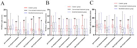
Figure 4: A. Comparison of WOMAC pain scores assessment among three groups of patients after treatment, B. Comparison of WOMAC stiffness scores assessment among three groups of patients after treatment, C. Comparison of WOMAC activity function scores assessment among three groups of patients after treatment (*P<0.05, WOMAC scores compared with the modified treatment group; #P<0.05, WOMAC scores compared with conventional treatment group).
WOMAC Stiffness Scores
In Group CT and Group MT, there were significant differences in WOMAC stiffness scores at all time points (all P < 0.001), and the WOMAC stiffness scores after treatment were significantly lower than those before treatment, with statistical significance (all P < 0.001).
The WOMAC stiffness scores of the C group was higher than that of the MT group at all time points after treatment, and the difference was statistically significant (all P < 0.001) (Figure 4B).
WOMAC Activity Function Scores
In Group CT and Group MT, there were significant differences in WOMAC activity function scores at all time points (all P < 0.001), and the WOMAC activity function scores after treatment were significantly lower than those before treatment, with statistical significance (all P < 0.001). In Group MT, the WOMAC activity function score after treatment was significantly lower than that in the 1st week after treatment, and the difference was statistically significant (1st month of post-treatment P = 0.038, 3rd month of post-treatment P = 0.032, 6th month of post-treatment P = 0.001) (Figure 4C).
Physical Component Summary (PCS)
The results of the repeated measures ANOVA demonstrated significant differences in PCS between patients in the MT group, CT group and the C group (P = 0.001). Additionally, significant differences were observed at each time point (P < 0.001), and a cross-over effect was identified between the groups and time (P < 0.001). The overall level of PCS in the MT group and CT group was higher than that in the C group after treatment. Three groups experienced a rise in PCS, but the rise was more pronounced in the MT group and CT group. The PCS scores of C group at all time points after treatment were worse than those of MT group and CT group, and the differences were statistically significant (all P < 0.001). The PCS at the 6th month after treatment, there were significant differences among the three groups, and the PCS in MT group were better than those in CT group (P < 0.001) (Figure 5A).
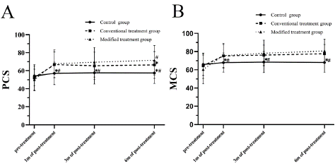
Figure 5: A. Variation diagram of the PCS scores of SF-36 scale before and after treatment (Score, SDX±). B. Variation diagram of the MCS scores of SF-36 scale before and after treatment (Score, SDX±). (*P<0.05, SF-36 scores compared with modified treatment group; #P<0.05, SF-36 scale compared with conventional treatment group).
Mental Component Summary (MCS)
The results of the repeated measures ANOVA demonstrated significant differences in MCS between patients in the MT group, CT group and the C group (P = 0.003). Additionally, significant differences were observed at each time point (P < 0.001), and a cross-over effect was identified between the groups and time (P < 0.001). The overall level of MCS in the MT group and CT group was higher than that in the C group after treatment. Three groups experienced a rise in MCS, but the rise was more pronounced in the MT group and CT group. The MCS scores of C group at the 3th and 6th month after treatment were worse than those of MT group and CT group, and the differences were statistically significant (all P < 0.001) (Figure 5B).
Dosage
In the MT group, the dose was celecoxib capsule 0 (0, 0) (mg), and the total dose was celecoxib capsule (0.2g) 24×200mg. In the CT group, the dose was celecoxib capsule 0 (0, 0) (mg), and the total dose was celecoxib capsule (0.2g) 34×200mg. In the C group, the average dose was celecoxib capsule 1 (1, 3) (mg), and the total dose was celecoxib capsule (0.2g) 107×200mg. the C group used more celecoxib capsule than the other two groups (P<0.001).
Adverse Reactions
There were no obvious adverse reactions and complications in the three groups.
Discussion
In this trial, rESWT was found to be an effective and safe modality for improving pain and decreasing disability in patients diagnosed with moderate knee OA. To our knowledge, this research first demonstrated the application of rESWT based on the soft tissue surgery theory could further improve the efficacy of rESWT for KOA.
The theory of soft tissue surgery, which was proposed by Professor Xuan ZhR in the 1980s, has been increasingly approved in China and guides the physicians to treat the musculoskeletal pain. Its core idea is that the pain is caused by aseptic inflammation and the pain improves by means of relaxation. The theory indicated that the aseptic inflammation was caused by the damage of the skeletal muscles, fascia, ligaments, joint capsules, synovium, adipose tissue outside the spinal canal, and adipose tissue inside the spinal canal in the human body. The theory indicates that the knee joint cartilage lacks innervation, and the wear and degeneration of cartilage is not the cause of joint pain, while aseptic inflammation of surrounding soft tissue plays an important role. This academic thought was also revealed in the report of Bokhari S [20]. At present, most studies are limited to the inflammation caused by tendon and ligament injuries around the joints, just like the CT group set up in this study. In addition, most research focused on the treatment energy, frequency, and disease staging, whereas research on the treatment site selection was rarely found. In the theory of soft tissue surgery, it is proposed that knee joint pain is usually related to the downward transmission of sterile inflammation in the thigh root and outer side of the hip. So, the root of the thigh and the lateral hip area were chosen to be treated with rESWT for further KOA improvement. The detection of tenderness points indicates the presence of sterile inflammation, and tenderness points or pain trigger points are increasingly being valued in pain diagnosis and treatment. In this study, attachment sites of the tensor fasciae, latae muscle, and adductor were highly sensitive to tenderness. A randomized single-blind study [21] demonstrated that strengthening the lateral and adductor muscles of the hip could improve knee joint load, pain, and function. In summary, increasing the lateral and adductor muscles of the hip and targeted treatment for tenderness points play an important role in improving KOA.
rESWT, as a non-invasive treatment method, not only owns the anti-inflammatory, releasing, and improving soft tissue blood supply effects on muscles with contracture injuries but also takes a certain repair effect. Based on the theory of soft tissue surgery, we have explored the use of rESWT to treat knee osteoarthritis and have achieved some encouraging results only in our unit. Now, more rigorous and scientific clinical trials are needed to reverify these results and popularize this new therapy method.
Due to the unique nature of shock wave therapy, it is almost impossible for therapists and patients to be double blind. So, in previous studies, the main measures were to reduce shock wave energy, fake therapy, or installing spacers in front of the treatment probe to reduce the deviation during the experimental process [22-24]. In order to ensure the success of the blind method, we added device debugging personnel to prevent the results of changing treatment parameters from being known by the therapist and used soundproof earphones to prevent different noises generated by handles without bullet heads during the treatment process. Finally, after blind testing, it was found to be effective. To our knowledge, this blind method that balances visual and auditory aspects has not yet been designed by the previous researchers and may serve as a reference for future shock wave therapy research.
At present, there is no consensus on the long-term efficacy of rESWT in treating KOA. Our research has shown that in most patients, a 4-week course of ESWT is superior to placebo in relieving pain and maintains a 6-month benefit trend. The repeated measurement analysis of variance for NRS, WOMAC total score, and SF-36 scale scores showed that the MT and CT groups at all time points after treatment were significantly improved than those before treatment (all P < 0.05), while the MT and CT groups had an advantage over the C group at all same time points after treatment (all P < 0.05). The above results proved that rESWT could alleviate the joint pain, reduce the joint stiffness, improve the joint function, and enhance the patient's quality of life, whether it is the local treatment or the holistic treatment based on the soft tissue surgery theory.
In addition, the NRS scores of the MT group were generally less than those of the CT group after treatment (all P < 0.05). It is worth mentioning that the NRS scores of the CT group showed a minor increase after 3 months of treatment. We considered that due to conduction of soft tissue damage in the hip and thigh roots, spasm of the soft tissue around the knee reoccurred, thereby resulting in the mild rebound of NRS scores at three months after treatment. At 3 months of post-treatment, the WOMAC pain scores in the MT group were prior to showing better than those in the CT group. This finding indicates that the pain reduction is a more sensitive indicator of efficacy in knee arthritis patients compared to the functional improvement. Like some non-drug treatment studies [25], both PCS and MCS improved in the ESWT group after treatment. Although there were no significant differences in MCS between the MT and CT groups at 6 months after treatment, significant differences may occur over a longer follow-up period based on the slope in the line graph. SF-36 has been widely used as an evaluation indicator for knee arthritis, but it is seldomly used to evaluate the efficacy of ESWT in treating knee arthritis. Chronic pain commonly takes a certain impact on the mental health of the subjects, and the poor mental health deteriorates the perception of pain of the subjects. The treatment of psychological problems plays an important role in pain improvement. The total psychological scores of the SF-36 scale can be used to quantify the mental and mental health problems caused by KOA and to evaluate the therapeutic effects from the mental dimension [26]. Based on the results of our study, this questionnaire scale is suitable for use here and is worth promoting. Similar to the abnormal situation of NRS, the total scores of WOMAC in the CT group showed an upward trend from 1 month after treatment to 3 months after treatment, while PCS showed a downward trend. However, the total scores of WOMAC in the MT group showed a downward trend at various time periods after treatment; the scores of PCS indicated an upward trend. At 1, 3, and 6 months after treatment, the WOMAC pain scores and WOMAC activity function scores further decreased. These results indicate that in the short term after treatment there are no significant differences of efficacy between the CT group and the MT group, but the long-term efficacy of the MT group is superior to that of the CT group. This finding is basically consistent with our preliminary experimental results [27]. For most KOA patients (excluding those with only tenderness points around the knee joint), if the treatment is limited to the knee joint but does not address the sterile inflammation in the primary lesion areas such as the lateral hip area and adductor muscle group, the long-lasting improvement may not be guaranteed. No serious adverse reactions or side effects occurred in the three groups except for minor local bruising in some patients during treatment, which proves that rESWT is safe and reliable and could be introduced for outpatient treatment.
Limitation
Patients in the control group only received the basic treatment such as health education, weight control, functional exercise, and remedial medication for pain, while during the rESWT treatment a treatment handle with the removed bullet was unable to produce a practical therapeutic effect. This resulted in poor treatment effectiveness for the control group patients, due to the higher numbers of the lost visits and excluded patients compared to the other two groups. Lack of objective indicators with high sensitivity evaluates the change in KOA. Our data covered only 6 months, and the sustained effects for longer duration remain unknown.
Conclusion
This study verified that rESWT for KOA was safe and effective. rESWT could not only relieve pain but also improve knee function and the life quality of patients. Based on the theory of soft tissue surgery, increasing treatment sites could further improve the therapeutic effects of rESWT.
Author Statements
Funding
This work was funded by the Fujian Provincial Department of Science and Technology (no. 2021Y0049).
Ethical Approval and Informed Consent Statements
This study was approved by the Medical Ethics Committee (Ethics number: K2021-04-015) and pre-registration was completed at the Chinese Clinical Trial Registry (registration number: ChiCTR2100052609).
References
- Tang X, Wang S, Zhan S, Niu J, Tao K, Zhang Y, et al. The Prevalence of Symptomatic Knee Osteoarthritis in China: Results from the China Health and Retirement Longitudinal Study. Arthritis & rheumatology (Hoboken, N.J.). 2016; 68: 648-653.
- Prieto-Alhambra D, Judge A, Javaid M, Cooper C, Diez-Perez A, Arden NK. Incidence and risk factors for clinically diagnosed knee, hip and hand osteoarthritis: influences of age, gender and osteoarthritis affecting other joints. Annals of the rheumatic diseases. 2014; 73: 1659-1664.
- Hunter D. Bierma-Zeinstra S. Osteoarthritis. Lancet (London, England). 2019; 393: 1745-1759.
- Hunter D, Schofield D, Callander E. The individual and socioeconomic impact of osteoarthritis. Nature reviews. Rheumatology. 2014; 10: 437-441.
- Ji Q, Wang P, He C. Extracorporeal shockwave therapy as a novel and potential treatment for degenerative cartilage and bone disease: Osteoarthritis. A qualitative analysis of the literature. Progress in biophysics and molecular biology. 2016; 121: 255-265.
- Nelson A, Allen K, Golightly Y, Goode AP, Jordan JM. A systematic review of recommendations and guidelines for the management of osteoarthritis: The chronic osteoarthritis management initiative of the U.S. bone and joint initiative. Seminars in arthritis and rheumatism. 2014; 43: 701-712.
- Matzkin E, Curry E, Kong Q, Rogers MJ, Henry M, Smith EL. Efficacy and Treatment Response of Intra-articular Corticosteroid Injections in Patients with Symptomatic Knee Osteoarthritis. The Journal of the American Academy of Orthopaedic Surgeons. 2017; 25: 703-714.
- Dhawan A, Mather R, Karas V, Ellman MB, Young BB, Bach BR, et al. An epidemiologic analysis of clinical practice guidelines for non-arthroplasty treatment of osteoarthritis of the knee. Arthroscopy: the journal of arthroscopic & related surgery: official publication of the Arthroscopy Association of North America and the International Arthroscopy Association. 2014; 30: 65-71.
- Fransen M, McConnell S, Harmer A, der Esch MV, Simic M, Bennell KL. Exercise for osteoarthritis of the knee: a Cochrane systematic review. British journal of sports medicine. 2015; 49: 1554-1557.
- Deyle G, Allen C, Allison S, Gill NW, Hando BR, Petersen EJ, et al. Physical Therapy versus Glucocorticoid Injection for Osteoarthritis of the Knee. The New England journal of medicine. 2020; 382: 1420-1429.
- Ko N, Chang C, Cheng C, Yu HK, Hu GC. Comparative Effectiveness of Focused Extracorporeal versus Radial Extracorporeal Shockwave Therapy for Knee Osteoarthritis-Randomized Controlled Study. International journal of environmental research and public health. 2022; 19: 9001.
- Ho K, Yang C, Lo H, Yeh HJ. Extracorporeal Shockwave Therapy with a Modified Technique on Tendon and Ligament for Knee Osteoarthritis: A Randomized Controlled Trial. American journal of physical medicine & rehabilitation. 2022; 101: 11-17.
- Liu Y, Wu C, Chen C, Zhang L, Xing G, Wu K, et al. Impact of soft tissue around the knee on the efficacy of extracorporeal shockwave therapy in knee osteoarthritis. Medicine. 2022; 101: e32334.
- Imamura M, Alamino S, Hsing W, Alfieri Marcon F, Schmitz C, Battistella LR, et al. Radial extracorporeal shock wave therapy for disabling pain due to severe primary knee osteoarthritis. Journal of rehabilitation medicine. 2017; 49: 54-62.
- Liao C, Tsauo J, Liou T, et al. Clinical efficacy of extracorporeal shockwave therapy for knee osteoarthritis: a systematic review and meta-regression of randomized controlled trials. Clinical rehabilitation. 2019; 33: 1419-1430.
- MM S. Efficacy of extracorporeal shock wave therapy versus mobilization with movement on pain, disability and range of motion in patients with knee osteoarthritis. Bull Fac Ph Th Cairo Univ. 2013; 18: 65-74.
- SS ES. The effect of shock wave therapy as a new modality for treatment of primary knee osteoarthritis. Egypt J Hosp Med. 2019; 75: 2092-2097.
- Joint Surgery Group, Orthopedics Branch, Chinese Medical Association Guidelines for Diagnosis and Treatment of Osteoarthritis (2018 Edition). Chin J Orthop. 2018; 38: 705-715.
- Li W, Xiao Z, Liu J. Deep learning-assisted knee osteoarthritis automatic grading on plain radiographs: the value of multiview X-ray images and prior knowledge. Quantitative imaging in medicine and surgery. 2023; 13: 3587-3601.
- Bokhari S. Tendonitis: the major cause of pain in osteoarthritis knee joint. Journal of Ayub Medical College, Abbottabad: JAMC. 2012; 24: 109-112.
- Bennell K, Hunt M, Wrigley T, Hunter DJ, Hinman RS. The effects of hip muscle strengthening on knee load, pain, and function in people with knee osteoarthritis: a protocol for a randomised, single-blind controlled trial. BMC musculoskeletal disorders. 2007; 8: 121.
- Zhang Y, Liu Y, Chou S, Weng H. Dose-related effects of radial extracorporeal shock wave therapy for knee osteoarthritis: A randomized controlled trial. Journal of rehabilitation medicine. 2021; 53: jrm00144.
- Kudo P, Dainty K, Clarfield M, Coughlin L, Lavoie P, Lebrun C. Randomized, placebo-controlled, double-blind clinical trial evaluating the treatment of plantar fasciitis with an extracoporeal shockwave therapy (ESWT) device: a North American confirmatory study. Journal of orthopaedic research: official publication of the Orthopaedic Research Society. 2006; 24: 115-123.
- Liu S, Wang L, Yang J. Instant analgesic effect of radial extracorporeal shock wave therapy on primary dysmenorrhoea according to functional magnetic resonance imaging: study protocol for a randomised placebo-controlled trial. Trials. 2020; 21: 164.
- Vaghela N, Mishra D, Patel J, Dani V. Promoting health and quality of life of patients with osteoarthritis of knee joint through non-pharmacological treatment strategies: A randomized controlled trial. Journal of education and health promotion. 2020; 9: 156.
- Robinson M, Edwards S, Iyengar S, Bymaster F, Clark M, Katon W, et al. Depression and pain. Frontiers in bioscience (Landmark edition). 2009; 14: 5031-5051.
- Li Yangmin, Wang Shuai, Zhang Aijun. Observation on the therapeutic effect of using divergent extracorporeal shock wave therapy for knee osteoarthritis following the theory of soft tissue surgery. Chinese Journal of Pain Science. 2022; 18: 807-813.