Case Report
Phys Med Rehabil Int. 2024; 11(5): 1242.
Rehabilitation Complications in An International Case of Dermatomyositis with Calcinosis Universalis
Khamisani A¹; Lemay D²; Thornton M³; Pontee N¹*
¹University of Miami/Jackson Memorial Hospital, USA
²University of South Florida Morsani College of Medicine, USA
³University of Miami Miller School of Medicine, USA
*Corresponding author: Pontee, N, MD, MS, Department of Physical Medicine and Rehabilitation, University of Miami, 1611 NW 12th Avenue, Miami, FL 33136, USA. Tel: (305)243-4567; Fax: (305)243-4650 Email: nlpontee@med.miami.edu
Received: October 10, 2024; Accepted: October 30, 2024 Published: November 06, 2024
Abstract
Dermatomyositis (DM) is a rare and intricate autoimmune disorder characterized by symmetric proximal muscle weakness and a spectrum of associated cutaneous manifestations. Approximately 20-25% of patients have the anti-Nuclear Matrix Protein 2 (NXP2) gene that codes for the anti-NXP2 autoantibody, which can create a nearly linear relationship between the onset of DM and calcinosis. A 21-year-old female patient from the Caribbean with aggressive dermatomyositis (NXP-2 antibody positive) was brought to a tertiary care hospital for impending respiratory failure and was subsequently admitted to an acute Inpatient Rehabilitation Facility (IRF), where her condition was complicated by calcinosis universalis. The patient was treated with calcium channel blockers, sodium thiosulfate injections, shockwave lithotripsy, and aggressive physical therapy to address her calcinosis-related pain and functional restriction. A multidisciplinary rehabilitation approach with various interventions was implemented, yielding improvement in functional results. The international component of this case added complexity, as it involved challenges related to geographic distance, care communication, and the availability of specialized care. This case provided a unique opportunity to manage the progression of a rare, complex disease.
Keywords: Dermatomyositis; Calcinosis Universalis; Rehabilitation; Functional
Case Presentation
A 21-year-old female with known NXP-2 Dermatomyositis (DM) presented to a US-based tertiary care hospital from the Caribbean for impending respiratory failure due to respiratory muscle weakness secondary to an acute dermatomyositis flare requiring intubation. She also received pulse methylprednisolone 220 mg intravenously daily, intravenous immunoglobulin for five days and five rounds of plasmapheresis as directed by Rheumatology. Her course was complicated by failure to wean from intubation due to bulbar weakness and difficulty with secretion management. Percutaneous gastrojejunostomy tube was placed for alimentation. Of note, one year prior, she was hospitalized at the same facility for 3 months for a similar clinical presentation and discharged to her home in the Caribbean but inadequate resources and follow up lead to her second flare.
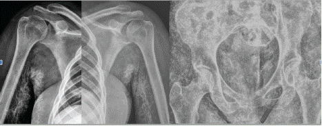
After clinical stabilization, she was admitted to an Inpatient Rehab Facility (IRF). The patient’s main complaints were weakness and pain around the shoulders and hips, dysphonia, and dysphagia. She was noted to have 2/5 strength in her lower extremities bilaterally and 3/5 strength in her upper extremities bilaterally,. Passive range of motion of her major joints induced pain and revealed severe restriction with abduction to about 20 degrees bilaterally and 30 degrees flexion bilaterally at the shoulders. She was maximum assist with bed mobility and sit to stands and could not ambulate. She had multiple, diffuse subcutaneous nodules seen on x-ray in all extremities bilaterally consistent with calcinosis universalis (Figure 1).

    Figure 1: (a) Right shoulder x-ray with extensive reticular calcifications
throughout bilateral axilla and soft tissue of arms/forearms. (b) Left shoulder
x-ray with extensive reticular calcifications throughout bilateral axilla and soft
tissue of arms/forearms. (c) Bilateral hip x-ray with diffuse innumerable soft
tissue calcifications indicating calcinosis cutis universalis.
She was placed on a calcium channel blocker, Amlodipine and administered intralesional sodium thiosulfate injections at 250mg/mL in the right upper extremity with a total of 9cc injected into the right posterior arm for her calcinosis. Shockwave lithotripsy was deferred due to lack of availability of subsequent therapy in her home country. She was also started on Pamidronate at 1mg/kg/day for 3 days every 3 months. Her rehab course was complicated by PEG tube malfunction leading to gastric perforation and delayed wound healing necessitating the initiation and continuation of Total Parenteral Nutrition (TPN).

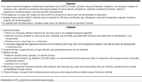
Table 1: Recommendations for the Management of Calcinosis Cutis Associated With ACTD [6].
By the end of her rehab stay, the patient had advanced to contact guard assist for sit to stand from wheelchair to rolling walker and was ambulatory with minimum assistance with a rolling walker. Prior to discharge her abduction in the right arm improved to 90 degrees and her left arm improved to 120 degrees. Flexion improved bilaterally to about 80 degrees bilaterally. Her pain reduced from 8/10 bilaterally to a 5 in her right arm and a 4 in her left.. No appreciable difference seen on repeat radiographs 3 weeks after injections Additionally, the patient was provided with day and night time use ankle foot orthoses for skin breakdown protection contracture prevention. She started Plaquenil 400 mg daily and Rituximab infusions during IRF stay per Rheumatology and Neurology consulting services jointly.
The inpatient physiatry team was integral to the effective coordination of care for the patient with the multiple subspecialty teams involved with her case including Neurology, Rheumatology, and her primary care provider. A home regimen of slow steroid taper with Prednisone 40mg with Trimethoprim/Sulfamethoxazole prophylaxis was ordered and the plan for monthly Intravenous Immunoglobulin (IVIG) infusions in the Caribbean was discussed with the patient's primary rheumatologist in her home country. The plan was to continue her care in the Caribbean with her primary care provider and to follow up at the American tertiary care center for further treatments and follow up with the subspecialty teams.
Discussion
In this case, we describe the clinical presentation and rehabilitation challenges of a patient with DM, including the classical myositis associated weakness, international status of the patient making care coordination difficult, and, most notably, severe soft tissue calcinosis. In patients with DM, the return of muscle strength and degree of disability following disease can be quite variable. One study observed that among patients with myositis, only 65% of surviving patients had normal strength and only 34% no or slight disability at the mean follow-up of 5 years, with no prognostic differences between myositis subtypes [1]. Thus, the rehabilitation of these patients can be a long and arduous process, especially when hindered by the cumulative effects of multiple past flares and other disease related complications, including a difficult nutritional status due to severe dysphagia and poor wound healing necessitating alternate alimentation.
Myositis-related calcinosis can cause significant pain, as in our patient, and restriction of range of motion and discomfort during therapies secondary to this extensive calcinosis posed significant roadblocks to the patient’s rehabilitation course. Such widespread calcinosis surrounding joints and limiting range of motion has been described in the literature, and several subtypes have been described based on structure and location of the calcific deposits [2]. Some patients may exhibit limited superficial calcinosis in a lace-like or nodular pattern that does not interfere with function, while the more severe side of the calcinosis spectrum can involve extensive sheetlike deposits that can be found even deeper into myofascial planes, which was likely the case for the patient we describe here. Our patient experienced calcinosis universalis, which presents a unique challenge by itself seeing that it has no established treatment plan for calcinosis other than treating the underlying disease [3].
Unfortunately, there is no gold standard treatment for calcinosis cutis to date. Patients with asymptomatic limited calcinosis may need no pharmacologic intervention at all [4]. The potential treatment options include medication and non-medication-based interventions as well as surgical and procedural approaches. In general, close attention should be paid to ensure that affected areas have adequate blood flow, and local trauma should be avoided as it can serve as a trigger for further calcinosis [4]. Additionally, warm salt water soaks may provide pain relief and assist in extrusion of the calcific deposits [4].
Surgical removal is an option for some patients, but our patient was not a good candidate for this intervention due to the widespread nature of the calcinosis. Other procedural interventions such as extracorporeal shock wave therapy has been studied with some patients benefiting in pain reduction and sonographic evidence of calcinosis regression, although the sample size was quite small [5].
Several medications have been studied in the treatment of calcinosis cutis. The calcium-channel blocker diltiazem is thought to exert therapeutic effects on calcinosis by reducing intracellular calcium influx in affected tissue [4]. One of the larger studies to date that looked at treatment of calcinosis cutis with diltiazem, 9 of 17 patients showed at least partial response to the medication [3]. As a relatively cheap medication that has more supporting data than many other medications used to treat calcinosis cutis, this is a common initial medication used for treatment. Bisphosphonates are theorized to work in calcinosis cutis by reducing bone turnover and inhibiting inflammatory cascades in macrophages [4]. One study in patients with juvenile subtype dermatomyositis showed calcinosis resolution in response to bisphosphonate therapy combined with aggressive immunosuppressive therapy in 4 of 6 patients [6]. Sodium thiosulfate, postulated to act by making calcium deposits more soluble, can be administered topically, intravenously, or intralesionally. Most data are currently limited to case series and retrospective reviews. One study showed clinical improvement in 5 of 5 patients who received intralesional sodium thiosulfate as determined by ultrasound guidance and pain assessment over several months [7].
Conclusion
This case of a 21-year-old female patient with aggressive (DM) and NXP-2 antibody positivity presented many challenges including respiratory failure, soft tissue calcinosis, and international healthcare coordination difficulties. The management of advanced calcinosis universalis presented significant rehabilitation barriers. The existing treatment options are based primarily on case series and retrospective studies with a small number of patients, highlighting the need for more definitive treatment options. The patient’s international status and complex presentation necessitated a multidisciplinary and international collaboration in order to transition care safely and optimize the patient’s outcome. Further research is required for treatment of dermatomyositis and its complications.
References
- Bronner IM, van der Meulen MFG, de Visser M, Kalmijn S, van Venrooij WJ, Voskuyl AE, et al. Long-term outcome in polymyositis and dermatomyositis. Ann Rheum Dis. 2006; 65: 1456-1461.
- Boulman N, Slobodin G, Rozenbaum M, Rosner I. Calcinosis in rheumatic diseases. Semin Arthritis Rheum. 2005; 34: 805-812.
- Balin SJ, Wetter DA, Andersen LK, Davis MDP. Calcinosis cutis occurring in association with autoimmune connective tissue disease: the Mayo Clinic experience with 78 patients, 1996-2009. Archives of dermatology. 2012; 148: 455–462.
- Elahmar H, Feldman BM, Johnson SR. Management of Calcinosis Cutis in Rheumatic Diseases. J Rheumatol. 2022; 49: 980-989.
- Blumhardt S, Frey DP, Toniolo M, Alkadhi H, Held U, Distler O. Safety and efficacy of extracorporeal shock wave therapy (ESWT) in calcinosis cutis associated with systemic sclerosis. Clin Exp Rheumatol. 2016; 34: 177-180.
- Tayfur AC, Topaloglu R, Gulhan B, Bilginer Y. Bisphosphonates in juvenile dermatomyositis with dystrophic calcinosis. Mod Rheumatol. 2015; 25: 615- 620.
- Tubau C, Cubiró X, Amat-Samaranch V, Garcia-Melendo C, Puig L, Roé- Crespo E. Clinical and ultrasonography follow-up of five cases of calcinosis cutis successfully treated with intralesional sodium thiosulfate. J Ultrasound. 2022; 25: 995-1003.