Case Report
Austin J Pulm Respir Med 2014;1(4): 1017.
Spontaneous Partial Resolution of a Giant Pulmonary Bulla
Ryland P Byrd Jr* and Thomas M Roy
Pulmonary and Critical Care Medicine Division, East Tennessee State University, USA
*Corresponding author: Ryland P Byrd Jr, Pulmonary and Critical Care Medicine Division, Quillen College of Medicine, East Tennessee State University, Veterans Affairs Medical Center 111-B, PO. Box. 4000, Mountain Home, TN 37684-4000, USA
Received: February 06, 2014; Accepted: July 31, 2014; Published: Aug 08, 2014
Abstract
Spontaneous partial resolution of giant pulmonary bullae occurs infrequently. The pathophysiology responsible for the natural elimination of giant bullae is not known with certainty. We report a patient who experienced spontaneous sub-total regression of his giant bulla following an infection. This observation suggests that airway inflammation and obstruction may play a role in the mechanism for spontaneous resolution and/or regression of giant bullae.
Keywords: Giant bulla; Spontaneous resolution; Pulmonary
Background
Bullous emphysema is a common consequence of the inhalation of combusted tobacco products. Multiple small bullae typically develop as a consequence of smoking tobacco products. The development of giant bullae is uncommon. Giant bullae typically progressively enlarge and, over time, cause compressive atelectasis of the adjacent pulmonary parenchyma [1,2]. Patients may experience increasing respiratory compromise as the giant bullae increase in size. Giant bullae rarely resolve spontaneously. Eleven cases of complete resolution and six cases with partial regression of giant bullae are recorded in the English literature [3-15]. We report a patient who experienced a sub-total resolution of a giant bulla following an infectious episode.
Case Presentation
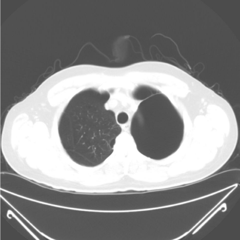
A 60 year-old male was referred for evaluation of abnormal computerized tomogram scanning (CT) of the chest. A CT two years earlier demonstrated a giant bulla in his left upper lobe (Figure 1).
Figure 1: Chest tomogram demonstrating a giant bulla of the left upper lobe prior to infection.
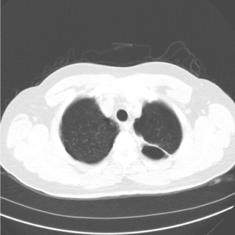
The CT at referral documented an air-fluid level in the giant bulla (Figure 2) and a small infiltrate proximal to the bulla. He had a cough productive of yellow sputum but he was no more short of breath than usual. The patient had a low-grade fever but otherwise his vital signs were normal. There were diffuse end-expiratory wheezes present bilaterally. Breath sounds were diminished in the left upper lobe. His white blood cell count was slightly elevated. He was prescribed oral antibiotics and continued on maximal inhaled therapy for chronic obstructive pulmonary disease (COPD). His medications included a scheduled inhaled long-acting beta-agonist, a long-acting anti-cholinergic agent, and corticosteroid. He use a short-acting inhaled beta-agonist as a rescue medication. An outpatient flexible fiber-optic bronchoscopy was scheduled. This procedure was performed without difficulty. No endobronchial lesion was identified.
Figure 2: Chest tomogram demonstrating an air-fluid level in the giant bulla of the left upper lobe.
The patient was known to have severe COPD. He had acquired this disorder due to his habit of inhaling the smoke from combusted tobacco products. He had a 35 pack-year history of cigarette smoking. His forced expiratory volume in one second (FEV1.0) was 1.18 liters or 36% of his predicted. His residual volume (RV) was 126% of predicted. He had undergone a resection of a giant bulla in his right upper lobe 15 years earlier. He had quit smoking cigarettes at that time. His alpha-1 anti-trypsin levels had been measured as normal previously.
The patient was followed regularly. The air-fluid level in the left upper lobe giant bulla resolved completely. In addition, the giant bulla in his left upper lobe regressed remarkably in size. This observation can best be appreciated in a CT scan obtained two years after the infectious episode (Figure 3). While he reported no change in his symptom of shortness of breath, his FEV1.0 and RV improved slightly to 1.49 liters or 40% of predicted and the residual volume decreased to 112% of predicted. There was nothing in the patient’s history or radiographs to suggest that he had experienced a pneumothorax that might explain the regression of his giant bulla.
Figure 3: Chest tomogram demonstrating sub-total regression of the left upper lobe giant bulla.
Discussion
By definition a giant pulmonary bulla occupies at least one third of the involved hemi-thorax. In smokers, giant bullae typically occur in the upper lobes and develop more often in men. Cigarette smoking is the leading cause of giant bullae. The pathophysiologic mechanism that results in the enlargement of the bulla is not well understood. However, the most widely held hypothesis is that giant bullae result from dilation of the airspaces distal to the terminal bronchioles due to a ball-valve effect in the more proximal airways. This check-valve phenomena causes increasing positive end expiratory pressures within the bullae promoting their gradual expansion [1,2,16].
Spontaneous reduction in size of a giant bulla occurs infrequently. Eleven cases of complete spontaneous resolution of giant bullae have been reported in the English medical literature (Table 1) [3-8,13-15]. Six cases of partial spontaneous regression of giant bullae have also been reported (Table 2) [8-11]. Our patient represents the seventh patient with a sub-total spontaneous resolution of a giant bulla. As with our patient, most of the other cases documented in the literature had bullae in their upper lobes.
    Gender
    Bulla    Location
    Reason for    Resolution/Regression
    Cigarette    Use
    Symptoms
    Pulmonary    Function Tests


    62 15
    Male
    LUL
    Intensification    of bronchodilator and anti-inflammatory therapy
    Current
    Improved
    Improved


    6414
    Male
    RUL
    Presumed    post-infectious
    Former
    Improved
    Normal


    553
    Male
    RUL
    Post-infectious
    Former
    Improved
    No change


    746
    Male
    LUL
    Post-infectious
    Current
    Improved
    NA


    475
    Male
    LUL
    Spontaneous    pneumothorax
    Former
    Improved
    Improved


    547
    Male
    Right
    Spontaneous    pneumothorax
    Current
    NA
    Improved


    704
    Male
    Right
    Benign    nodule
    Former
    Improved
    Improved


    598
    Male
    RUL
    Post-infectious
    NA
    NA
    NA


    57 8
    Male
    RUL
    Post-infectious
    NA
    NA
    NA


    Age
    Male
    RUL
    Post-infectious
    NA
    NA
    NA


    5713
    Male
    LUL
    NA
    NA
    NA
    NA
Table 1: Patient demographics complete resolution of giant bullae.
    Age (years)
    Gender
    Bulla    location
    Reason for    Resolution/Regression
    Cigarette    Use
    Symptoms
    Pulmonary    Function Tests


    60
    Male
    LUL
    Infection
    Former
    Stable
    Improved


    5 11
    Male
    RUL
    Intensification    of bronchodilator and anti-inflammatory therapy
    Current
    Improved
    Improved


    599
    Male
    LUL
    Adenocarcinoma
    Current
    NA
    NA


    7512
    Female
    LUL
    Presumed    spontaneous pneumothorax
    Current
    Improved
    No change


    6410
    Male
    RUL
    NA
    Non-smoker
    NA
    NA


    2410
    Male
    RUL
    NA
    Former
    NA
    NA


    2510
    Male
    RUL
    NA
    NA
    NA
    NA
Table 2: Patient demographics with partial regression of giant bullae.
Most of the patients described in the English literature with spontaneous resolution or regression of their giant bullae have been males (Table 1 & 2). Of the 18 cases reported, 17 were men. The only female has been reported experienced a partial regression of her bulla [12]. The greater use of tobacco products by men in the past is likely responsible for this observation rather than an actual gender bias. It is probably a reporting bias. Because women are more susceptible to the development of smoking-related COPD than men [16], it seems unlikely that women would be protected from the development of giant bullae due to their gender. As the percentage of females with COPD increases, giant bullae and the spontaneous resolution of bullae will undoubtedly be observed more frequently in women. In support of this notion, an elderly female with a partial spontaneous regression of a right upper lobe giant bulla was reported recently [12].
The pathophysiologic mechanism resulting in spontaneous resolution or regression of giant bullae is not well understood. Most commonly, spontaneous resolution and regression of the bullae have been attributed to an infectious process [3,6,8]. Five of the patients reported in the English medical literature had an air-fluid level within the giant bullae prior to the subsequent disappearance of the bullae. It is theorized that airway inflammation in association with the infected bullae results in closure of the communication between the airway and the bullae [8]. The gases within the now non-communicating space are slowly absorbed. The absorption of the gases results in loss of volume and ultimately the collapse of the giant bullae. Our patient’s history is consistent with this proposed mechanism.
An association between lung cancer and giant bullae is well established [17-21]. One patient reported in the literature had partial regression of a left upper lobe bulla due to obstruction of the airway from adenocarcinoma of the lung [9]. There is also a report of a patient whose giant bulla resolved due to obstruction of the communicating airway by a benign nodule [4]. Due to these observations, patients with spontaneous resolution or regression of giant bullae should undergo flexible fiber-optic bronchoscopy to visualize the airways and to rule out an obstructing endobronchial neoplasm. Flexible fiberoptic bronchoscopy in our patient failed to identify an endobronchial lesion.
Two patients have experienced complete spontaneous resolution of their giant bullae after spontaneous pneumothoraces. In each of these patients, it was the giant bullae that ruptured and caused the pneumothoraces [5,7]. Both patients were successfully treated with chest tube thoracostomy. Evacuation of the air from the pleural space resulted in re-expansion of the lung, without reappearance of the giant bullae. A spontaneous pneumothorax was suspected historically in a third patient with partial regression of her giant bulla [12]. It has been proposed that a check-valve mechanism allowed the pressures to increase in the giant bullae until they ruptured resulting in pneumothoraces. The airways in these patients likely closed the ball-valve segment causing the adjacent lung to re-expand without further leakage of gases into the pleural space. There was no reason to suspect that the giant bulla in our patient had ruptured or that a pneumothorax had developed.
There have been two reports of regression of a giant bullae following intensification of inhaled bronchodilator and anti-inflammatory medications. One patient experiences a partial regression of the giant bulla [11]. The other patient had complete resolution of his giant bulla [15]. Moreover, some of the patients reported in the literature had ceased smoking cigarettes prior to the resolution or regression of their giant bullae [3-6,10,14]. These observation suggests that the removal of the airway irritant of tobacco smoke and control of airway inflammation may have played a role in the resolution or regression of the giant bullae. Whether smoking cessation in these patients resulted in a decrease in airway inflammation and thereby relieved a check-valve or was coincidental is purely speculative. These observations, however, reinforce the concept that smoking cessation is an important health improvement measure in all patients, including those with giant bullae. Our patient had quit smoking 15 years prior to the infection is his bulla. In addition, he had been on maximal inhaled therapy, including inhaled corticosteroid, for several years prior to the regression of his giant bulla. It, therefore, seems less likely that these interventions played a role in the regression of his giant bulla.
Eight of the patients reported in the literature enjoyed a decrease in their symptoms of COPD following spontaneous resolution or regression of their giant bulla [3-6,11,12,14,15]. In addition, six patients had a documented improvement in their pulmonary function tests [4,5,7,11,15]. In four of the patient, pulmonary function tests improved dramatically after resolution or regression of their giant bullae [4,5,11,15]. As might be expected, each of these four patients also experienced improvement in their respiratory symptoms. Two of the patients improved symptomatically despite having no change in their measured pulmonary function [3,12]. Unfortunately, our patient’s dyspnea remained stable, despite a slight improvement in his measures of lung function. The reason his shortness of breath did not improve is probably a reflection of his severe COPD.
The natural history of a giant bulla is typically gradual enlargement over time. Giant bullae often compress normal lungs as they enlarge. Patients with giant bullae occupying 30-50% of the hemi-thorax who have atelectatic normal adjacent lung are often considered for bullectomy. The surgical resection of the giant bullae may allow for re-expansion of the compressed lung with subsequent improvement in symptoms and measures of lung function [22]. Spontaneous resolution and regression can be thought of as an auto-bullectomy and may offer the same advantage to the patients.
Conclusion
Spontaneous resolution and regression of giant bullae are unusual events. Most often the resolution and regression follows an infectious event. However, because of the association of lung cancer and giant bullae, the patient’s airways should be directly visualized to rule out a neoplasm obstructing the airway. The medical literature suggests that, at least in some patients, a reversible airway process may play a role in the formation of their bullae. Smoking cessation and intensification of inhaled medications should be instituted in all patients with giant bullae and may mitigate against the giant bullae. These relatively simple measures may avert the need for a surgical bullectomy.
References
- Stone DJ, Schwartz A, Feltman JA. Bullous emphysema. A long-term study of the natural history and the effects of therapy. Am Rev Respir Dis. 1960; 94: 493-507.
- Boushy SF, Kohen R, Billig DM, Heiman MJ. Bullous emphysema: clinical, roentgenologic and physiologic study of 49 patients. Dis Chest. 1968; 54: 327-334.
- Wahbi ZK, Arnold AG. Spontaneous closure of a large emphysematous bulla. Respir Med. 1995; 89: 377-379.
- Bradshaw DA, Murray KM, Amundson DE. Spontaneous regression of a giant pulmonary bulla. Thorax. 1996; 51: 549-550.
- Ridgeway NA, Ginn DR. Rupture and spontaneous resolution of a giant bulla with improvement in airways obstruction. Tenn Med. 1998; 91: 431-432.
- Millar EA, d'A Semple P. Spontaneous closure of a large emphysematous bulla. Respir Med. 1996; 90: 120-121.
- Satoh H, Suyama T, Yamashita YT, Ohtsuka M, Sekizawa K. Spontaneous regression of multiple emphysematous bullae. Can Respir J. 1999; 6: 458-460.
- DOUGLAS AC, GRANT IW. Spontaneous closure of large pulmonary bullae: a report on three cases. Br J Tuberc Dis Chest. 1957; 51: 335-338.
- Saito H, Okuno M. Spontaneous regression of a bulla with the development of adenocarcinoma of the lung. Intern Med. 1999; 38: 439-441.
- Orton DF, Gurney JW. Spontaneous reduction in size of bullae (autobullectomy). J Thorac Imaging. 1999; 14: 118-121.
- Park HY, Lim SY, Park HK, Park SY, Kim TS, Suh GY, et al. Regression of giant bullous emphysema. Intern Med. 2010; 49: 55-57.
- Scarlata S, Cesari M, Caridi I, Chiurco D, Antonelli-Incalzi R. Spontaneous resolution of a giant pulmonary bulla in an older woman: role of functional assessment. Respiration. 2011; 81: 59-62.
- Satoh H, Ishikawa H, Ohtsuka M, Sekizawa K. Spontaneous regression of pulmonary bullae. Australas Radiol. 2002; 46: 106-107.
- Shanthaveerappa HN, Mathai MG, Byrd RP Jr, Fields CL, Roy TM. Spontaneous resolution of a giant pulmonary bulla. J Ky Med Assoc. 2001; 99: 533-536.
- Byrd RP Jr, Roy TM. Spontaneous resolution of a giant pulmonary bulla: what is the role of bronchodilator and anti-inflammatory therapy? Tenn Med. 2013; 106: 39-42.
- Sørheim IC, Johannessen A, Gulsvik A, Bakke PS, Silverman EK, DeMeo DL, et al. Gender differences in COPD: are women more susceptible to smoking effects than men? Thorax. 2010; 65: 480-485.
- Stern EJ, Webb WR, Weinacker A, Müller NL. Idiopathic giant bullous emphysema (vanishing lung syndrome): imaging findings in nine patients. AJR Am J Roentgenol. 1994; 162: 279-282.
- Stoloff IL, Kanofsky P, Magilner L. The risk of lung cancer in males with bullous disease of the lung. Arch Environ Health. 1971; 22: 163-167.
- Goldstein MJ, Snider GL, Liberson M, Poske RM. Bronchogenic carcinoma and giant bullous disease. Am Rev Respir Dis. 1968; 97: 1062-1070.
- Venuta F, Rendina EA, Pescarmona EO, De Giacomo T, Vizza D, Flaishman I, et al. Occult lung cancer in patients with bullous emphysema. Thorax. 1997; 52: 289-290.
- Tsutsui M, Araki Y, Shirakusa T, Inutsuka S. Characteristic radiographic features of pulmonary carcinoma associated with large bulla. Ann Thorac Surg. 1988; 46: 679-683.
- Nickoladze GD. Functional results of surgery for bullous emphysema. Chest. 1992; 101: 119-122.


