
Case Report
Austin J Pulm Respir Med 2015; 2(4): 1036.
Bronchoscopy as “Treatment Adjunct” in Management of Endobronchial Tuberculosis: Case Report and Review of Literature
Upadhyay S*
Department of Pulmonary Medicine, Rama Medical College Hapur, India
*Corresponding author: Upadhyay Sushil, Department of Pulmonary Medicine, Rama Medical College Hapur, Ghaziabad, India
Received: November 21, 2015; Accepted: December 16, 2015; Published: December 31, 2015
Abstract
A case of young postnatal female with endobronchial tuberculosis leading to complete collapse of one lung is presented. The patient did not respond even after 2months of ATT until the lumen of left lung was recreated and supportive collection was drained by bronchoscopic intervention.
The case highlights the role of bronchoscopy in management of acute complication of active pulmonary tuberculosis with endobronchial component causing complete collapse of lung.
Keywords: Endobronchial tuberculosis; Bronchoscopy; Tracheobronchial tree; Bronchiectasis; Bronchostenosis
Introduction
Endobronchial Tuberculosis (EBTB) is defined as tuberculous infection of the tracheobronchial tree with microbial and histopathological evidence [1]. Endobronchial Tuberculosis occurs in about 10–40% of patients with active tuberculosis [2]. It is more common in pediatric population [3], young adults(less than 35 years old [4] and has predilection for women [5]).
Airway obstruction and lobar or even total lung collapse as complication of pulmonary tuberculosis is a known phenomenon. In such cases endoluminal infection is most commonly present although hilar lymphnodes may occasionally be implicated in causing extrinsic compression of bronchus [6].
Diagnosis of endobonchial tuberculosis is frequently difficult because of non specific symptoms and insensitivity of chest skiagram [7] and sputum examination [8]. Computerized tomography [9] and bronchoscopy [10] play a pivotal role and are complimentary to each other in solving the diagnostic dilemma as well as prognostication of endobronchial tuberculosis and defining the role of surgery in such cases.
Whereas, role of bronchoscopy in diagnosis of EBTB is undisputed, the role bronchoscopic intervention in treating obstructive complications of endobronchial tuberculosis is not well documented in literature.
Case Report
27 year old female presented with profound and persistent non productive cough, left sided chest discomfort, breathlessness on exertion and fever off and on. She had been referred by her primary care physician as she was not improving with ATT given for last 2months.She had been suffering for last 3months.she had delivered her first baby 2months back. Chest skiagram done a week after delivery was reported having left upper lobe infiltrates. On examination there was diminished breath sound on left side chest. She maintained spo2 of 94% on room air. Rroutine blood biochemistries were normal. Repeat CXR (Figure 1) showed opaque left hemithorax with ipsilateral mediastinal shift. CT thorax (Figure 2) confirmed complete collapse of left lung with cutoff of left main bronchus. In view of no response to ATT and progressive clinical and radiological worsening possibility of malignancy was considered. Bronchoscopy was advised for further evaluation. Fibreoptic bronchoscopy revealed (Figure 3) nodular and ulcerated mucosa from mid trachea to Left Main Bronchus (LMB). LMB lumen was completely occluded by white slough covering the underlying angry looking nodular, ulcerated and irregular mucosa. This being the reason of no ventilation in the left lung. Right Main Bronchus (RMB) and right bronchial tree was normal in appearance.
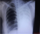
Figure 1: CXR PAV (at presentation).
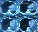
Figure 2: CT thorax (complete left lung collapse).
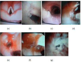
    Figure 3:  (a) Bronchoscopy; (b) Trachea; (c) LMB occluded; (d) Biopsy; (e)
LMB post biopsy; (f) Balloon dilatation; (g) LMB post dilatation.
The forceps was passed through bronchoscope and punch biopsies were taken. The granulation tissue was cautiously nibbled with the forceps. After some digging into the tissue a pinhole opening could be created. Balloon catheter was passed through this small opening and balloon was inflated multiple times. After several attempts of ballon dilatation the lumen could be widened a little. However bronchoscope could not be negotiated across this hole. Lot of pus like material was aspirated from left lung. At this point of time option of placing a stent was discussed with family if suggested by repeat skiagrams.
The bronchial washings were negative for AFB and aerobic culture was sterile. The biopsy revealed granuloma and caseation. Patient was continued with Antitubercular Treatment (ATT). Oral steroid was given along with nebulized streptomycin and budesonide BID. She perceived significant relief in cough and defervescence of fever was noted.
Chest X-ray repeated next day (Figure 4) of bronchoscopy showed partial aeration in left lung. She was discharged to home.
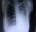
Figure 4: CXR day after FOB.
Repeat Chest X-ray at one month follow up (Figure 5) showed complete expansion of left lung. Steroid (oral and inhaled) was stopped as well as pyrazinamide (after 3months).
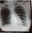
Figure 5: CXR 1month post FOB.
She completed the course of ATT given for nine months and is doing well post treatment.
Discussion
Endobronchial Tuberculosis (EBTB) is defined as tuberculous infection of the tracheobronchial tree with microbial and histopathological evidence. Morten [11] was the first to describe endobronchial tuberculosis in 1698.
Endobronchial tuberculosis is more common in pediatric population (41.7%-43% of active PTB) [12]. In adults, EBTB occurs in about 10–40% of patients with active tuberculosis [2]. More than half the cases of endobronchial TB occur in patients aged less than 35 years old. Fifteen percent of cases have been reported in elderly [13]. EBTB has predilection for young women [14] which is usually explained by easy implantation of organisms from infected sputum, since women do not generally expectorate sputum possibly because of their sociocultural factors. The present case is a young female who caught the illness during perinatal period.
Five potential mechanisms have been suggested for the development of endobronchial infection due to M. Tuberculosis [15]: (1) direct extension from adjacent parenchymal focus; (2) implantation of organisms from the infected sputum; (3) hematogenous dissemination; (4) lymph node erosion into the bronchus and (5) through lymphatic drainage from parenchyma to the peribronchial region.
EBTB usually starts as a submucosal tubercle those progresses to ulcer formation. Granulation tissue that is generally formed looks like polypoid mass. These ulcers heal by fibrosis. Bronchial stenosis and strictures are irreversible, relatively common and delayed complications of EBTB. Bronchostenosis may develop in 60% to 95% cases and may even involve the main stem bronchi. Bronchiectasis is also a common complication of EBTB and most commonly develops as paracicatricial process, secondary to pulmonary destruction and fibrosis (traction bronchiectasis). Bronchiectasis is typically asymptomatic and usually occurs in the upper lobes [16].
The degree of fibrostenosis depends on the depth of the ulcer [17]. In up to 90% cases, there is radiographic evidence of obstruction with segmental atelectasis, occurring most frequently in the right middle lobe and anterior segment of the right upper lobe [18]. This is notable that our patient had isolated involvement of left main bronchus starting from mid of trachea and right bronchial tree was completely spared.
In patients with complete lung collapse and non response to adequate trial of chemotherapy lung cancer is most important differential diagnosis especially in microbiologically unconfirmed cases. However, other differential diagnoses are simple inflammation, sarcoidosis, bronchial asthma, atelectasis, foreign body aspiration, pneumonia and endobronchial actinomycosis [19].
In the present case, with no symptomatic relief (viz. persistent cough, fever and breathlessness) even after two months of ATT bronchogenic cancer was thought of cancer as foremost alternative possibility and workup to rule out the same was initiated.
Bronchoscopy plays key role in the diagnosis of EBTB, producing more than 90 percent yield on smear as well as on culture. Even if biopsy fails to supply tangible results, the bronchoscopic changes, supported by clinical and radiological findings, may be sufficient to establish the diagnosis of EBTB [17].
A widely accepted classification defining EBTB by fibreoptic bronchoscopy has the following seven subtypes: (i) actively caseating (ii) edematous-hyperemic (iii) fibrostenotic (iv) tumorous (v) granular (vi) ulcerative and (vii) nonspecific bronchitis [20]. The present case had bronchoscopic features consistent with actively caseating and ulcerative type of EBTB.
CT scan is also a useful adjunct to direct endoscopic visualization. CT accurately depicts the bronchial abnormality in 93% to 100% cases. CT findings include [21] isolated segmental bronchial narrowing with concentric wall thickening (41%-43%), complete endobronchial obstruction (32%) and extrinsic obstruction by adjacent adenopathy (23% - 50%).
The prognosis of actively caseating type, edematous-hyperemic and tumorous type EBTB is grave, resulting in fibrostenosis in up to two thirds of patients. The prognosis is good for granular and nonspecific bronchitic type EBTB. Antituberculous chemotherapy is effective in controlling the infection, but does not prevent residual Bronchostenosis [5,14].
In edematous hyperemic EBTB, the bronchial lumen is constantly narrowed due to severe mucosal swelling with surrounding hyperemia, occasionally causing complete obstruction of the airway with lung collapse [10]. However our patient had actively caseating type of EBTB with complete occlusion of left main bronchus lumen preventing aeration and leading to complete collapse of left lung. This presentation is rare to find in literature.
Endoscopic treatment for this kind of obstruction is not welldocumented in the literature. In the present case, we first tried debridement of the occlusive caseous necrotic tissue with the forceps. Pin hole size opening was thus created. This was followed by repeated balloon dilatations resulting in widening and recreation the LMB lumen. Copious purulent exudates drained out from the LMB. Airway patency and aeration of left lung was achieved as reflected in the post procedure chest X-ray. Remarkable symptomatic relief was noted following this intervention with resolution of cough, fever and breathlessness. Bronchoscopic dilation, therefore, seems to be an effective strategy in the initial management for this condition.
Role of bronchoscopic stenting in this condition remains to be defined. Endobronchial stents have been used successfully in the past to reduce the risk of restenosis in adult patients [22]. However, complications associated with bronchoscopic stenting, such as mucolization, stent migration, crusting and granulation, can cause significant morbidity necessitating repeated bronchoscopic interventions [23].
Use of oral steroid in EBTB, in addition to standard chemotherapy seems to prevent the complication of EBTB such as bronchostenosis to some extent [24]. Verhaeghe et al. have demonstrated rapid healing and complete resolution of endobronchial tuberculosis by local endoscopic injection of corticosteroids in different sittings [25]. Recent studies have demonstrated that aerosolized streptomycin and corticosteriods in addition to conventional chemotherapy may lead to faster healing of ulcerative lesions in EBTB and also help to prevent bronchial stenosis [26]. We used inhaled streptomycin 0.5gm and budesonide 0.5mg in twice daily dose for four weeks based on above experience and found it to useful adjunct to standard chemotherapy and it worked well.
Conclusion
Bronchoscopy in endobronchial tuberculosis has scope not only in diagnosis but has an important role in management of its complications thereby changing the prognosis and outcome of treatment.
References
- Hoheisel G, Chan BKM, Chan CHS, Chan KS, Teschler H, Costabel U. Endobronchial tuberculosis: diagnostic features and therapeutic outcome. Respiratory Medicine. 1994; 88: 593-597.
- Tetikkurt C. Current perspectives on endobronchial tuberculosis. Pneumon. 2008; 3: 239-245.
- Chan S, Abadco DL, Steiner P. Role of flexible fiberoptic bronchoscopy in the diagnosis of childhood endobronchial tuberculosis. Pediatr Infect Dis J. 1994; 13: 506-509.
- Ip MS, So SY, Lam WK, Mok CK. Endobronchial tuberculosis revisited. Chest. 1986; 89: 727-730.
- Lee JH, Park SS, Lee DH, Shin DH, Yang SC, Yoo BM. Endobronchial tuberculosis: clinical and bronchoscopic feature in 121 cases. Chest. 1992; 102: 990-993.
- Papagiannopoulos KA, Linegar AG, Harris DG, Rossouw GJ. Surgical management of airway obstruction in primary tuberculosis in children. Ann Thorac Surg. 1999; 68: 1182-1186.
- Fraser RG, Pare JAP, Pare PD. Diagnosis of Diseases of the Chest. WB Saunders, Philadelphia, PA, USA. 1988; 2.
- Aggarwal AN, Gupta D, Joshi K, Behera D, Jindal SK. Endobronchial involvement in tuberculosis: a report of 24 cases diagnosed by flexible bronchoscopy. Journal of Bronchology. 1999; 6: 247-250.
- Lee KS, Yoon JH, Kim TK, Kim JS, Chung MP, Kwon OJ. Evaluation of tracheobronchial disease with helical CT with multiplanar and three dimensional reconstruction: correlation with bronchoscopy. Radiographics. 1997; 17: 555-567.
- Chung HS, Lee JH, Han SK. Classification of endobronchial tuberculosis by the bronchoscopic features. Tuberculosis and Respiratory Diseases. 1991; 38: 108-115.
- Hudson EH. Respiratory tuberculosis: Clinical diagnosis. Heaf ERG, editor. In: Symposium on Tuberculosis. London :Cassell and Co. 1957: 321-464.
- Chan S, Abadco DL, Steiner P. Role of flexible fiberoptic bronchoscopy in the diagnosis of childhood endobronchial tuberculosis. Pediatr Infect Dis J. 1994; 13: 506-509.
- Van den Brande PM, Van de Mierop F, Verbeken EK, Demedts M. Clinical spectrum of endobronchial tuberculosis in elderly patients. Arch Intern Med. 1990; 150: 2105-2108.
- Lee JH, Chung HS. Bronchoscopic, radiologic and pulmonary function evaluation of endobronchial tuberculosis. Respirology. 2000; 5: 411-417.
- Smart J. Endo-bronchial tuberculosis. Br J Tuberc Dis Chest. 1951; 45: 61-68.
- Aggarwal AN, Gupta D, Joshi K, Behera D. Endobronchial involvement in tuberculosis: A report of 24 cases diagnosed by fibreoptic bronchoscopy. J Bronchol. 1999; 6: 247-250.
- Smith LS, Schillaci RF, Sarlin RF. Endobronchial tuberculosis. Serial fiberoptic bronchoscopy and natural history. Chest. 1987; 91: 644-647.
- Frostad S. Segmental atelectasis in children with primary tuberculosis. Am Rev Tuberc. 1959; 79: 597-605.
- Lee SH, Shim JJ, Kang EY, Lee SY, Jo JY, In KH, et al. Endobronchial actinomycosis simulating endobronchial tuberculosis: a case report. J Korean Med Sci. 1999; 14: 315-318.
- Chung HS, Lee JH. Bronchoscopic assessment of the evolution of endobronchial tuberculosis. Chest. 2000; 117: 385-392.
- Lee KS, Kim YH, Kim WS, Hwang SH, Kim PN, Lee BH. Endobronchial tuberculosis: CT features. J Comput Assist Tomogr. 1991; 15: 424-428.
- Wan IY, Lee TW, Lam HC, Abdullah V, Yim AP. Tracheobronchial stenting for tuberculous airway stenosis. Chest. 2002; 122: 370-374.
- Jacobs JP, Quintessenza JA, Botero LM, van Gelder HM, Giroud JM, Elliott MJ, et al. The role of airway stents in the management of pediatric tracheal, carinal, and bronchial disease. Eur J Cardiothorac Surg. 2000; 18: 505-512.
- Takahashi N, Horie T. [Medical treatment for bronchial stenosis due to endobronchial tuberculosis]. Kekkaku. 1999; 74: 885-889.
- Verhaeghe W, Noppen M, Meysman M, Monsieur I, Vincken W. Rapid healing of endobronchial tuberculosis by local endoscopic injection of corticosteroids. Monaldi Arch Chest Dis. 1996; 51: 391-393.
- Rikimaru T, Koga T, Sueyasu Y, Ide S, Kinosita M, Sugihara E, et al. Treatment of ulcerative endobronchial tuberculosis and bronchial stenosis with aerosolized streptomycin and steroids. Int J Tuberc Lung Dis. 2001; 5: 769-774.