Case Report
Austin J Pulm Respir Med 2016; 3(2): 1042.
Alveolar Hemorrhage in Adult Onset Henoch Schonlein Purpura: An Uncommon Presentation
Masood U*, Sharma A, Syed W and Manocha D
Department of Internal Medicine, SUNY Upstate Medical University, USA
*Corresponding author: Masood U, Department of Internal Medicine, SUNY Upstate Medical University, 750 East Adams Street, Syracuse, NY, 13204, USA
Received: May 19, 2016; Accepted: June 02, 2016; Published: June 03, 2016
Abstract
Henoch-Schonlein Purpura (HSP) is a systemic vasculitis that infrequently occurs in adults. While rheumatological and gastrointestinal complications are common, lung involvement is a rare complication. We present a case of 77 year old female who presented to the hospital with hemoptysis. Concomitantly, she was found to have purpuric lesions on her legs and abdomen. Bronchoscopic evaluation revealed evidence of Diffuse Alveolar Hemorrhage (DAH). Skin biopsy of the lesion confirmed the patient to have leukocytoclastic vasculitis on histology consistent with HSP. Patient was managed with steroids in addition to supportive management leading towards the resolution of her symptoms. This case is unique as it presents a rare complication of HSP as the initial presentation i.e. pulmonary disease causing DAH. Furthermore, adult onset HSP is also an uncommon occurrence. It is very important to recognize DAH early in HSP as it holds a high mortality rate.
Keywords: Henoch-schonlein purpura; Adult onset; Vasculitis; Alveolar hemorrhage
Abbreviations
HSP: Henoch-Schonlein Purpura; DAH: Diffuse Alveolar Hemorrhage; CT: Computerized Tomography
Introduction
Henoch-Schonlein Purpura also known as IgA vasculitis is a systemic vasculitis that commonly occurs in children. It affects many organ systems, manifesting with symptoms that include the classical tetrad of non-thrombocytopenic purpura, arthritis/arthralgias, abdominal pain, and renal disease [1]. Pulmonary involvement is a rare complication of the disease [2,3]. We report a case of a 77 year old female who presented to the hospital with hemoptysis and was found to have alveolar hemorrhage as a complication of adult onset HSP.
Case Report
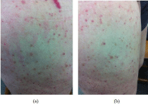
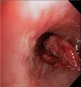
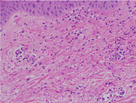
A 77 year old female with past medical history significant for hypothyroidism and transient ischemic attack presented to the hospital with hemoptysis. She initially went to a rural hospital where Computerized Tomography (CT) of the chest showed ground glass opacities in the right middle and lower lobes. She was transferred for possible bronchoscopic evaluation. On admission, she reported 3 episodes of moderate amount of hemoptysis in the last 3 days. She also reported a purpuric rash on her legs and abdomen which appeared 2 days ago (Figure 1). The rash started on her legs and then spread to her lower abdomen. On admission, she required 2 L of oxygen via nasal cannula to maintain her oxygen saturations above 92%. Rests of her vitals were within normal limits. Physical examination was pertinent for palpable purpuras on both legs and lower abdomen and mild bi-basilar lung crackles. All of the coagulation and hematological parameters were within normal limits. Her basic metabolic panel revealed a normal kidney function. Bronchoscopy was performed next demonstrating edema in right and left lower lobes, petechial lesions in the right main-stem bronchus and a blood clot in the right lower lobe (Figure 2). The clot was successfully removed using cryotherapy without any evidence of residual abnormal mucosa or bleeding beneath the clot. Bronchoalveolar lavage done during the bronchoscopy yielded bloody fluid suggestive of diffuse alveolar hemorrhage. Punch biopsy of the purpuric lesions revealed dense neutrophilic infiltrate in the epidermis and the superficial dermis with scattered eosinophils on microscopy, consistent with leukocytoclastic vasculitis (Figure 3). Immunofluorescence studies revealed fine granular deposition of IgA, C3, fibrin and lambda chains in small vessels of the papillary dermis consistent with immunoglobulin A (IgA) vasculitis (Figure 4). Rheumatology was consulted and patient was started on methyl prednisone (1 g daily for 3 days) in addition to the ongoing supportive management with close monitoring and intravenous hydration. A detailed autoimmune work up was found to be negative except for a mildly elevated level of C reactive protein (Table 1). Her skin lesions improved and hemoptysis resolved over the next few days. She was discharged after a 5 day hospital course with a 30 day prednisone taper (starting at 60 mg daily) and a follow up with rheumatology. Patient was seen in rheumatology clinic after 4 weeks and denied any recurrent signs or symptoms.

Figure 1: Purpuric lesions on the left (a) and right (b) thigh.

Figure 2: Bronchoscopic image showing clot in the right lower lobe.

    Figure 3:  High power (40 x) H and E stain showing leukocytoclastic vasculitis
characterized by fragmentation of neutrophil nuclei (karyorrhexis).

    Figure 4:  Immunofluorescence staining showing C3 (a), fibrin (b) and IgA (c)
deposition within small vessels of the papillary dermis.
    Test    (Ab = Antibody, Ag = Antigen)
    Result


    Anti-nuclear    Ab
    Negative


    Anti-centromere    Ab
    5 U/ml


    Anti-double-stranded    DNA Ab
    16 IU/ml


    Anti-glomerular    basement membrane Ab
    Negative


    Anti-histone    Ab
    13 U/ml


    Anti-Jo-1    Ab
    77 U/ml


    Anti-neutrophilic    cytoplasmic Ab
    Negative


    Rheamatoid    factor
    7 IU/ml


    Anti-ribonucleoprotein    Ab
    9 U/ml


    Scl-70    Ab
    5 U/ml


    Anti-smith    Ab
    6 U/ml


    Anti-SSA    (Ro) Ab
    19 U/ml


    Anti-SSB    (La) Ab
    10 U/ml


    C3 level
    132    mg/dL


    C4 level
    24 mg/dL


    IgA    level
    159    mg/dL


    Cryoglobulins
    Negative


    C-    Reactive Protein level
    4 mg/L


    Estimated    Sedimentation Sate
    28 mm/hr
Table 1: Autoimmune work up.
Discussion
HSP is a systemic vasculitis that is immune mediated in nature. The characteristic finding of HSP is leukocytoclastic vasculitis with IgA deposition within the affected organs. While the exact cause of HSP is unknown, various infections and chemical triggers have been associated with it. Studies do show genetic, immunological and environmental factors that may be playing a role in pathogenesis of the disease. Whereas 90 percent of the cases of HSP occur in children, 10 percent of the cases occur in adults [4]. The disease also seems to occur more frequently in the male gender. In terms of ethnicities, Blacks seem to be affected less commonly by HSP when compared to Asians and Caucasians [5]. Both in children and adults, HSP is characterized by palpable purpuras without thrombocytopenia and coagulopathy, arthritis/arthralgia, abdominal pain and renal disease. Compared to children, adults tend to have less incidence of gastrointestinal complications such has intussusception but are at increased risk of developing significant renal involvement [6]. As HSP is a self limited disease, treatment is geared towards controlling the symptoms and decreasing the risk of complications. It generally consists of supportive management involving adequate hydration, rest, and symptomatic relief of pain. Steroids are sometimes utilized for pain control and also seem to be useful in renal disease. Other drugs such as cyclophosphamide are sometimes used in practice but there is insufficient data on their effectiveness [7].
Pulmonary involvement in HSP is a rare occurrence. It occurs more often in adult onset HSP and usually presents either as Diffuse Alveolar Hemorrhage (DAH), usual interstitial pneumonia or interstitial fibrosis [8,9]. The presence of DAH is associated with high mortality and morbidity. Mortality rate of DAH in HSP is estimated to be 27.8% [10]. Management of DAH in HSP is generally supportive though steroids and cyclophosphamide have shown some benefit in slowing down the progression of disease and preventing further potentially life threatening episodes of DAH [10]. While steroids alone can be helpful in mild pulmonary disease, severe disease can require both steroids and cytotoxic therapy [3]. In our particular case, the DAH was mild and supportive measures with steroid therapy were sufficient in treating it.
This case emphasizes recognizing HSP as a potential cause of DAH. As DAH in HSP can be associated with high mortality, it is important to recognize it early to provide timely treatment. Furthermore, our case establishes pulmonary disease as a potential presentation of HSP rather than the usual organ manifestations mentioned above.
Acknowledgement
Daniel Zaccarini, MD for creating the histopathology slides.
References
- Jennette JC, Falk RJ, Bacon PA, Basu N, Cid MC, Ferrario F, et al. 2012 revised International Chapel Hill Consensus Conference Nomenclature of Vasculitides. Arthritis Rheum. 2013; 65: 1-11.
- Sim YS, Choi MY. A Case of Pulmonary Hemorrhage Associated with Henoch-Schönlein Purpura. Tuberc Respir Dis. 2009; 67: 226-228.
- Toussaint ND, Desmond M, Hill PA. A patient with Henoch-Schönlein purpura and intra-alveolar haemorrhage. NDT Plus. 2008; 1: 167-170.
- Piram M, Mahr A. Epidemiology of immunoglobulin a vasculitis (Henoch- Schönlein): current state of knowledge. Curr Opin Rheumatol. 2013; 25: 171- 178.
- Gardner-Medwin JM, Dolezalova P, Cummins C, Southwood TR. Incidence of Henoch-Schönlein purpura, Kawasaki disease, and rare vasculitides in children of different ethnic origins. Lancet. 2002; 360: 1197-1202.
- Blanco R, Martinez-Taboada VM, Rodriguez-Valverde V, Garcia-Fuentes M, Gonzalez-Gay MA. Henoch-Schonlein purpura in adulthood and childhood: two different expression of the same syndrome. Arthritis Rheum. 1997; 40: 859.
- Szer IS. Gastrointestinal and renal involvement in vasculitis: management strategies in Henoch-Schönlein purpura. Cleve Clin J Med. 1999; 66: 312- 317.
- Markus HS, Clark JV. Pulmonary haemorrhage in Henoch-Schönlein purpura. Thorax. 1989; 44: 525-526.
- Nadrous HF, Yu AC, Specks U, Ryu JH. Pulmonary involvement in Henoch- Schönlein purpura. Mayo Clin Proc. 2004; 79: 1151-1157.
- Rajagopala S, Shobha V, Devaraj U, D’Souza G, Garg I. Pulmonary hemorrhage in Henoch-Schönlein purpura: case report and systematic review of the English literature. Semin Arthritis Rheum. 2012; 42: 391-400.