Research Article
Austin J Pulm Respir Med 2017; 4(1): 1048.
The Stress Tests with the Designation of Oxygen in the Blood in the Evaluation of the Idiopathic Pulmonary Fibrosis
Jeznach A¹* and Kozielski J²
¹The Internal Department of 116 Military Hospitals, Poland
²Medical University of Silesia, Katowice, Poland
*Corresponding author: Andrzej Jeznach, The Internal Department of 116 Military Hospitals, Poland
Received: September 26, 2016; Accepted: March 09, 2017; Published: March 16, 2017
Abstract
Background: The aim of this work is to characterize how important is effort research with blood gas measurement to estimate IPF (Idiopathic Pulmonary Fibrosis) course with reference to standards of this estimate that are spirometry research, diffusive capacity of lungs research, HRCT (High Resolution Computed Tomography)
Methods: In the research, there were included patients who had at the same time spirometry research, diffusive capacity of lungs research, effort research with blood gas measurement, before and after effort, HRCT of lungs. The received dates were compared with the dates of the next hospitalization, whereas all above mentioned researches were conducted. The criterion of including in research was existence of fibrosis of lungs stated in HRCT of chest in patients with IPF. The diagnosis of acute interstitial fibrosis of lungs was established on the basis of clinic characteristics and additional researches conducted in Lung Diseases and Tuberculosis Clinic in Zabrze. All dates were received on the basis of archival documentation of Lung Diseases and Tuberculosis Clinic in Zabrze.
18 patients with IPF were examined. 10 females and 8 males. Aged since 33 to 72.
Results: The conducted researches showed that the changes in HRCT appear together with the changes of suppleness of oxygen in effort and FVC (forced vital capacity) among patients with IPF. For patients with interstitial changes in lungs valuable researches are (in order) estimate of PaO2 (partial pressure of the oxygen in arterial blood) in effort TAU = 0,7(value of tau-Kendall statistic); p= 0,012 (value of Pearson’s chi square statistic) estimate of FVC (TAU = 0,280; p= 0,011). In this case the value of DLCO (diffusing capacity or transfer factor of the lung for carbon monoxide) in rest was not statistically essential (p=0,2).
Conclusion: On the basis of above results the measurement of PaO2 is suggested, in the highest effort as the main research in progression of fibrosis of tissue monitoring of patients with IPF.
Keywords: Exertional tests; Idiopathic pulmonary fibrosis
Introduction
Stress tests are a tool to assess the limits and exercise tolerance mechanisms. They provide knowledge on the functional reserve systems involved in the response to exercise. They are based on the principle that a system such as respiratory or cardiovascular fails easier when subjected to effort [1]. In this way, you can seek the cause of shortness of breath when clinical examination and applied rest tests did not explain its ethnology. The tests provide information that cannot be obtained by conducting the rest test [2-6].
There is a need to identify practical measures to diagnose and monitor the course of Idiopathic Pulmonary Fibrosis. The research is being conducted on the determination of the optimal exponent of the fibrosis that occur in the lung parenchyma [7]. High Resolution Computed Tomography is now considered a sensitive tool to identify changes on the tissue level and one of the best non-invasive methods to assess the course of IPF [8]. The nature and location of interstitial changes determine the stage changes. At the same time the importance of research assessment exercise with blood gases before and after exercise in patients with idiopathic pulmonary fibrosis is emphasized. The aim of the study is to determine to what extent the exercise test with measurement of blood gases is useful to assess the progress of IPF in relation to the standards of this assessment that is spirometry, lung diffusion capacity test, HRCT.
Material and Methodology
Material
18 patients (10 women, 8 men, aged from 33 to 72 years) who had been admitted to the Department of Tuberculosis and Lung Diseases in Zabrze because of IPF were examined. The study included patients who had spirometry, lung diffusion capacity test, exercise test with marking blood gases before and after exercise and HRCT lung performed simultaneously. The obtained data were compared with data from another hospitalization, during which the above tests were performed again. The criterion for inclusion in the study was the presence of disseminated pulmonary fibrosis diagnosed by chest HRCT study. The diagnosis of interstitial pulmonary fibrosis was determined on the basis of clinical features and additional studies conducted in the clinic. Patients reported a history of progressive exertion dyspnea without the exposure to organic, inorganic, or pharmacological agents of pulmonary fibrosis. The average patient follow-up was 2 years, the longest eight years, the shortest 5 months.
Methodology
Stress tests included a test on an ergometer bicycle Ergo medic Monark 839E Company (Germany) according to the protocol of Wasserman approved by ERS (European Respiratory Society) [9] and the ATS (American Thoracic Society) [10]. The determination of blood gas levels were carried out after sampling arterialized blood from the earlobe before exercise and at the peak of exercise. Gas analysis was performed by using AVL Compact I (Austria). 4 mmHg was recognized as the margin of error in case of determining the level of oxygen in the blood. In view of the above, the improvement or deterioration compared to the previous exercise test with marking the blood gases, was established when the difference in level of oxygen pressure was higher than 4mmHg before and after exercise. In the single study for the deterioration or improvement of the level difference oxygen tension greater than 4mmHg before and after exercise was established. The criteria recommended for interstitial diseases ATS [10] were adopted.
Pulmonary function tests included measurement of diffusion capacity for carbon monoxide and measurement of forced vital capacity. Diffusion capacity for carbon monoxide (DLCO) was measured in one breath technique using a measuring apparatus Mes Profile Graphics (USA) according to the ERS and ATS [11,12]. In the study of diffusion capacity of the gas as an improvement or deterioration in relation to the previous study it was decided to be an increase or decrease in DLCO by 15%. Diffusing capacity tests were not performed in [13] patients due to the lack of cooperation during the examination.
The spirometry was performed by using Transfer screen II Jaeger (Germany) according to the recommendations of the ERS. In the spirometry for the improvement or deterioration in relation to the previous studies it was an increase or decrease in Forced Vital Capacity (FVC) by 200ml and / or a decrease in FVC by 12% predicted. The criteria of the European Respiratory Society (ERS), the American Thoracic Society (ATS) were adopted and guidelines of Polish Respiratory Society (PTCHP). HRCT studies were evaluated by two independent radiologists who were not aware of the clinical diagnosis. HRCT was evaluated by a radiologist evaluating image control test for the specific study from the past. The HRCT examination was performed with the use of Siemens Somatom AR (Germany) device using a high-resolution algorithm.
Most of the patients received treatment in the form of steroids or immuno modulation drugs during their follow-up. Of the 18 subjects enrolled in the study 6 patients did not take drugs during the followup, [14] received steroid with an immuno modulation, 8 took only steroid.
Statistical analysis
Statistical studies were performed using SPSS. In the first stage, the value of the independence test based on the Chi square χ2 statistics was used to investigate the existence of a link between the results of the various tests performed for the description of ILD-Interstitial Lung Diseases. In this study, the differences at the significance level of p <0.05 was considered statistically significant.
In the second stage for the characteristics measured on an ordinal scale that have a specific significance of a relationship, the value of TAU-Kendall statistics was set, saying about the strength of the relationship.
Results
Coexistence of changes in HRCT, FVC, DLCO, blood oxygen tension (PO2) at rest and exercise in the course of the follow-up of patients in each group are shown below in graphical form. The most optimal configuration of events, providing a high coexistence is the laying of the events on the selected diagonal passing through the points: (progression, progression), (unchanged, unchanged) and (regression, regression). The smaller the number of events outside the contracting diagonal passing through the points (progression, progression), (unchanged, unchanged) and (regression, regression), the higher the co-existence of changes in individual groups.
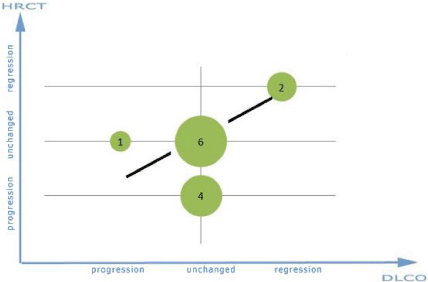
Comparison of changes in the gas diffusing capacity with changes in HRCT in idiopathic pulmonary fibrosis
Figure 1 Graph of changes in the gas diffusion capacity in comparison to changes in HRCT in idiopathic pulmonary fibrosis.

    Figure 1: Graph of changes in the gas diffusion capacity in comparison to
changes in HRCT in idiopathic pulmonary fibrosis.
For the selected diagonal passing through the points (progression, progression), (unchanged, unchanged), and (regression, regression) there are two clusters of events stances 6 and 2, which is at a total number of events (1 + 4 + 6 + 2 = 13) 61% of the events compliant and provides a strong coexistence of these events.
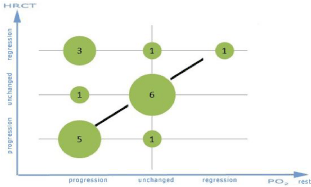
Comparison of changes in oxygen tension at rest with changes in HRCT in idiopathic pulmonary fibrosis
Figure 2 Graph of changes in the oxygen tension at rest in comparison to changes in HRCT in idiopathic pulmonary fibrosis.

    Figure 2: Graph of changes in the oxygen tension at rest in comparison to
changes in HRCT in idiopathic pulmonary fibrosis.
For the selected diagonal passing through the points (progression, progression), (unchanged, unchanged), and (regression, regression) there are three clusters of events stances, respectively 5.6 and 1, which at a total number of events of (6 + 5 + 3 + 1 + 1 + 1 + 1 = 18) is 66% of the events compliant and provides a strong coexistence of these events.
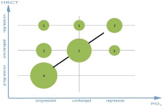
Comparison of changes in the oxygen pressure at the peak of effort with changes in HRCT in idiopathic pulmonary fibrosis
Figure 3 Graph of changes in the oxygen tension at the peak of effort in comparison with changes in HRCT in idiopathic pulmonary fibrosis.

    Figure 3: Graph of changes in the oxygen tension at the peak of effort in
comparison with changes in HRCT in idiopathic pulmonary fibrosis.
For the selected diagonal passing through the points (progression, progression), (unchanged, unchanged), and (regression, regression) there are three clusters of events stances respectively 6, 5 and 2, which at a total number of events of (6 + 5 + 2 + 2 + 1 + 1 + 1 = 18) is 72% of the events compliant and provides a strong coexistence of these events.
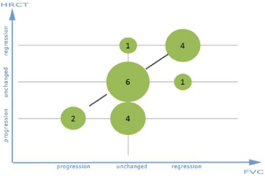
Comparison of changes in vital capacity of gases to changes in HRCT in idiopathic pulmonary fibrosis
Figure 4 Graph of changes in forced vital capacity compared with changes in HRCT in idiopathic pulmonary fibrosis.

    Figure 4: Graph of changes in forced vital capacity compared with changes
in HRCT in Idiopathic pulmonary fibrosis.
For the selected diagonal passing through the points (progression, progression), (unchanged, unchanged), and (regression, regression) there are three clusters of events with a dose rate of 2, 6 and 4, which as the total number of events of (2 + 6 + 4 + 4 + 1 + 1 = 18) is 66% of the events compliant and provides a strong coexistence of these events in Table 1.
    PaO2
    PaO2 at the peak of effort
    FVC
    DLCO


    at rest


    HRCT
    P=0,332
    P=0,012
    P=0,011
    P=0,2


    FVC
    P=0,566
    P=0,340

    P=0,025


    DLCO
    P=0,063
    P=0,856


      DLCO: Diffusing Capacity or Transfer Factor of the Lung    for Carbon Monoxide; FVC: Forced Vital Capacity;    HRCT: High Resolution Computed Tomography; IPF: Idiopathic Pulmonary Fibrosis;    P: Value of Pearson’s Chi Square Statistic; PaO2: Partial Pressure of    the Oxygen in Arterial Blood
Table 1: Tests Chi –square PaO2
In the first stage, the value of the independence test based on the Chi square χ2 statistics was used to investigate the existence of a link between the results of the various tests performed for the description of interstitial lung disease (ILD). In this study, the differences at the significance level of p <0.05 were considered statistically significant. Next, the measurement of the strength of the association between the above mentioned variables was performed in Table 2.
    PaO2 at    the peak of effort
    FVC
    DLCO


    HRCT
    TAU=0,7
    HRCT
    TAU=0,28
    FVC
    TAU=0,5


    DLCO:    Diffusing Capacity or Transfer Factor of the Lung for Carbon Monoxide; FVC-    Forced Vital Capacity; HRCT: High Resolution Computed Tomography; PaO2: Partial    Pressure of the Oxygen in Arterial Blood
Table 2:  Table of TAU-Kendall statistics for the variables between which the
relation at a significance level of p <0.05 has been found.
Summary of the results
- The results confirmed the relation between HRCT and changes in oxygen pressure at the peak of effort during exercise testing in patients with idiopathic pulmonary fibrosis.
- The strength of the relation between the results of oxygen in the blood after exercise and the results of the HRCT of the chest is greater than between the results of forced vital capacity and HRCT results in a population of patients with idiopathic pulmonary fibrosis.
- The relation between the results of forced vital capacity and the results of the HRCT of the chest in a population of patients with interstitial lung diseases was confirmed.
- The strength of the relation between the results of forced vital capacity and performance HRCT is weaker than between the results of oxygen in the blood after exercise and HRCT in a population of patients with idiopathic pulmonary fibrosis.
- The relationship between the results of forced vital capacity and diffusion capacity gas results in a population of patients with idiopathic pulmonary fibrosis was confirmed.
- The strength of the relation between the results of forced vital capacity and performance gas diffusion capacity is weaker than between the results of oxygen in the blood after exercise and HRCT, but greater than between the results of forced vital capacity and HRCT in a population of patients with idiopathic pulmonary fibrosis.
- The relationship between HRCT results and changes in gas diffusion capacity in patients with idiopathic pulmonary fibrosis was not confirmed.
Discussion
HRCT is currently the best non-invasive method of assessing the lung structures used in the differentiation of pulmonary fibrosis from inflammatory changes, and determining the extent of disease. The characteristics of fibrosis in this study are the image of the reticulum and honeycomb mesh of sub pleural location. Frequent repetition of this examination exposes the patient to X-rays and increases the economic cost of medical treatment [15].
In developed in November 2010 and published in early 2011 ATS and ERS position on recognition of IP the role of this research and advance in image interpretation lungs are emphasized. The specificity of HRCT image is currently available to identify histopathological pattern of common Interstitial Pneumonia (UIP). It is believed that the presence of clinical features, such as: unexplained chronic exertional dyspnoea, chronic cough, bilateral crackles at the base of the lung fields, rod-shaped fingers radiological and such radiographic features in HRCT as the image of the reticulum and honeycomb mesh of sub pleural location is sufficient for the diagnosis of IPF, and performing an open lung biopsy in the diagnosis does not have to be necessary [1-4,16,17]. The diagnosis should be established by a team of experts made up of a pulmonologist, radiologist, and pathologist. Its role is also to exclude other causes of the observed non IPF lung fibrosis [5,16]. These include, inter alia, pulmonary fibrosis due to chronic allergic alveolitis, occupational exposure, systemic diseases, drug reactions.
The high diagnostic value of HRCT has been confirmed among other studies by Orense and et al. These authors compared the sensitivity of HRCT, spirometry, gas diffusion capacity test, stress test with the designation of blood gases in the detection of idiopathic pulmonary fibrosis. The disease was confirmed by biopsy of the lung. They found that despite the high 88% reduction in test sensitivity of HRCT diagnosis of patients with unexplained dyspnoea pulmonary function tests, both at rest and at effort should be performed to determine the extent and degree of pulmonary fibrosis. The valid HRCT does not exclude early and clinically active fibrotic lung tissue. The authors found a significant difference in DLCO between the 2 groups of patients, i.e. patients with normal and abnormal HRCT image with features of interstitial lung disease. The average DLCO in the group with abnormal HRCT was 46.1 +/- 2.3% predicted; when patients with normal HRCT average value was 65.7 +/- 3.8%. TLC did not differ between the groups with normal and abnormal HRCT with features of interstitial lung disease. Average TLC in the group with abnormal HRCT was 77.0 +/- 3.6% predicted, while the average of patients with normal HRCT was 68.6 +/- 9.5% predicted. Exercise tests performed in our patients showed a strong correlation between the size of desaturation during exercise, and the severity of fibrosis in HRCT [6]. The average value of the saturation in the group with abnormal HRCT during exercise was 87.6 +/- 1.1%, while for patients with normal HRCT standard saturation was 96.3 +/- 1.1%.
In Xaubet’s study in which 39 patients with IPF lung confirmed by biopsy were tested, the extent of change in HRCT during diagnosis showed a moderate, but statistically significant, correlation with FVC (r = 0.46, p = 0.003), and DLCO (r = 0.40, p = 0.003). Xaubet and colleagues observed the patients for an average of seven months (from 4 to 11 months) and found a significant correlation between FVC and DLCO, and the image HRCT in idiopathic interstitial fibrosis. It was also shown that the rate of change in HRCT in the course of IPF is correlated with changes in DLCO to similar extent (r = 0.57, p = 0.01) as a change in FVC (r = 0.51, p = 0.01) [2,18].
The reduction of amount of properly functioning pulmonary alveoli is responsible for reduction of FVC in the first place. This is due to obliteration of bubbles, filling them through effusion associated with inflammation, edema and cellular infiltrates. At the beginning of interstitial lung disease independent of the ethnology occurs alveolitis involving the accumulation of immune cells and mediators of inflammation in response to a stimulus damaged. If there is an imbalance of destructive processes, and repair or stimulus runs continuously it comes to structural changes. The consequence thereof is impaired gas exchange, and disturbances in production of collagen leading to changes in the mechanical properties of lung parenchyma [18].
The studies have shown that due to the nature of the relationship between changes in lungs and its vastness, in some patients the resting study, despite the observed changes in the lungs of fibre may be correct. Gas exchange abnormalities in ILD typically present themselves as hypoxemia with increased alveolar-capillary gradient for oxygen tension. These disorders are increasing even during an increased demand for oxygen system, i.e. during exercise. Hypoxemia affects many patients with IPF while resting, but only in some patients it comes to its occurrence during exercise. The mechanism leading to disruption of gas exchange is impaired diffusion of structural abnormalities associated with capillary barrier, and disorders of ventilation to perfusion ratio (V/Q) due to regional changes in ventilation caused by mechanical disorders of the lung parenchyma disorders. The disorders of ratio V/Q have already physiologically inhomogeneous character. During exercise, these disorders may increase. The research by which mentioned gas exchange abnormalities can be confirmed, are primarily arterial blood gases and measuring arterial oxygen saturation at rest and exercise, the alveolar-capillary test of the difference in oxygen pressure (PA-AO2), and a study of lung diffusion capacity for carbon monoxide (DLCO) [18]. My research has shown that in 7 patients, when the decrease in PaO2 followed along with the severity of fibrosis in HRCT, FVC and DLCO were normal. Agusti et al demonstrated a correlation between low (average DLCO, 52% predicted) ability of diffusion of gases at rest and arterial hypoxemia during exercise in patients with IPF. They found, in addition to the correlation between DLCO and hypoxemia during exercise, the correlation between the increase in pulmonary vascular resistance and exercise. Pulmonary vascular resistance was calculated as the difference between the average pulmonary artery pressure and average pulmonary capillary wedge pressure divided by the cardiac output. The increase in vascular resistance was due to the loss of vascular fibrosis in their past and their destruction in response to hypoxia. Crystal suggests coexistence of these two processes with functional predominance of vascular lesions in the early stages of IPF and anatomical during disease progression [19]. Augusti and et al found that the main cause of hypoxemia at rest was the disorder V/Q ratio. During exercise, they observed in patients the decrease in PaO2 at constant V/Q. There was a decrease in oxygen diffusion capacity, which according to these researchers spoke in favor of the concept of the alveolar-capillary block as the cause of hypoxemia [20].
Proper exercise test result is likely to exclude the interstitial disease in patients with normal resting functional indices and normal chest X-ray image. In my study, I have shown a strong correlation between the progression or regression of fibrosis in HRCT, and the deterioration or improvement in PaO2 during the exercise test (p = 0.012). Statistically significant (p <0.05) relationship between the severity of pulmonary fibrosis in HRCT, and a decrease in PaO2 at the peak of exercise, I have shown in a population of patients diagnosed with idiopathic pulmonary fibrosis. The measure of TAU significance on a scale from 0.0 to 1.0 for the population of patients with idiopathic pulmonary fibrosis was 0,700, while the measure of the significance of TAU between changes in FVC and HRCT was 0.28. Therefore, as it is clear from my research, it is appropriate to perform marking PaO2 at the peak of exercise, as a measure of lung tissue fibrosis progression in a population of patients with idiopathic pulmonary fibrosis.
Fulmer et al in patients with IPF, in which the diagnosis was based on research histopathological, have demonstrated a correlation between the parameters of gas exchange during exercise, i.e. the pressure of oxygen in the blood capillary, CO2 production, alveolarcapillary gradient and severity of fibrosis and inflammation of the lung parenchyma. Resting tests of vital capacity and diffusion capacity did not correlate either with the severity of fibrosis or the severity of inflammatory infiltration [21]. Erbes and colleagues retrospectively studied 99 patients with histologically confirmed IPF performing functional tests and stress tests of the blood gas mark to determine the prognostic value of these studies [22].
They found that the deterioration in terms of total lung capacity (TLC) is prognostic for IPF, whereas DLCO, reducing PAO2 during exercise is not associated with a difference in the survival of these patients. In search of valuable tools for forecasting and assessment of interstitial lung disease, efforts are made to determine the strength of the relationship between HRCT and lung function tests such as spirometry, and measurement of diffusing capacity of gases. Wallert retrospectively studied 121 patients with histologically confirmed IPF. He compared FVC and DLCO at rest, resting PaO2, P(AAO 2) at the peak of exercise during the 6-minute walking test and desaturation during a 6-minute walking test, trying to find the most sensitive manifestation IPF exponent in function tests. He said that the abnormal gas exchange is present in patients with normal FVC. Incorrect FVC was found in 61% of patients. He considers to be the most sensitive diagnostic method of gas exchange abnormalities in patients with IPF measurement of DLCO at rest (sensitivity 97.5%), then P(A-AO2) at the peak of exercise (sensitivity 92.5%), desaturation during a 6-minute walking test (sensitivity 83%.)
In my attempt I showed a relationship between changes in FVC and DLCO. The statistical significance between changes DLCO and FVC at rest (p = 0.025) in the population of patients with IPF was confirmed. The measure of TAU significance was 0,500. In this case, the coexistence of changes in both studies in the course of IPF can be stated. The results of these studies are complementary and changes in DLCO FVC accompany changes in the course of fibrosis in the lung tissue of patients with IPF.
Changes in DLCO showed no correlation with changes in HRCT (p = 0.2) in a population of patients with idiopathic pulmonary fibrosis. This means that changes in DLCO did not imitate changes in HRCT in these patients at a given level of significance (p = 0.05). On the basis of these results, I think that marking DLCO during rest to monitor the course of fibrotic lung tissue is not a valuable diagnostic method in these patients.
No relationship between the type, length of treatment, or gender, and long-term improvement or stabilization in the results of functional or radiographic appearance was shown. In similar studies conducted in the past taking medicine had little impact on survival in patients with ILD compared to patients not receiving therapy [23].
Taking into account my test results and the literature it should be assumed that the assessment of disease progression in patients with idiopathic pulmonary fibrosis the valuable functional study are in the order: the assessment of PaO2 (TAU = 0.7; p = 0.012), the assessment of FVC at rest (TAU = 0.28; p = 0.011). In this case, the value of DLCO at rest was not statistically significant (p = 0.2).
It should be noted, however, that the study group of patients with IPF was small that is why functional studies on the usefulness of monitoring fibrosis in ILD requires further calculations and comparisons.
Summary
The aim of this work is to characterize how important is effort research with blood gas measurement to estimate IPF course with reference to standards of this estimate that are spirometry research, diffusive capacity of lungs research, HRCT.
In the research, there were included patients who had at the same time spirometry research, diffusive capacity of lungs research, effort research with blood gas measurement, before and after effort, HRCT of lungs. The received dates were compared with the dates of the next hospitalization, whereas all above mentioned researches were conducted. The criterion of including in research was existence of fibrosis of lungs stated in HRCT of chest in patients with IPF. The diagnosis of acute interstitial fibrosis of lungs was established on the basis of clinic characteristics and additional researches conducted in Lung Diseases and Tuberculosis Clinic in Zabrze. All dates were received on the basis of archival documentation of Lung Diseases and Tuberculosis Clinic in Zabrze.
18 patients with IPF were examined. 10 females and 8 males. Aged since 33 to 72.
The conducted researches showed that the changes in HRCT appear together with the changes of suppleness of oxygen in effort and FVC among patients with IPF. For patients with interstitial changes in lungs valuable researches are (in order) estimate of PaO2 in effort (TAU = 0,7; p= 0,012) estimate of FVC (TAU = 0,280; p= 0,011). In this case the value of DLCO in rest was not statistically essential (p=0,2).
On the basis of above results the measurement of PaO2 is suggested, in the highest effort as the main research in progression of fibrosis of tissue monitoring of patients with IPF.
Conclusion
In patients with a diagnosis of IPF for the assessment of disease progression defined by HRCT study, the most important role is played by the study of oxygen pressure during exercise.
Another type of functional tests for the assessment of disease progression in patients with IPF is the monitoring of forced vital capacity.
References
- Hunninghake GW, Zimmerman MB, Schwartz DA, King TE Jr, Lynch J, Hegele R, et al. Utility of a lung biopsy for the diagnosis of idiopathic pulmonary fibrosis. Am J Respire Crit Care Med. 2001; 164: 193-196.
- Fell CD, Martinez FJ, Liu LX, Murray S, Han MK, Kazerooni EA, et al. Clinical Predictors of a Diagnosis of Idiopathic Pulmonary Fibrosis. Am J Respire Crit Care Med. 2010; 181: 832-837.
- Staples CA, Müller NL, Vedal S, Abboud R, Ostrow D, Miller RR. Usual interstitial pneumonia: correlation of CT with clinical, functional, and radiologic findings. Radiology. 1987; 162: 377-381.
- Lama VN, Flaherty KR, Toews GB, Colby TV, Travis WD, Long Q, et al. Prognostic Value of Desaturation during a 6-Minute Walk Test in Idiopathic Interstitial Pneumonia. Am J Respir Crit Care Med. 2003; 168: 1084-1090.
- Kus J. Sródmiąższowe choirboy pluc- podstawowa klasyfikacja i zarys postepowania diagnostycznego. Postepy nauk medycznych. 2011; 04: 256- 259.
- Orens JB, Kazerooni EA, Martinez FJ, Curtis JL. Martinez F. The Sensitivity of HRCT in Detecting Idiopathic Pulmonary Fibrosis Proved by Open Lung Biopsy, A Prospective Study. Chest. 1995; 108; 109-115.
- Thomeer M, Grutters JC, Wuyts WA, Willems S, Demedts MG. Clinical use of biomarkers of survival in pulmonary fibrosis Respiratory Research. 2010; 11: 89.
- Oren A, Sue DY, Hansen JE, Torrance DJ, Wasserman K. The role of exercise testing in impairment evaluation. Am Rev Respire Dis. 1987; 135: 230-235.
- Roca J, Whipp BJ, Agusti AGN, Anderson SD, Casaburi R, Cotes CF, et al. Clinical exercise testing with reference to lung diseases:indications, standardization and interpretation strategies. Eur Respire J. 1997; 10: 2662- 2689.
- Miller MR, Crapo R, Hankinson J, Brusasco V, Burgos F, Casaburi R, et al. General considerations for lung function testing. Series “ATS/ERS Task Force: Standardization of Lung Function Testing”. Eur Respire J. 2005; 26: 153-161.
- Wise RA, Teeter JG, Jensen RL, England RD, Schwartz PF, Giles DR, et al. Standardization of the Single-Breath Diffusing Capacity in a Multicenter Clinical Trial. Chest. 2007; 132: 1191-1197.
- Xaubet A, Agustí C, Luburich P, Roca J, Montón C, Ayuso MC, et al. Pulmonary Function Tests and CT Scan in the Management of Idiopathic Pulmonary Fibrosis. Am J Respire Crit Care Med. 1998; 158: 431-436.
- Weisman I, Zeballos R. Cardiopulmonary exercise testing. Pulmonary Critical Care Update Series. 1995; 11: 1-9.
- Ortega F, Monte Mayor T, Sanchez A, Cabello F. Castillo J Role of cardiopulmonary exercise testing and the criteria used to determine disability in patients with severe chronic obstructive pulmonary disease. Am J Respire Crit Care Med. 1994; 150: 747-751.
- Hartley PG, Galvin JR, Hunninghake GW, Merchant JA, Yagla SJ, Speak Man SB, et al. High-resolution CT-derived measures of lung density are valid indexes of interstitial lung disease, J Appl Physiol. 1994; 76: 271-277.
- Raghu G, Collard HR, Egan JJ, Martinez FJ, Behr J, Brown KK, et al. Evidence-based Guidelines for Diagnosis and Management. Am J Respire Crit Care Med. 2011; 183: 788-824.
- Hunninghake GW, Lynch DA, Galvin JR, Gross BH, Müller N, et al. Findings Are Strongly Associated With a Pathologic Diagnosis of Usual Interstitial Pneumonia. Chest. 2003; 124: 1215-1223.
- Crystal RG, Fulmer JD, Roberts WC, Moss ML, Line BR, Reynolds HY. Idiopathic pulmonary fibrosis. Clinical, histologic, radiographic, cytology, and biochemical aspects. Ann Intern Med. 1976; 85: 769-788.
- Morelli S, Ferrate L, Sgreccia A, Eleuteri ML, Perrone C, De Marzio P, et al. Pulmonary hypertension is associated with impaired exercise performance in patients with systemic sclerosis. Scand J Rheumatol. 2000; 29: 236-242.
- Agustí AG, Roca J, Gea J, Wagner PD, Xaubet A, Rodriguez-Roisin R. Mechanisms of gas-exchange impairment in idiopathic pulmonary fibrosis. Am Rev Respire Dis. 1991; 143: 219-225.
- Fulmer JD, Roberts WC, von Gal ER, Crystal RG. Morphologic-Physiologic Correlates of the Severity of Fibrosis and Degree of Cellularity in Idiopathic Pulmonary Fibrosis. The Journal of Clinical Investigation. 1979; 33: 665-676.
- Wagner P, DantzkerD, Dueck R, de PoloJ, Wasserman K, West J. Distribution of ventilation-perfusion ratios in patients with interstitial lung disease. Chest. 1976; 69: 256-257.
- King TE Jr, Tooze JA, Schwarz MI, Brown KR, Cherniack RM. Predicting survival in idiopathic pulmonary fibrosis: scoring system and survival model. Am J Respire Crit Care Med. 2001; 164: 1171-1181.