Clinical Image
Austin J Pulm Respir Med 2017; 4(1): 1049.
A Case of Disseminated Tuberculosis with Mediastinal Widening
Hashim Z and Kumar N*
Department of Pulmonary Medicine, Sanjay Gandhi Post Graduate Institute of Medical Sciences, India
*Corresponding author: Nishith Kumar, Department of Pulmonary Medicine, Sanjay Gandhi Post Graduate Institute of Medical Sciences, India
Received: February 16, 2017; Accepted: March 09, 2017; Published: March 16, 2017
Clinical Image
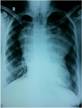
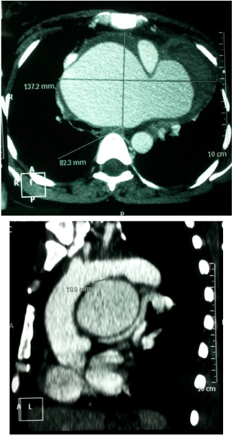
A 26 year old female patient, unmarried known case of disseminated tuberculosis on anti tubercular treatment since two months presented to our OPD with complaints of worsening dyspnea, hoarseness of voice and progressive swelling over neck for past one month. Physical examination revealed an averagely built young female in moderate respiratory distress with engorged neck veins. An upright PA chest radiograph (Figure 1) and computed tomographic scan (with contrast) of the chest (Figures 2a, 2b) are shown.

Figure 1: An upright PA chest radiograph.

Figure 2: Computed tomographic scan (with contrast) of the chest.
Evaluate at the radiograph and suggest the probable diagnosis based on clinico-radiological finding. Tubercular pseudoaneurysm of arch of aorta.
Discussion
Tuberculosis may cause wide array of clinical and radiological presentation due to its disseminative property by contiguity or hematogenously. Tubercular pseudoaneurysm of aorta is a rare finding with a high mortality rate. We present a case of 26 year old female who was admitted with complaints of breathlessness, cough, back ache and loss of appetite from past 2 months. Chest radiology was suggestive of bilateral miliary mottling. Patient was also having lytic lesion on sacral bone from which Ultra Sound guided pus aspiration was done. Pus came positive for AFB and MTB culture. Her bronchoalveolar lavage sample was positive for MTB. Patient was started on anti tubercular treatment (isoniazid, rifampicin, pyrazinamide and ethambutol) and discharged. Patient presented to our OPD after two months with complaints of worsening dyspnea, hoarseness of voice and progressive swelling over neck for past one month. A chest X ray disclosed a new onset left retrocardiac opacity. A Computed Tomographic (CT) scan with intravenous contrast medium indicated the presence of pseudo aortic aneurysm of arch of aorta. Tuberculous aortic aneurysms are prone to rupture abruptly thus the patient was referred to department of cardiovascular thoracic surgery for surgical management.
In 1962, Volini et al. [1] described the pathogenesis and complications of tuberculosis of the aorta. Tuberculous aortic aneurysms are of false variety, and may occur by direct spread of tuberculous lesion into the aorta wall. Combined medical and surgical management offers the best chance of cure. Surgical repair is warranted in all symptomatic patients because of fear of rupture [2].
In conclusion, the radiological images illustrate findings of a relatively rare disease entity seen as a complication of one of the commonly encountered infectious disease. Tuberculous pseudoaneurysm of arch of aorta can easily be mistaken for a lung lesion or a mediastinal mass. Early recognition is very important as surgical resection, anti tubercular therapy along with regular followup provides the best chances for successful management [3].
References
- Volini FI, Olfield RC, Thompson JR, Kent G. Tuberculosis of the Aorta. JAMA. 1962; 181: 78-83.
- Yavuz S, Eris C, Toktas F, Turk T. eComment. Aortic aneurysms secondary to tuberculosis. Interact Cardiovasc Thorac Surg. 2013; 17: 743-744.
- Golzarian J, Cheng J, Giron F, Bilfinger TV. Tuberculous pseudoaneurysm of the descending thoracic aorta: successful treatment by surgical excision and primary repair. Tex Heart Inst J. 1999; 26: 232-235.