Clinical Image
Austin J Pulm Respir Med 2017; 4(1): 1051.
Self-Induced Pulmonary Granulomatosis
Indramohan P1* and Bhanot N2
1Department of Geriatrics, University of Pittsburgh Medical Center, St. Margaret's Hospital, Pittsburgh, USA
2Department of Infectious Diseases, Allegheny Health Network Pittsburgh, USA
*Corresponding author: Indramohan P, Department of Geriatrics, University of Pittsburgh Medical Center, St. Margaret's Hospital, Pittsburgh, USA
Received: May 11, 2017; Accepted: June 22, 2017; Published: June 29, 2017
Case Scenario
A 32-year-old male with history of Intravenous Drug Abuse (IVDA) underwent an uneventful surgical debridement of a chronic lumbar wound infection. Antibiotics and pain medications (oxycodone and oxycontin) were prescribed. Post operatively the patient developed 3 isolated episodes of acute hypoxic respiratory failure requiring transient intubation and ventilator support.
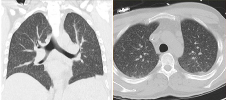
CT scans during the first 2 episodes were unremarkable. CT scan of his chest done during the third episode is shown below. Interestingly, a syringe with white powder like material was also found by his bedside during the last episode, which was sent for testing and returned positive for oxycodone, which he was receiving in the hospital for back pain.
Describe the findings on the CT scan on Figure 1?
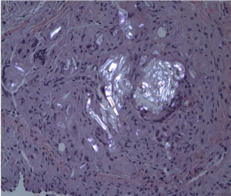
Describe the histopathological findings on Figure 2?
Based on the above information from his personal history, CT scan and histopathological findings, what is your clinical diagnosis?
What is your differential diagnosis for bilateral pulmonary nodules?
CT shows diffuse pulmonary nodules < 3 mm in size, in peri-bronchovascular distribution, involving all the lobes.
Histopathology shows foreign body granuloma with birefringent polarisable crystalline material consistent with pulmonary granulomatosis.
The patient’s history of IVDA, presence of lung nodules by pathological analysis, and his recent use of recreational drug use confirmed the clinical diagnosis of foreign body granulomatosis [1].
Differential diagnosis for bilateral diffuse pulmonary nodules include rheumatologic process, infectious process, such as a fungal infections, Tuberculosis, Nocardiosis, malignant causes, septic emboli, silicosis , hypersensitivity pneumonitis and less common causes such as amyloidosis, AVMs and hamartomas [2].

Figure 1: Non- contrast CT showing bilateral diffuse pulmonary nodules.

    Figure 2: Polarized view of the nodules in 20x showing foreign body
granuloma with birefringent polarizable crystalline material.
Answers with explanations:
References