Clinical Image
Austin J Pulm Respir Med. 2021; 8(3): 1078.
Pulmonary Amyloidosis with Multiple Cystic Lesions with Central Calcifications
Forster C1,2*, Bénière C3, Perentes JY1,2 and Christodoulou M1
¹Service of Thoracic Surgery, Hospital of Sion, Av. Grand-Champsec 80, 1951 Sion, Switzerland
²Service of Thoracic Surgery, Lausanne University Hospital, Rue du Bugnon 46, 1011 Lausanne, Switzerland
³Service of Pathology, Hospital of Sion, Av. Grand-Champsec 80, 1951 Sion, Switzerland
*Corresponding author: Céline Forster, Service of Thoracic Surgery, Hôpital de Sion, Av. Grand-Champsec 80, 1951 Sion, Switzerland
Received: July 19, 2021; Accepted: August 04, 2021; Published: August 11, 2021
Clinical Image
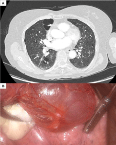
A non-smoker 80-year old woman with a past medical history of inactive and untreated systemic lupus erythematous diagnosed in 1983 and polymyalgia rheumatica treated with prednisone (3 mg once daily) since 2018, was referred to our emergency department because of left-sided chest pain and dyspnoea. She presented no cough, weight loss or fever and there was no history of Sjogren’s syndrome. Complete blood count was unremarkable and no inflammatory syndrome was observed. A chest CT-scan revealed multiple diffuse cystic parenchymal lesions with thin walls and central nodular calcifications in both lungs (Figure 1A). The sputum culture was negative for mycobacterium tuberculosis, legionella pneumophilia, mycoplasma pneumonia, chlamydia pneumonia, coronavirus, echinococcosis and aspergillus. Anti-Nuclear Antibody (ANA) and anti-Ku tests were positive whereas anti-neutrophil cytoplasmic antibody (ANCA) and anti-nucleoprotein tests were negative. The preoperative pulmonary function tests showed a FEV1 of 78% and a DLCO of 73% of the predicted values.

    Figure 1: Title: (A) Preoperative CT-scan of the lungs showing multiple bilateral
cysts with thin walls and central macrocalcifications. (B) Thoracoscopic
exploration revealing multiple vascularized cysts and macrocalcifications.
A surgical biopsy of the largest lesion consisting of a medial segmentectomy of the middle lobe by Video-Assisted Thoracoscopy (VATS) was performed. Perioperative status showed multiple diffuse vascularized cysts with macrocalcifications (Figure 1B). Postoperative course was uneventful with chest tube removal on Postoperative (PO) day 3 and home discharge on PO day 4.
The histopathological analysis revealed mainly perivascular and intersitital amyloid deposits with focal ossification. The Congo Red stain showed apple green birefringence in polarized light (Figure 2). Preferential Kappa or Lambda light chain staining could not be demonstrated by immunohistochemistry. Amyloid typing by immunofluorescence was inconclusive. There was an inconspicuous bronchus-associated lymphoid tissue with polyclonality demonstrated by PCR, which did not present any characteristics for a marginal zone lymphoma neither for lymphocytic interstitial pneumonia. In the absence of a significant anomaly of the plasmatic protein electrophoresis and clinical signs for systemic amyloidosis, a bone marrow biopsy was not performed. A diagnosis of localized pulmonary amyloidosis of undertermined subtype and without histologic evidence of underlying cause was retained [1-3].

    Figure 2: Title: Histological assessment of the resected pulmonary segment
revealing partially calcified perivascular amyloid deposits (Congo Red,
polarized light, x 100).
During the follow-up, the patient did not show any respiratory or infectious complication. The 3 months postoperative pulmonary functions showed a FEV1 of 79% and a DLCO of 67% of the predicted values. The cardiac MRI did not show any sign of cardiac amyloidosis.
Statements
Funding statement
This work received no specific grant from any funding agency in the public, commercial or not-for-profit sectors.
Contributorship statement
C.F, C.B, J.Y.P and M.C. contributed to the planning and writing of the article. C.B provided figures. All authors reviewed the article.
References