Research Article
Austin J Radiat Oncol & Cancer. 2015; 1(3): 1013.
Quality Assurance of Simultaneous Treatment of Multiple Targets Planned with Mono Isocenter using three Dimensional Conformal Radiotherapy (3DCRT) Technique
Suman Kumar Putha1, Saxena PU1, Banerjee S1, Challapalli Srinivas1*, Vadhiraja BM2, Arun Kumar ES1, Sridhar Chinthamani3 and Dinesh Pai K1
1Department of Radiotherapy & Oncology, Kasturba Medical College Hospital, Mangalore, India
2Department of Radiation Oncology, Manipal Hospital, Bangalore, India
3Department of Radiation Oncology, Father Muller Oncology Center, Mangalore, India
*Corresponding author: Challapalli Srinivas, Professor in Medical Radiation Physics, Department of Radiotherapy and Oncology, Kasturba Medical College Hospital, India
Received: November 02, 2015; Accepted: December 01, 2015; Published: December 02, 2015
Abstract
Objective: The purpose of this study was to conduct quality assurance (QA) of plans of four patients having multiple metastatic lesions (targets) simultaneously treated with mono isocentric three dimensional conformal radiotherapy.
Material and Methods: Patient geometry was simulated with two/three water equivalent phantom shaving ionization chamber (IC) sleeves (IC-1, IC-2 & IC-3 as if targets are in different locations of patient).QA plans were generated using mono isocenter technique with a dose prescription of 3.0 Gy to the targets for point dose verification. Plan evaluations was done using dose volume histogram (DVH) in terms of maximum, mean doses to target, conformity index (CI) and homogeneity index (HI). A two dimensional ion chamber array detector was used for fluence verification.
Results: Calculated maximum dose (Gy), mean dose (Gy), CI and HI values with standard deviation around the targets in all QA plans were 3.09±0.02, 3.03±0.02, 0.96±0.03 and 0.04±0.03 respectively. Measured point doses to all lesions were within ±2.0% of the computed dose in all QA plans. A pass percentage of 97% was obtained with the set criteria of 3mm distance to agreement and 3% dose difference for fluence verification around the targets in QA plans.
Conclusion: Treatment execution of multiple targets simultaneously with mono isocenter can reduce positional errors and delivery time.
Keywords: Quality assurance; Multiple targets; Mono isocenter; Conformal radiotherapy
Introduction
Solid tumours in pelvic / vertebral region may present with multiple metastases (targets) necessitating their simultaneous radio therapeutic treatment. Such treatments are usually executed using modern radiotherapy techniques such as Intensity Modulated Radiotherapy (IMRT), Stereotactic Body Radiation Therapy (SBRT), Stereotactic Radio Surgery (SRS), rapid arc, Volumetric Arc Radiotherapy (VMAT), cyberknife® and tomotherapy. In convention, multiple targets can be treated individually with a set of beams having their own isocenter. Treatment planning and execution of multiple targets results in prolongation of treatment time (starting from pretreatment Quality Assurance (QA), patient positioning, and setup corrections for every target treatment and treatment delivery) [1]. Another method is to have a common isocenter, around which the gantry rotates and delivers the radiation to multiple target sites, one at a time. This method can be an alternative in 3DCRT treatments for multiple lesions using mono isocenter instead of multiple isocentric technique. Many investigators have verified the dosimetric quality of a common isocentric plan to treat multiple tumours especially in brain metastatic lesions [2,3]. In another study, the quality of target coverage and dose conformity with mono isocentric VMAT-SRS plans to Dynamic Conformal Arc Therapy (DCAT) was compared [4]. Several authors have studied the validity of mono isocentric plans and concluded that it could be a better option of treatment delivery with less systematic errors [5-7]. Marks et al have confirmed through their investigation that three dimensional conformal radiotherapy (3DCRT) technique can be a possible alternative to radio surgery with fixed shaped coplanar or non-coplanar techniques with wedged radiation fields having beams conformed to irregular shaped intracranial lesions, as the goal of both the techniques is to achieve better dose conformity [8]. Similar logic can be used to treat multiple lesions simultaneously with different/single beam sets conformed to different lesion sites elsewhere extra cranially which can be less error prone compared to multiple isocentric treatment plans since the treatment plans with multiple isocenter are time consuming and may attribute many uncertainties in setup and positioning resulting in large systematic errors in the treatment delivery. Planning of these techniques with 3DCRT using Treatment Planning System (TPS), requires a logical approach (different beam sets conformed to multiple lesions sharing a common isocenter, having different weight points, the feasibility/flexibility to use different wedge angles using motorized wedge option, to obtain better conformal dose coverage [9-11]. The objective of this study is to validate a mono isocentric plan generated by 3DCRT technique in terms of dose conformity and coverage for the treatment of multiple metastatic lesions using composite point dose method and Two Dimensional (2D) ion chamber array detector.
Methods and Materials
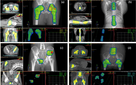
Four patients having multiple metastatic lesions (targets) which are covered in the region of Multi Leaf Collimator (MLC) were selected for this study. Clinical descriptions of individual cases as shown in (Figure 1) are a) Carcinoma of lung with bilateral hip bone and femoral metastases where both targets lie along the transverse plane, b) Carcinoma right lung with vertebral metastases where both targets lie along longitudinal plane, c) Renal cell carcinoma with pubic and ace tabular metastases where both targets lies in different planes and d) Carcinoma of penis post partial penectomy with bilateral inguinal & one vertebral metastatic lesions where the inguinal targets are in different plane with respect to the vertebral target. A dose of 30 Gy in 10 fractions was prescribed to the 100% isodose line that is covering the targets. All patients were planned for palliative radiotherapy using mono isocenter 3DCRT technique. CMS XiO® (Elekta Ltd, Crawly, UK) version 4.80.02 Treatment Planning System (TPS) utilizes Clarkson, convolution, superposition and fast superposition algorithms. However, superposition algorithm was used for dose calculations. Treatments were executed with medical linear accelerator (M/s Elekta Compact), using 6 MV (Mega voltage) photon beam at a dose rate of 350 MU/min with 40 pair multi leaf collimator (MLC) leaves (projected leaf width 1.0 cm at isocenter) arranged in two banks and also having motorized wedge facility.

    Figure 1:  95% isodose distribution that covers around targets of a representative patient with (a) bilateral hip bone and femoral metastases where both targets lie
along the transverse plane (b) vertebral metastases where both targets lie along longitudinal plane (c) pubic and ace tabular metastases where both targets lies
in different planes and (d) bilateral inguinal & one vertebral metastatic lesions where the inguinal targets are in different plane with respect to the vertebral target.
Quality Assurance (QA)
a) TPS QA
To evaluate the dosimetric performance of the TPS with threedimensional dose calculation algorithm using the basic beam data measured for 6 MV X-rays, simple test cases (involve simple field arrangements as well as the presence of a low-density material in the beam to resemble an air in-homogeneity) to complex ones (the presence of in-homogeneity, beam modifiers or beam modifiers with asymmetric fields) were created according to the Technical Report Series-430 (TRS 430) [12] in a homogeneous water phantom. Absolute dose measurements were performed for the each case with the MU calculation given by the TPS and the measured dose is compared with the corresponding calculated dose values. A percentage difference maximum of 1.98% and 4.54% for all simple and complex test cases were observed respectively. This ensures that the dosimetric calculations performed by the TPS are within the accuracy of ±5% which is very much warranted in patient dose delivery as per ICRU [13]. Point dose measurements were performed for a variety of square fields and off-centered planes, i.e. [5 cm × 5 cm, 2 cm], [7 cm × 7 cm, 3 cm], [10 cm × 10 cm, 4 cm], [13 cm × 13 cm, 5 cm], [15 cm × 15 cm, 6 cm]. The average of the four off-center dose points in the cross-plane and in-plane directions was used as the mean off center dose value in both measurements and calculations. The accuracy of dose calculation in the off centered plane was investigated, presenting within ±1.5%.
b) Phantom arrangements for simulation
For point dose verification (Composite dosimetry) of above generated plans, same patient geometry was simulated by three water equivalent phantoms [two identical water phantoms (having dimensions of 30 cm × 15 cm × 15 cm each) which are routinely used for beam quality index measurements (usually called as D10/20 phantom) and a solid water equivalent slab phantom (Model: SP34, M/s Iba dosimetry, Germany) consisting of 30 slabs (each slab dimensions of 30cm×30cm×1cm)].
All phantomshada provision of inserting 0.65 cc farmer type ionization chamber (IC) sleeve. They were arranged in four different combinations in order to generate four QA plans that simulate actual patients target geometry. Computed Tomography (CT) scans were acquired along with ICs placed inside the sleeves of the phantoms and the Digital Imaging and Communication in Medicine (DICOM) images were transferred to the contouring station (CMS Focal Sim). The chamber positions were contoured as IC-1, IC-2 & IC-3 in the scanned images which simulated the targets in an actual patient. Isocenter was chosen at the centre of combined target that was generated with a 5 mm margin encompassing both IC-1 & IC-2 (in two targets case) and IC-1, IC-2 & IC-3 (in three targets case). Contoured CT data set were transferred to CMS Xio TPS for beams placement and dose calculations.
c) Beam placements and dose calculations
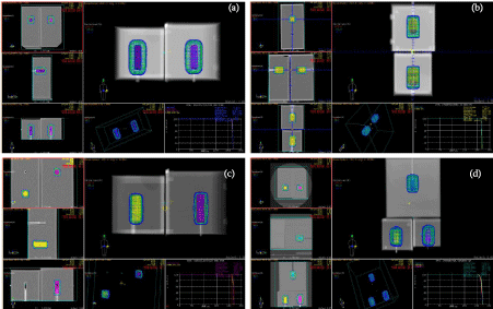
A group of four main beams with gantry angles A group of four main beams with gantry angles 00, 900,1800 and 2700 were placed taking centre of combined target as isocenter in all QA plans. Beams were conformed to the respective targets (ICs). In order to obtain uniform dose distribution around the targets, appropriate beam weights, weight points (placed inside the targets) and different wedge angles were chosen. Beam weights were adjusted until the optimum coverage and acceptable hot spots were achieved. A dose of 3.0 Gy was prescribed to the 100% isodose line that is covering all the targets. By viewing the 105% dose cloud in a beam’s eye view projection of the treatment fields, subfields were designed by blocking the volume of targets receiving greater than 105% of the prescribed dose, and the beam weightage was adjusted among sub and main fields in order to achieve the uniform dose distribution. (Figure 2) shows95% isodose distribution that covers around targets of a QA plan of corresponding patient with (a) where both targets lie along the transverse plane (b) where both targets lie along longitudinal plane (c) where both targets lie in different planes and (d) where the inguinal targets are in a different plane with respect to the vertebral target.

    Figure 2:  95% isodose distribution that covers around targets in QA Plans of corresponding patient with (a) where both targets lie along the transverse plane (b)
where both targets lie along longitudinal plane (c) where both targets lies in different planes and (d) where the inguinal targets are in different plane with respect
to the vertebral target.
d) Plan evaluation
Plan evaluation was done using dose volume histogram (DVH) in terms of conformity index (CI) and homogeneity index (HI), maximum and mean doses (Dmax and Dmean) to target. Conformity index and homogeneity index were calculated using following relations [14].
Conformity index (CI)
CI is defined as the ratio of TVRI to TV
CI = TVRI/TV
Where, TVRI = target volume covered by the reference Isodose and TV = target volume
Homogeneity index (HI)
HI is defined as the ratio of difference of D2% to D98% vs D50%for the PTV.
HI= (D2%– D98%)/ D50%
Where, D2% to D98% vs D50%correspond to the dose delivered to 2%, 50% and 98% of target volumes respectively.
e) Point dose verification
The generated QA plans were exported to Mosaiq® record and verification system and were scheduled for point dose verification. All measurements were carried out with phantoms and ICs placed inside the sleeves which were connected to the electrometers (Model: DOSE1, Supplied by M/s Iba, Germany). Scheduled QA plans were executed under linear accelerator and the charge collected (M) from each electrometer was converted to absorbed dose after applying correction factors of temperature & pressure (KTP), polarization(Kpol), saturation (KSat), beam quality (KQ, Qo) and calibration factor(NDW) of the IC’s.
f) Two dimensional (2D) dose verification
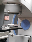
A two dimensional (2D) ion chamber array detector (Model: I’mRT MatriXX, M/s Iba dosimetry, Germany) was used for planar dose verification. This device consists of a 1020 vented ion chamber array detectors arranged in 32 χ 32 grids. The each chamber volume is 0.08 cm3 with the height of 5mm and diameter of 4.5 mm. The maximum dose rate detectable by the detectors is 5 Gy/min and minimum of 0.1Gy/min. The bias voltage required for the matrix system is 500±30V. The maximum field of view is 24 × 24 cm2. The matrix device can be directly connected to computer via standard Ethernet interface to acquire the measurements. The I’mRT MatriXX device with 5 cm solid water phantom (SP-34) positioned above it was scanned with 2 mm CT slice thickness. In order to verify the TPS generated plan, a verification plan was produced with CT data of the detector system to estimate the fluence. In the verification plan, all gantry and collimator angles were set to zero degrees and exported to the scanned detector system with the detector plane positioned at isocenter. Generated verification plan was exported and executed using Mosaiq® record and verification system for planar dose verification with I’mRT MatriXX device. The beam central axis was made perpendicular to the I’mRT MatriXX measurement level at the center of the measurement area during the measurement (Figure 3). By executing the verification plan, the cumulative fluence at the detector plane was calculated and transferred to the OmniPro software for comparison. Dose distributions obtained with this device was rescaled at 0.1 cm resolution using OmniPro IMRT analyzing software. All measured fluence was compared with TPS dose plane by 2D gamma evaluation using 3% dose difference and 3 mm distanceto- agreement (DTA) criteria. Also the percentage of the evaluated dose points passing the gamma index was kept at a limit of greater than or equal to 95%.

Figure 3: 2D dose verification with I’MatriXX under Linac.
Results
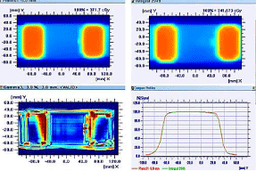
Plan evaluation parameters (dose minimum, maximum, mean, CI & HI) from DVH to all targets in all QA plans are shown in (Table 1). As observed, Dmax, Dmean, CI and HI values with standard deviation around the targets in all QA plans were 3.09±0.02 Gy, 3.03±0.02 Gy, 0.96±0.03 and 0.04±0.03respectively.Point dose measurements to all ICs were obtained using NDW based formalism [15] and compared with the calculated values from TPS in all QA plans are shown in (Table 2). It was observed that the percentage deviation of measured dose obtained for all targets were within ±2.0% against calculated values from TPS in all QA plans. A pass percentage of 97% was obtained with the set criteria of 3mm distance to agreement (DTA) and 3% dose difference for fluence verification around the targets in QA plans (1to3) where the targets are covering in the maximum field of view of I’mRT MatriXX device. However we could not perform 2D verification in case of the QA plan 4 since all three targets were not in the maximum field of view of the I’mRT MatriXX device. (Figure 4) represents 2D fluence verification using I’mRT MatriXX™ device of QA plan 1 where the targets were along transverse plane
    QA Plan
    a
    b
    c
    d


    Targets (IC’s)
    IC-1
    IC-2
    IC-1
    IC-2
    IC-1
    IC-2
    IC-1
    IC-2
    IC-3


    Dmax (Gy)
    3.07
    3.07
    3.07
    3.08
    3.08
    3.07
    3.12
    3.12
    3.12


    Dmean (Gy)
    3.03
    3.03
    3.04
    3.04
    3.02
    3.01
    3.08
    3.04
    3.02


    CI
    0.99
    0.98
    0.99
    1.00
    0.94
    0.93
    1.00
    0.92
    0.93


    HI
    0.03
    0.03
    0.00
    0.00
    0.03
    0.03
    0.03
    0.07
    0.10
Table 1: Plan Evaluation Parameters from DVH of all QA plans.
    Measured    Mean dose
      (Gy)= M × TCF@
    TPS    calculated Mean
      dose    (Gy)
    Percentage    Deviation
      (%)


    QA    Plan
    IC-1
    IC-2
    IC-3
    IC-1
    IC-2
    IC-3
    IC-1
    IC-2
    IC-3


    a
    2.97
    2.99
    NA
    3.03
    3.03
    NA
    1.98
    1.32
    NA


    b
    3.07
    3.01
    NA
    3.04
    3.04
    NA
    -0.99
    0.99
    NA


    c
    3.02
    3.07
    NA
    3.02
    3.01
    NA
    0.00
    -1.99
    NA


    d
    3.11
    3.10
    3.06
    3.08
    3.04
    3.02
    -0.97
    -1.97
    -1.32


    @TCF: Total Correction Factor (NDW × KTP × Kpol × KSat× KQ,Qo)14.
      NA:    Not Applicable to the QA plan.
Table 2: Percentage deviation between measured and TPS calculated dose.

Figure 4: Evaluation of 2D fluence verification of QA Plan 1 using I’mRT MatriXX device.
Discussion
In the present study, treatment planning of multiple targets simultaneously treated with common isocenter using 3DCRT technique was studied and the delivery accuracy was checked in terms of point dose and fluence measurements. Earlier a similar kind of attempt has been made by several investigators for the treatment of multiple intracranial lesions using highly sophisticated state of art of radiotherapy with algorithms based on inverse planning [1-7].
Potter et al investigated the possibility of simultaneous treatment of multiple tumor sites that can share one isocenter without sacrificing the quality of dosimetry by using Micro-Multi Leaf Collimator (mMLC) consists of 96 tungsten leaves aligned in four banks commissioned for Stereotactic Radio Surgery (SRS). They concluded that the best method found is to share a common isocenter, but treat the targets individually which reduces the QA and treatment time significantly, and achieved the similar dose coverage as the conventional technique [1]. Luxton et al., demonstrated the feasibility of conformal treating elongated targets to an approximately homogeneous dose using a single isocenter methodology in a head phantom [2]. They concluded that the standardized single isocenter treatment plans with the isocenter at the centre of the target can achieve good conformation of the dose distribution to targets elongated along any of the principal axes and located anywhere in the brain. In this present study, all the QA plans were simulated created in the same manner and good conformity & homogeneity was achieved as observed from the point and fluence dose verifications. Clark et al evaluated the relative plan quality of single-isocenter vs. multi-isocenter volumetric modulated arc therapy (VMAT) for radio surgical treatment of multiple central nervous system metastases by creating the VMAT plans using Rapid Arc technology for treatment of simulated patients with three brain metastases by means of various configurations as single-arc/singleisocenter, triple-arc (non-coplanar)/single-isocenter, and triple-arc (coplanar)/triple-isocenter configurations which were normalized to deliver 100% of the 20-Gy prescription dose to all lesions [3]. Their results suggest that VMAT radio surgery for multiple targets using a single isocenter can be efficiently delivered. Huang et al proposes single-isocenter VMAT is promising for SRS in the treatment of multiple brain metastases that was able to achieve comparable dose conformity, target coverage, and quality of coverage to conventional dynamic conformal arc therapy (DCAT) and 3DCRT plans with significantly superior delivery efficiency [4]. The mean CI for DCAT/3DCRT & VMAT plans was 1.75 ± 0.31 & 1.32 ± 0.2 in patients with 2 lesions respectively in their study. A conformity index equal to 1 corresponds to ideal conformation. A conformity index greater than 1 indicates that the irradiated volume is greater than the target volume and includes healthy tissues. If the conformity index is less than 1, the target volume is only partially irradiated. If the conformity index is situated between 1 and 2, treatment is considered to comply with the treatment plan; an index between 2 and 2.5, or 0.9 and 1, is considered to be a minor violation, and an index less than 0.9 or more than 2.5 is considered to be a major violation [12]. In the present study, the mean CI (from QA plans) is found to be 0.96 ± 0.03.
Conclusion
Our investigation of dosimetric performance and treatment delivery efficiency suggests that simultaneous treatment of multiple targets with single isocenter in 3DCRT technique is a better option. The results of composite point dosimetry in this study were in agreement with the TPS calculated dose, at the same time achieving the required coverage as in other sophisticated techniques and higher state of art equipment in the field of Radiotherapy. This technique can be further implemented with different doses to individual targets in same the plan that can significantly help in radiobiological control of gross and distant lesions (if any). Evaluation of 3DCRT with higher end treatment modalities with more number of patients (having multiple targets) treated by mono-isocentric technique is the scope of further study.
References
- Potter L, Lian J, Morris D, et al Treating multiple tumors simultaneously with a 4-bank mMLC.
- Luxton G, Jozsef G. Single isocenter treatment planning for homogeneous dose delivery to nonspherical targets in multiarc linear accelerator radio surgery, 1995; 31: 635-643.
- Clark GM, Popple RA, Young PE, Fiveash JB. Feasibility of single iso-center volumetric modulated arc radiosurgery for treatment of multiple brain metastases, 2010; 76: 296-302.
- Huang C. Treatment of multiple brain metastases using stereotactic radio surgery with single isocenter volumetric arc therapy: Comparison with conventional dynamic conformal arc and static beam stereotactic radio surgery, 2012.
- Ebert MA, Zavgorodni SF, Kendric LA, et al., Multi-isocenter stereotactic radiotherapy: implications for target dose distributions of systematic and random localization errors, 2001; 51: 545-554.
- Shrtaus N, Schifter D, Alani S, et al., Stereotactic Treatment of multiple Targets using single Isocenter: Planning, Dosimetric and Deliver advantages. Med Phys 2011; 38: 3395.
- VanderSpek L, Wang J, Alksne J, Murphy KT. Single fraction, single isocenter intensity modulated radiosurgery (IMRS) for multiple brain metastases: Dosimetric and early clinical experience. Int J Radiat OncolBiol Phys 2007; 69: 265.
- Marks LB, Sherouse GW, Das S, et al., Conformal radiation therapy with fixed shaped coplanar or non-coplanar radiation beam bouquets: a possible alternative to radio surgery. Int J Radiat OncolBiol Phys 1995; 33: 1209-1219.
- Ramani R, O'Brien PF, Davey P, et al., Implementation of multiple isocenter treatment for dynamic radiosurgery. Br J Radiol. 1995; 68:731–735.
- Sherouse GW. A mathematical basis for selection of wedge angle and orientation. Med Phys 1993; 20: 1211-1218.
- Sherouse GW, Bourland JD, Reynolds K, et al., Virtual Simulation in the clinical setting: some practical considerations. Int J Radiat Oncol Biol Phys 1990; 19: 1059-1065.
- Andreo P, Cramb J, Fraass BA, Ionescu-Farca F, Izewska J, Levin V, et al., Commissioning and quality assurance of computerized planning systems for radiation treatment of cancer, 2004.
- ICRU report 83. Prescribing, recording, and reporting photon-beam Intensity-Modulated Radiation Therapy, J ICRU.2010; 10: 1-106.
- Feuvret L, Noël G, Mazeron JJ, et al., Conformity Index: A Review. Int J Radiat Oncol Biol Phys 2006; 64: 333-342.
- An international code of practice for dosimetry based on absorbed dose to water IAEA Tech. Series No.398, Absorbed dose determination in external beam radiotherapy. International atomic energy agency, Vienna: IAEA; 2000.