Case Report
Austin J Radiol 2023; 10(1) 1208.
Extremely Rapid Progression of Oral Cavity Carcinosarcoma
Berrada Kenza*, El Harass Yahya, Amalik Sanaa, Latib Rachida and Omor Youssef
National Institute of Oncology, Mohammed V University in Rabat, Rabat, Morocco.
*Corresponding author: Berrada Kenza National Institute of Oncology, Mohammed V University in Rabat, Rabat, Morocco.
Received: November 30, 2022; Accepted: January 03, 2022; Published: January 09, 2023
Abstract
Carcinosarcoma is a very rare malignant tumor of the salivary glands including accessory salivary gland, associating malignant mesenchymal and epithelial tissue. It usually occurs after 50 years of age. Its prognosis is very poor. We present a case of carcinosarcoma having evolved extremely rapidly.
Introduction
Carcinosarcoma is the only true malignant mixed tumor since it associates a malignant mesenchymal contingent with a malignant epithelial contingent [1]. It is a very rare tumor, whose prevalence among malignant tumors of the main salivary glands is 0.16 to 0.2% [1]. It develops in 70% of de novo cases and in 30% from a pre-existing pleomorphic adenoma. Its prognosis is very pejorative [2]. Carcinosarcoma can also develop from an accessory salivary gland [3]. The age of occurrence of this type of lesion is usually between 50 and 70 years [4]. We report the case of a 64-year-old man who had a carcinosarcoma whose rapid evolution did not allow optimal management
Case report
A 64-year-old man, with cerebral vascular arrest in 2015 and chronic smoking, presented to the maxillofacial service for lip swelling of rapid and brutal installation.
The history of the disease goes back 2 months from the diagnosis when the patient presented a palatine lesion rapidly increasing in volume, invading the upper right lip and reaching about 10cm, which motivated the patient to carry out several consultations on an outpatient basis.
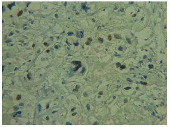
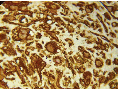
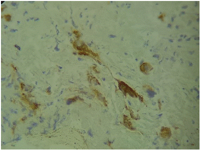
The patient consulted the maxillofacial service where he benefited from a biopsy, the pathological analysis of this granuloma concluded to a chondrosarcoma. Immunomarkings showed P63, EMA, AML, h caldesmone, and vimentin expression (Figure 1).

Figure 1a: Pathological slide showing the expression of P63.

Figure 1b: Pathological slide showing the expression of VIMENTINE.

Figure 1c: Pathological slide showing the expression of EM.
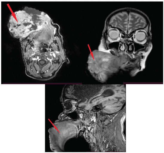
A head and neck MRI showed a voluminous exophytic process centered on the right jugal region invading the right mandibular body, without locoregional cervical lymphadenopathy
The surgery was recovered from the mandibular invasion, a palliative chemotherapy was then decided (Figure 2).

    Figure 2: Axial, coronal and saggital MRI sections showing a voluminous

exophytic process centered on the right jugal region.
The evolution was extremely raid, the patient passe away few days after the diagnosis.
Discussion
Carcinosarcoma is a rare tumour expressing biphasic features (mesenchymal and epithelial) with distinguished hallmarks of malignancy [4]. Instead of its previous names, pseudosarcoma, spindle cell carcinoma and sarcomatoid carcinoma, Virchow first coined the term “Carcinosarcoma” [4]. Amongst head and neck region, it commonly involves salivary glands, larynx, oral cavity, hypo pharynx, pyriform fossa, Sino nasal tract and oropharynx [5].
The occurrence of oral cavity carcinosarcoma in a 64-yearold patient is not exceptional. The age of occurrence of carcinosarcomas is located after 50 years for the quasi-totalitis ´ of the lesions described [5].
The diagnosis is suspected clinically first and then confirmed histologically. The purpose of imaging is to make the assessment of locoregional extension and post-therapeutic monitoring.
Carcinosarcoma is known to have grave prognosis. Lethality has been as high as 60% overall and 42% in 30months.The outcome has been reported to be influenced by gross morphology and size of the tumour, location, carcinomatous differentiation, depth of invasion and stage of the disease. Local recurrence and metastasis are common [6]. Metastasis is most common in lungs Primary surgery followed with adjuvant radiotherapy remains the cornestone of treatment carcinosarcoma.
References
- Stojadinovic S, Reinert S, Philippou ST, Machtens E. Carcinosarcoma of the mandibular mucosa: the first report. Int J Oral Maxillofac Surg. 2002; 31: 444-447.
- Thamilselvi R, Subramaniam PM, Shivarudrappa AS, Venugeethan A, Sinha P. Sarcomatoid salivary duct carcinoma of minor salivary gland: a rare case. Indian J Pathol Microbiol. 2010; 53: 808-810.
- King OH. Carcinosarcoma of accessory salivary gland. First report of a case. Oral Surg Oral Med Oral Pathol. 1967; 23: 651-659.
- Furuta Y, Nojima T, Terakura N, Fukuda S, Inuyama Y. A rare case of carcinosarcoma of the maxillary sinus with osteosarcomatous differentiation. Au-ris Nasus Larynx. 2001; 28: S127-S129.
- Olsen KD, Lewis JE, Suman VJ. Spindle cell carcinoma of the larynx and-hypopharynx. Otolaryngology-Headand Neck Surgery. 1997; 116: 47-52.
- Goellner JR, Devine KD,Weiland LH. Pseudosarcoma of the larynx. Am J Clin Pathol. 1973; 59: 312-326.